
IBD top toe Aida Kapic Lunder MD Research
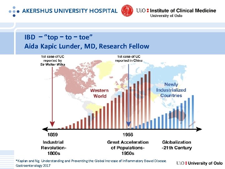
IBD – ”top – toe” Aida Kapic Lunder, MD, Research Fellow *Kaplan and Ng. Understanding and Preventing the Global Increase of Inflammatory Bowel Disease. Gastroenterology 2017
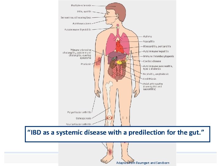
“IBD as a systemic disease with a predilection for the gut. ” Human touch and empathy – with professional skill Adapted from Baumgart and Sandborn
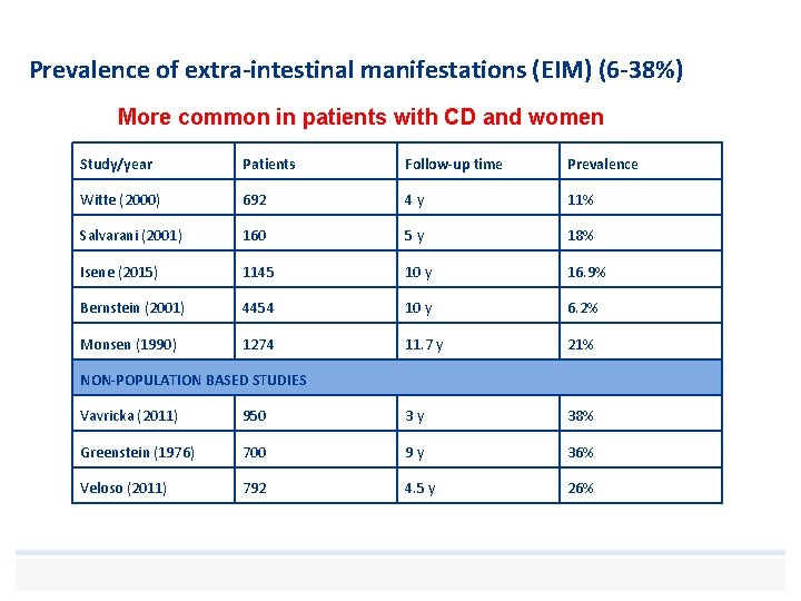
Prevalence of extra-intestinal manifestations (EIM) (6 -38%) More common in patients with CD and women Study/year Patients Follow-up time Prevalence Witte (2000) 692 4 y 11% Salvarani (2001) 160 5 y 18% Isene (2015) 1145 10 y 16. 9% Bernstein (2001) 4454 10 y 6. 2% Monsen (1990) 1274 11. 7 y 21% NON-POPULATION BASED STUDIES Vavricka (2011) 950 3 y 38% Greenstein (1976) 700 9 y 36% Veloso (2011) 792 4. 5 y 26%
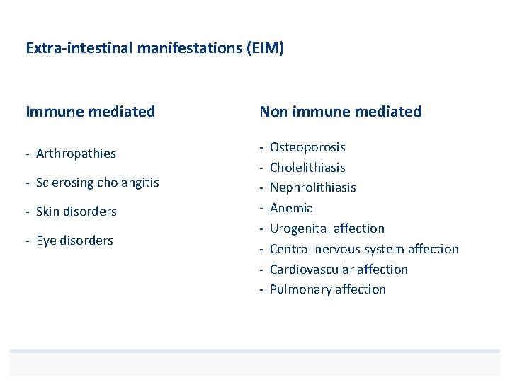
Extra-intestinal manifestations (EIM) Immune mediated Non immune mediated - Arthropathies - - Sclerosing cholangitis - Skin disorders - Eye disorders Osteoporosis Cholelithiasis Nephrolithiasis Anemia Urogenital affection Central nervous system affection Cardiovascular affection Pulmonary affection
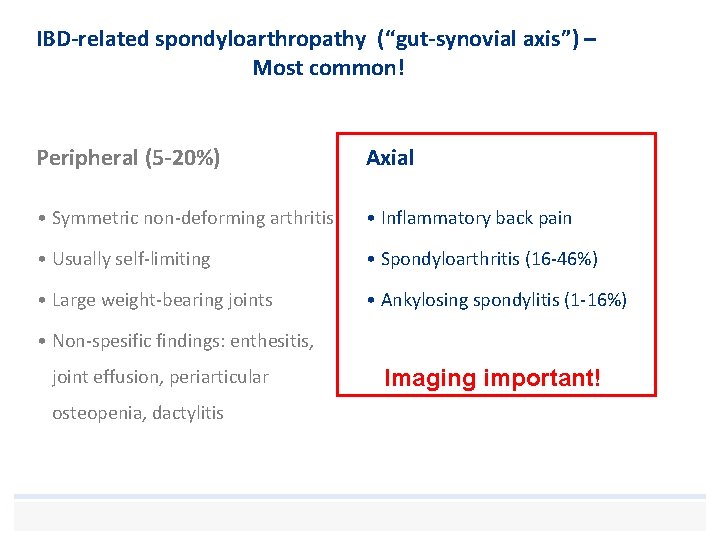
IBD-related spondyloarthropathy (“gut-synovial axis”) – Most common! Peripheral (5 -20%) Axial • Symmetric non-deforming arthritis • Inflammatory back pain • Usually self-limiting • Spondyloarthritis (16 -46%) • Large weight-bearing joints • Ankylosing spondylitis (1 -16%) • Non-spesific findings: enthesitis, joint effusion, periarticular osteopenia, dactylitis Imaging important!
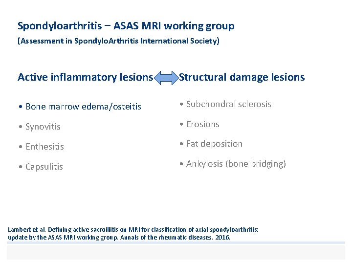
Spondyloarthritis – ASAS MRI working group (Assessment in Spondylo. Arthritis International Society) Active inflammatory lesions Structural damage lesions • Bone marrow edema/osteitis • Subchondral sclerosis • Synovitis • Erosions • Enthesitis • Fat deposition • Capsulitis • Ankylosis (bone bridging) Lambert et al. Defining active sacroiliitis on MRI for classification of axial spondyloarthritis: update by the ASAS MRI working group. Annals of the rheumatic diseases. 2016.
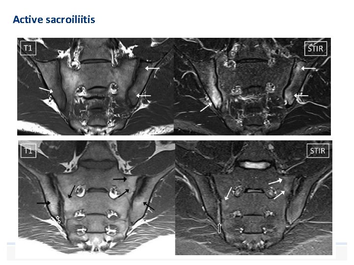
Active sacroiliitis
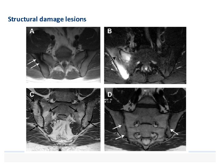
Structural damage lesions
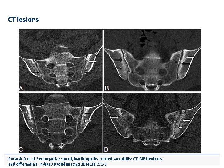
CT lesions Prakash D et al. Seronegative spondyloarthropathy-related sacroiliitis: CT, MRI features and differentials. Indian J Radiol Imaging 2014; 24: 271 -8
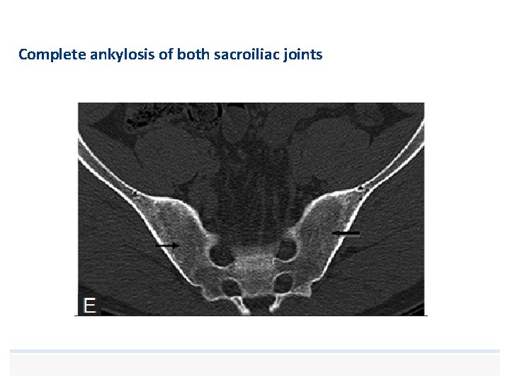
Complete ankylosis of both sacroiliac joints
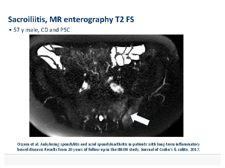
Sacroiliitis, MR enterography T 2 FS • 57 y male, CD and PSC Ossum et al. Ankylosing spondylitis and axial spondyloarthritis in patients with long-term inflammatory bowel disease: Results from 20 years of follow-up in the IBSEN study. Journal of Crohn's & colitis. 2017.
Ankylosing spondylitis (1 -16%) • Diagnosed when both the • clinical criteria (back pain, stiffness and/or limitation of motion) and • the imaging criteria for sacroiliitis (grade 2 or more bilaterally or grade 3 -4 unilaterally) • are met.
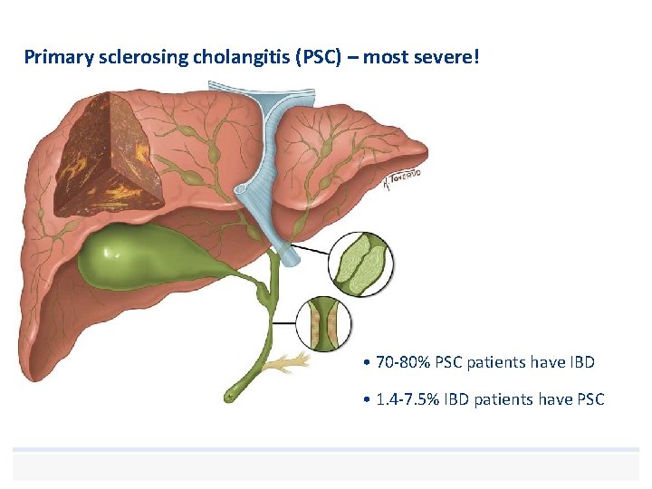
Primary sclerosing cholangitis (PSC) – most severe! • 70 -80% PSC patients have IBD • 1. 4 -7. 5% IBD patients have PSC
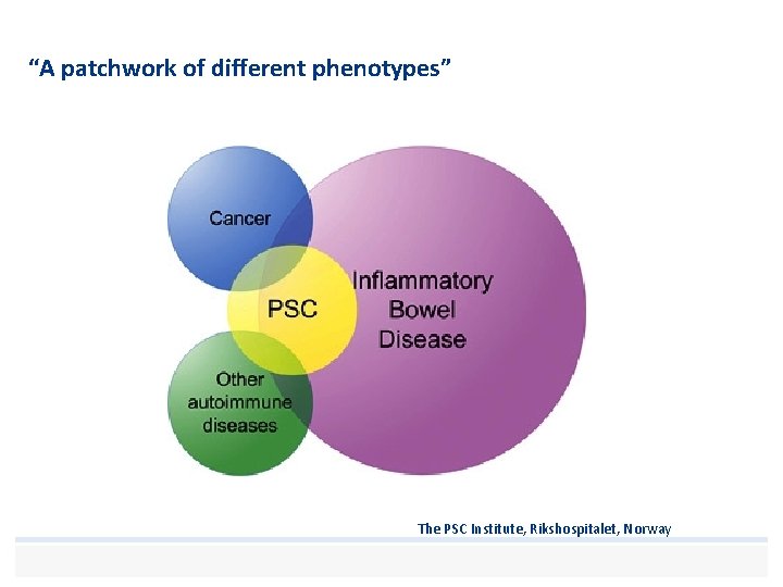
“A patchwork of different phenotypes” The PSC Institute, Rikshospitalet, Norway
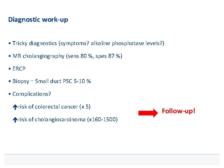
Diagnostic work-up • Tricky diagnostics (symptoms? alkaline phosphatase levels? ) • MR cholangiography (sens 80 %, spes 87 %) • ERCP • Biopsy – Small duct PSC 5 -10 % • Complications? risk of colorectal cancer (x 5) risk of cholangiocarcinoma (x 160 -1500) Follow-up!
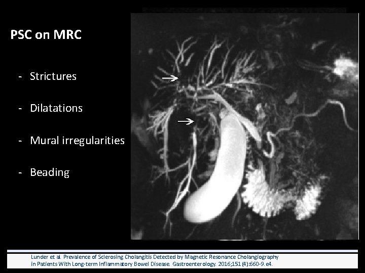
PSC on MRC - Strictures - Dilatations - Mural irregularities - Beading Lunder et al. Prevalence of Sclerosing Cholangitis Detected by Magnetic Resonance Cholangiography in Patients With Long-term Inflammatory Bowel Disease. Gastroenterology. 2016; 151(4): 660 -9. e 4.
CCA – 5 -15 % life time incidence in patients with PSC! • Overlapping imaging findings • Look for: – Focal thickening – Focal Biliary dilatation – Capsular retractions – Portal vein invasion – Fibrous tissue in CCA delayed enhancement
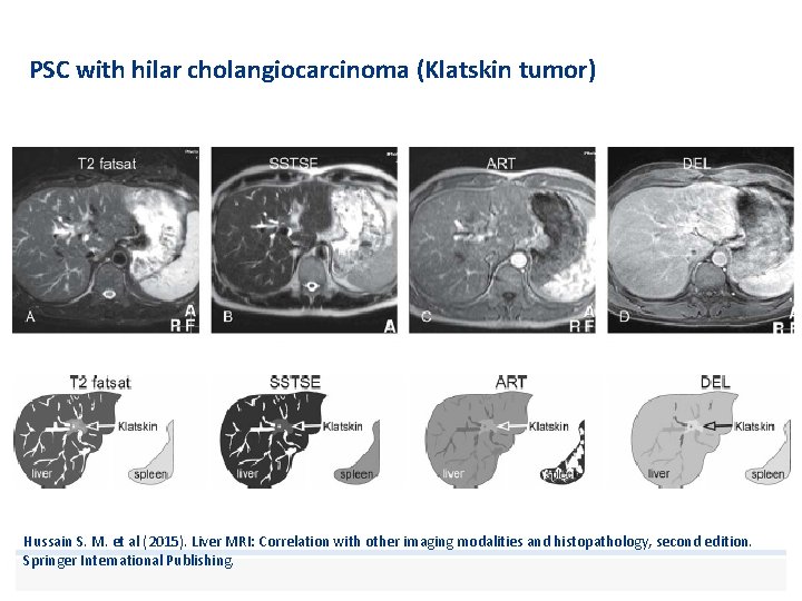
PSC with hilar cholangiocarcinoma (Klatskin tumor) Hussain S. M. et al (2015). Liver MRI: Correlation with other imaging modalities and histopathology, second edition. Springer International Publishing.
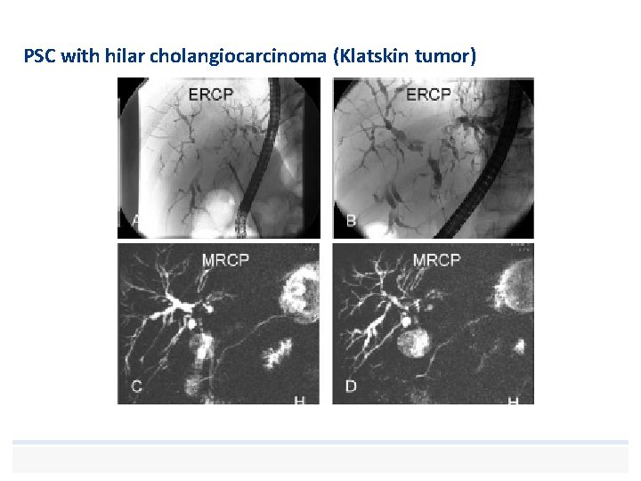
PSC with hilar cholangiocarcinoma (Klatskin tumor)
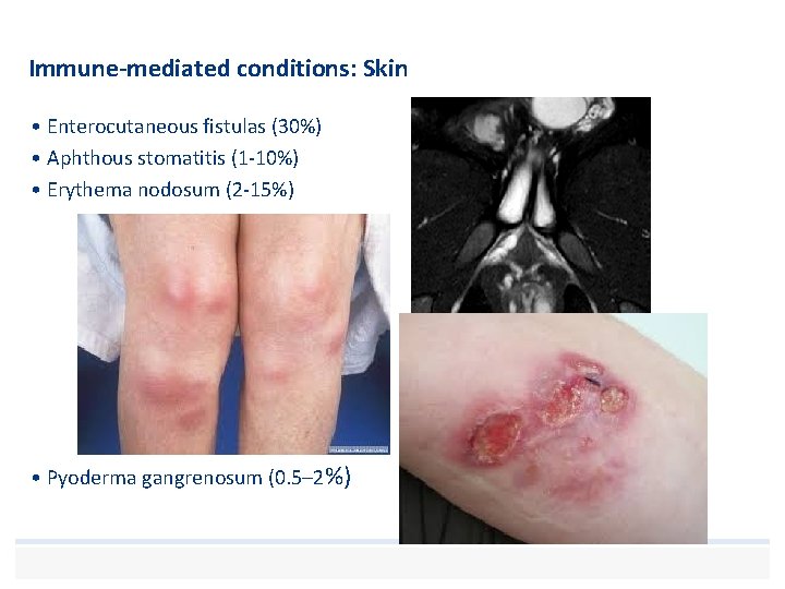
Immune-mediated conditions: Skin • Enterocutaneous fistulas (30%) • Aphthous stomatitis (1 -10%) • Erythema nodosum (2 -15%) • Pyoderma gangrenosum (0. 5– 2%)
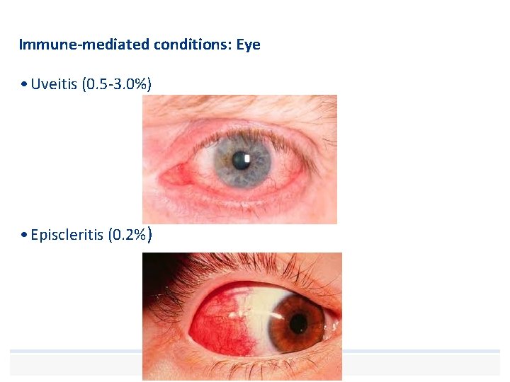
Immune-mediated conditions: Eye • Uveitis (0. 5 -3. 0%) • Episcleritis (0. 2%)
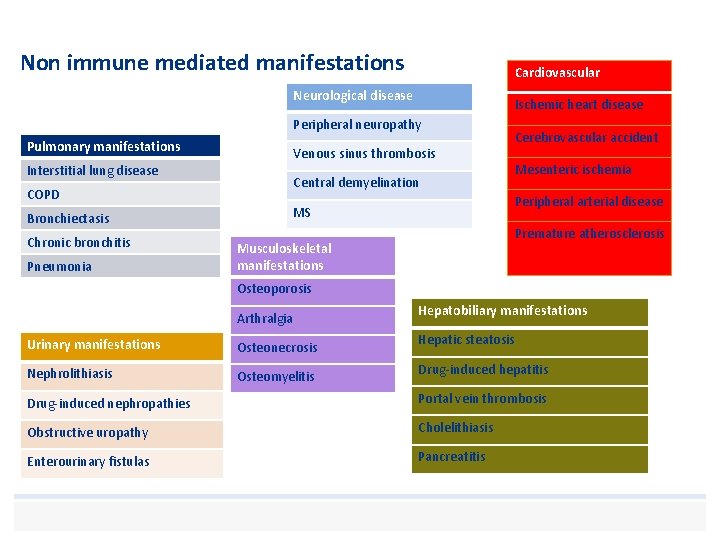
Non immune mediated manifestations Cardiovascular Neurological disease Ischemic heart disease Peripheral neuropathy Pulmonary manifestations Venous sinus thrombosis Interstitial lung disease Central demyelination COPD Chronic bronchitis Pneumonia Mesenteric ischemia Peripheral arterial disease MS Bronchiectasis Cerebrovascular accident Premature atherosclerosis Musculoskeletal manifestations Osteoporosis Arthralgia Urinary manifestations Osteonecrosis Nephrolithiasis Osteomyelitis Hepatobiliary manifestations Hepatic steatosis Drug-induced hepatitis Drug-induced nephropathies Portal vein thrombosis Obstructive uropathy Cholelithiasis Enterourinary fistulas Pancreatitis
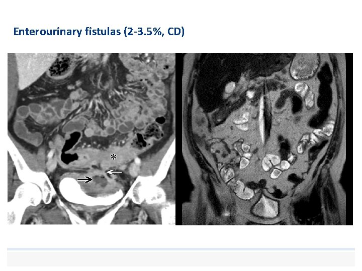
Enterourinary fistulas (2 -3. 5%, CD)
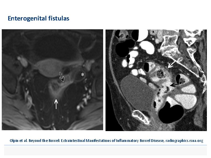
Enterogenital fistulas Olpin et al. Beyond the Bowel: Extraintestinal Manifestations of Inflammatory Bowel Disease, radiographics. rsna. org
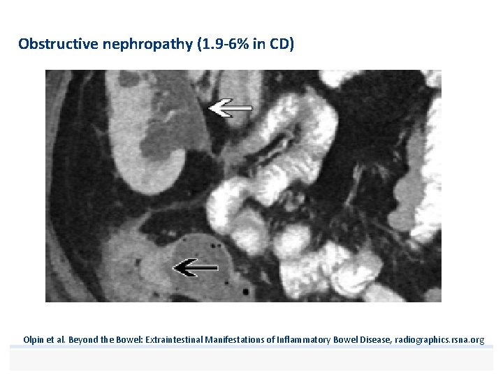
Obstructive nephropathy (1. 9 -6% in CD) Olpin et al. Beyond the Bowel: Extraintestinal Manifestations of Inflammatory Bowel Disease, radiographics. rsna. org
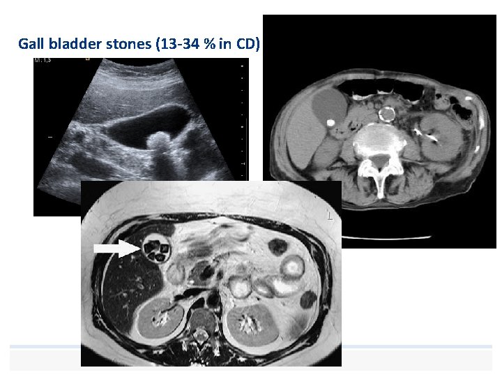
Gall bladder stones (13 -34 % in CD)
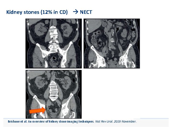
Kidney stones (12% in CD) NECT Brisbane et al. An overview of kidney stone imaging techniques. Nat Rev Urol. 2016 November.
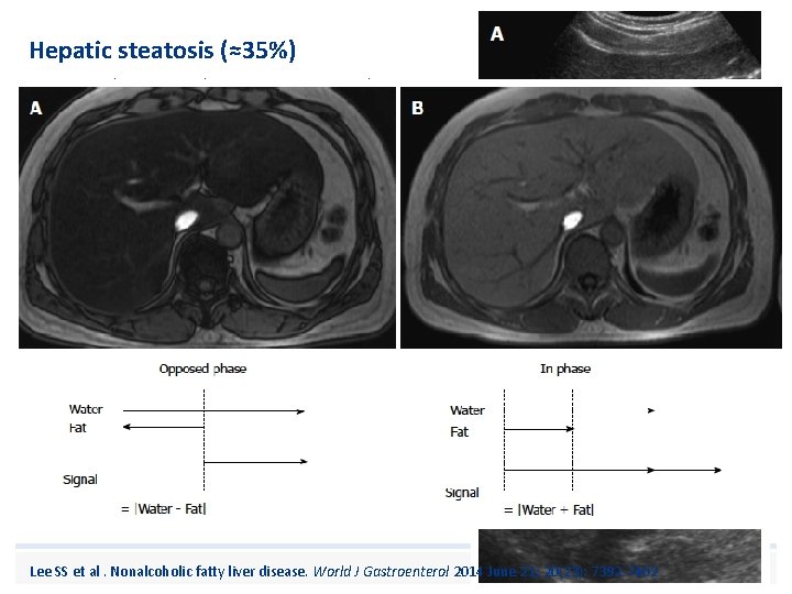
Hepatic steatosis (≈35%) • Due to chronic malnutrition, protein loss, corticosteroid therapy? Lee SS et al. Nonalcoholic fatty liver disease. World J Gastroenterol 2014 June 21; 20(23): 7392 -7402
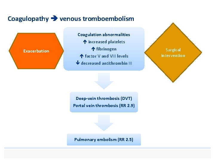
Coagulopathy venous tromboembolism Coagulation abnormalities increased platelets Exacerbation fibrinogen factor V and VIII levels decreased antithrombin III Deep-vein thrombosis (DVT) Portal vein thrombosis (RR 2. 9) Pulmonary embolism (RR 2. 5) Surgical intervention
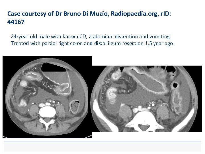
Case courtesy of Dr Bruno Di Muzio, Radiopaedia. org, r. ID: 44167 24 -year old male with known CD, abdominal distention and vomiting. Treated with partial right colon and distal ileum resection 1, 5 year ago.
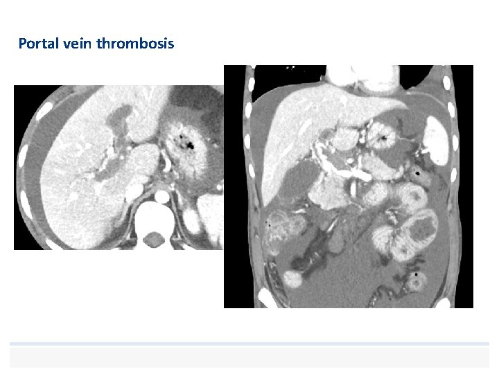
Portal vein thrombosis
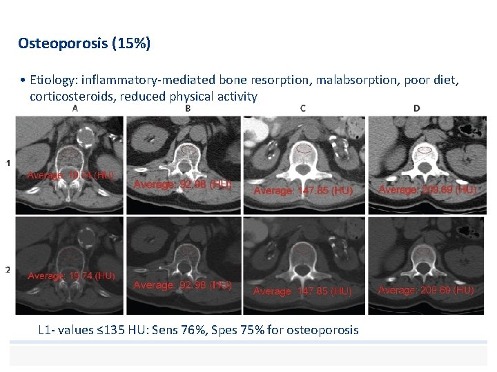
Osteoporosis (15%) • Etiology: inflammatory-mediated bone resorption, malabsorption, poor diet, corticosteroids, reduced physical activity • Underdiagnosed. . . L 1 - values ≤ 135 HU: Sens 76%, Spes 75% for osteoporosis
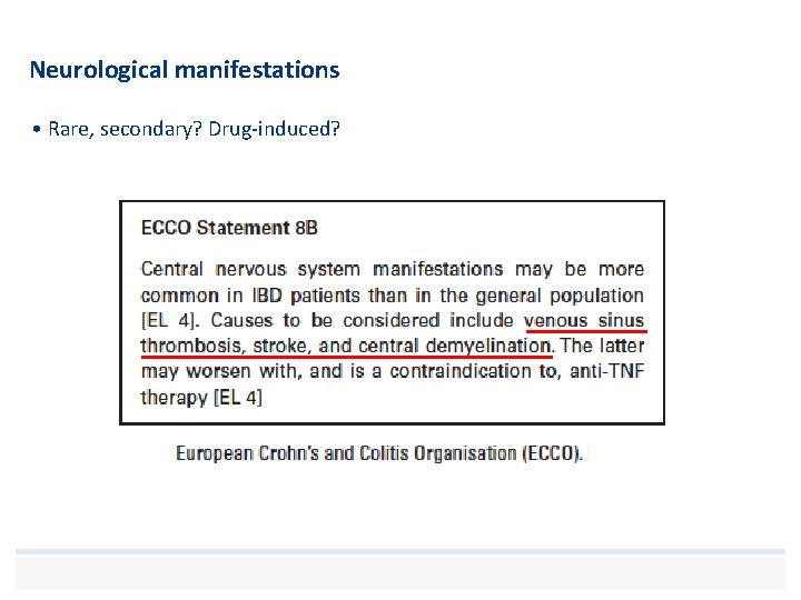
Neurological manifestations • Rare, secondary? Drug-induced?
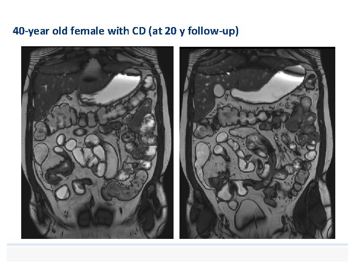
40 -year old female with CD (at 20 y follow-up)
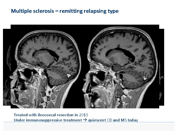
Multiple sclerosis – remitting relapsing type Treated with ileocoecal resection in 2013 Under immunosuppressive treatment quiescent CD and MS today
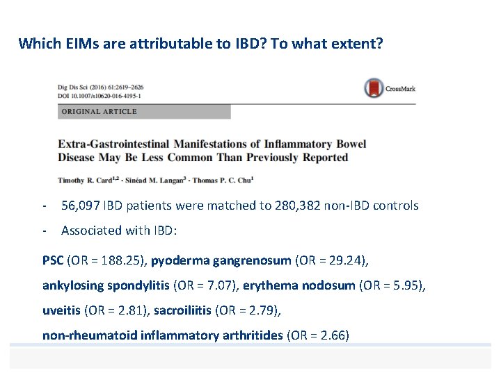
Which EIMs are attributable to IBD? To what extent? - 56, 097 IBD patients were matched to 280, 382 non-IBD controls - Associated with IBD: PSC (OR = 188. 25), pyoderma gangrenosum (OR = 29. 24), ankylosing spondylitis (OR = 7. 07), erythema nodosum (OR = 5. 95), uveitis (OR = 2. 81), sacroiliitis (OR = 2. 79), non-rheumatoid inflammatory arthritides (OR = 2. 66)
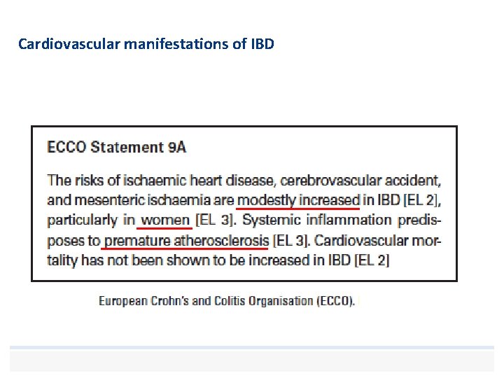
Cardiovascular manifestations of IBD
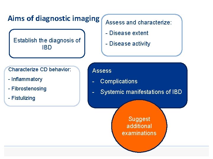
Aims of diagnostic imaging Assess and characterize: - Disease extent Establish the diagnosis of IBD - Disease activity Characterize CD behavior: Assess - Inflammatory - Complications - Fibrostenosing - Fistulizing - Systemic manifestations of IBD Suggest additional examinations
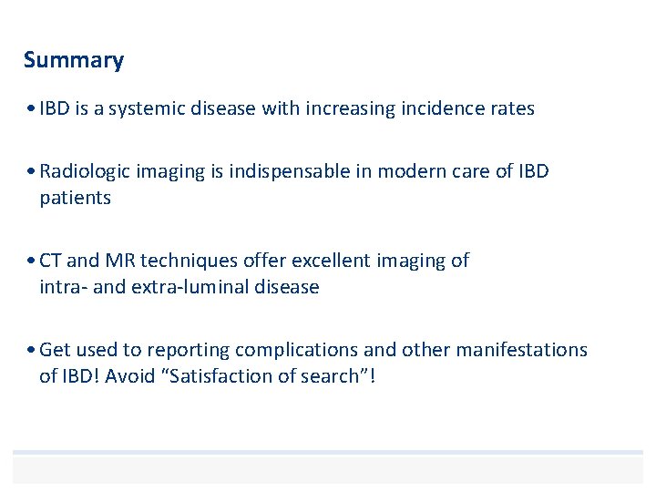
Summary • IBD is a systemic disease with increasing incidence rates • Radiologic imaging is indispensable in modern care of IBD patients • CT and MR techniques offer excellent imaging of intra- and extra-luminal disease • Get used to reporting complications and other manifestations of IBD! Avoid “Satisfaction of search”!
References • Card TR, Langan SM, Chu TP. Extra-Gastrointestinal Manifestations of Inflammatory Bowel Disease May Be Less Common Than Previously Reported. Digestive diseases and sciences. 2016; 61(9): 2619 -26. • Harbord M, Annese V, Vavricka SR, Allez M, Barreiro-de Acosta M, Boberg KM, et al. The First European Evidence-based Consensus on Extra-intestinal Manifestations in Inflammatory Bowel Disease. Journal of Crohn's & colitis. 2016; 10(3): 239 -54. • Kaplan GG, Ng SC. Understanding and Preventing the Global Increase of Inflammatory Bowel Disease. Gastroenterology. 2017; 152(2): 313 -21. e 2. • Baumgart DC, Sandborn WJ. Crohn's disease. Lancet (London, England). 2012; 380(9853): 1590 -605. • Olpin et al. Beyond the Bowel: Extraintestinal Manifestations of Inflammatory Bowel Disease, radiographics. rsna. org • Pickhardt PJ, Pooler BD, Lauder T, del Rio AM, Bruce RJ, Binkley N. Opportunistic Screening for Osteoporosis Using Abdominal Computed Tomography Scans Obtained for Other Indications. Ann Intern Med. 2013; 158: 588– 595. doi: 10. 7326/0003 -4819158 -8 -201304160 -00003 • Brisbane et al. An overview of kidney stone imaging techniques. Nat Rev Urol. 2016 November.
References • Ossum AM, Palm O, Lunder AK, Cvancarova M, Banitalebi H, Negard A, et al. Ankylosing spondylitis and axial spondyloarthritis in patients with long-term inflammatory bowel disease: Results from 20 years of follow-up in the IBSEN study. Journal of Crohn's & colitis. 2017. • Lambert RG, Bakker PA, van der Heijde D, Weber U, Rudwaleit M, Hermann KG, et al. Defining active sacroiliitis on MRI for classification of axial spondyloarthritis: update by the ASAS MRI working group. Annals of the rheumatic diseases. 2016. • Radiopaedia • Lunder AK, Hov JR, Borthne A, Gleditsch J, Johannesen G, Tveit K, et al. Prevalence of Sclerosing Cholangitis Detected by Magnetic Resonance Cholangiography in Patients With Long-term Inflammatory Bowel Disease. Gastroenterology. 2016; 151(4): 6609. e 4. • Hussain, S. M. et al (2015). Liver MRI: Correlation with other imaging modalities and histopathology, second edition. Springer International Publishing. • Prakash D et al. Seronegative spondyloarthropathy-related sacroiliitis: CT, MRI features and differentials. Indian J Radiol Imaging 2014; 24: 271 -8 • Lee SS et al. Nonalcoholic fatty liver disease. World J Gastroenterol 2014 June 21; 20(23): 7392 -7402
Thanks to the Abdominal Radiology Section!
Thank you! Human touch and empathy – with professional skill
- Slides: 43