The Upper Extremity Commonly Encountered Injuries Rhonda Feldman


")


 – think about “Frozen Shoulder”")


- Slides: 54
The Upper Extremity Commonly Encountered Injuries Rhonda Feldman, MSS, MHS, PA-C
Common Etiologies SHOULDER PAIN
Shoulder Pain • 16 - 34% of general population suffers from shoulder pain • Rotator Cuff pathology comprises significant portion • 11. 2 cases per 1000 patients per year (source – Up. To. Date)
Shoulder Landmarks
AP Shoulder
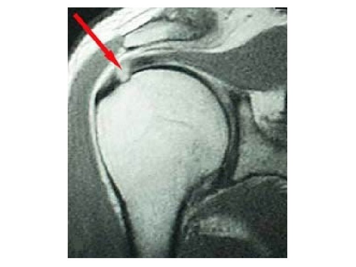
Rotator Cuff • Comprised of 4 tendons – supraspinatus, infraspinatus, subscapularus, teres minor • Inflammation of surrounding bursa will mimic the pain of a sprain (tear) • Mechanism of Injury – reaching up or out, especially in a weight bearing situation or falling with a strike to elbow or outstretched arm
Rotator Cuff Pathology Impingment Tear/Sprain
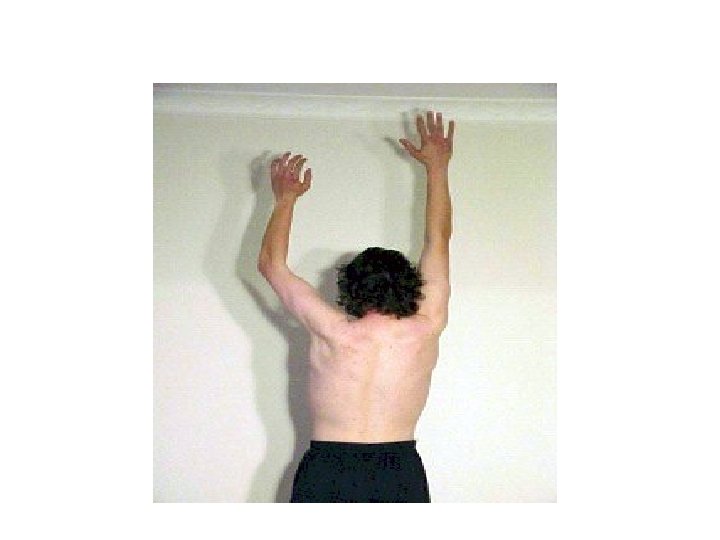
Clinical Signs/Symptoms • • Achy Shoulder and Arm Loss of Strength (marked with tear) Pain with reaching out or across Decreased range of motion with persistent pain will lead to “Frozen Shoulder”
Diagnosis of RC Tear • First Step – Clinical Exam – Strength Testing • 2 nd Step – X-ray – will not show tear, but will rule out bony problem (avascular necrosis, fx, dislocation) • 3 rd Step – MRI – no need to MRI if strength is 5/5 – try conservative approach first – PT, steroid injection
Clinical Keys to Diagnosis • Make sure you are testing true strength and not allowing patient’s poor effort to influence measurement • If strength is maintained, RC tear is tiny (nonsurgical) or there is no tear
Red Flags • Severely diminished Range of Motion (ROM) – think about “Frozen Shoulder” • RC sprains (tears) hurt, but Frozen Shoulder is agonizing! • Numbness, tingling, loss of sensation – it’s not the RC – think cervical nerve issue • Loss of tone, atrophy of muscle – think neurological issue
Diagnostic Pearl Hawkins Test – eliciting pain with this test indicates that the rotator cuff is the etiology of Shoulder Pain. Further muscular testing will indicate which tendon is involved or whether pain is from inflammation, not tear
What’s This?
Anterior Dislocation
Shoulder Dislocation • The Shoulder is the most common joint in the body to dislocate • Most common dislocation is an anterior dislocation of the humeral head • Dislocations may range from subluxation to full • Repeated dislocations result in chronic instability
Brachial Plexus • Definition – a network of nerves that communicates signals from the spine to innervate the upper extremity • Common injuries – birth, trauma, tumors or inflammation
Common Mechanism of Injury for Brachial Plexus
Diagnosis • Suspicion from patient history – vehicle accident, stab, gunshot, inflammatory process, compression • Review of Systems reveals pain, loss of sensation and strength, paralysis • Clinical Exam reveals biceps weakness, loss of reach, obvious loss of function in infant • Nerve testing, CT or MRI
Red Flags • Difficult to determine which injury will spontaneously heal • Paralysis means immediate neurosurgical evaluation • If it looks like a BPI, but history supports no trauma – it may be inflammatory – this means you must determine what is the cause of the inflammation – is something happening systemically?
AC Separation
Acromioclavicular Separation
AC Separation Diagnosis • History – acute injury, common in athletes, common in falls • Clinical Exam – obvious deformity at distal clavicle, pain on palpation (may be minor) • Neurological, Sensory, and Vascular exam is intact • X-ray to confirm
Medial & Lateral Epicondylitis ELBOW PAIN
Lateral Epicondylitis
Medial Epicondylitis
Diagnosis Of Epicondylitis • History – review onset of pain – and ask about recent activity – this is typically an over-use issue • Will describe increased pain with grasping • Exam – very tender over epicondyle
Arm & Elbow • Over-use very common etiology • Trauma – dislocations & radial head fractures most common
It’s All About the Angles
AP Elbow
Lateral Elbow
Lateral Epicondylitis
Medial Epicondylitis
Clinical Pearl Pressing down on index and long finger with arm in extension causes pain in the area of the lateral epicondyle
Olecranon Bursitis Swelling and fluid accumulation of the bursa lying over the olecranon. May become very large, may be painful or not. Very commonly seen in primary care.
Radial Head Fracture Very Common Fracture Commonly Missed! Pain with palpation over radial head Pain with supination and pronation
Carpal Tunnel Syndrome, CMC OA WRIST AND HAND COMPLAINTS
Structure – X-ray
Hand Wrist Complaints • Carpal Tunnel Syndrome the most common entrapment neuropathy – affects 3 -6% of general population (ref – American Family Physician) • CMC Thumb Osteoarthritis – very common, women>men, typically wear and tear • Contractions – common, often without injury or clear etiology
Carpal Tunnel Syndrome
Clinical Pearls – Testing for CTS Tinels Sign Phalens Test
CMC Osteoarthritis Loss of Joint Space Destruction of Bony Articulating Surfaces May include cyst formation Pain and decreased range of motion are clinical hallmarks
Trigger Finger
Dupuytren’s Contracture
Boxer’s Fracture Most Common Fracture of the Metacarpals Mechanism of injury – striking blow with fist Clinical hallmarks – loss of knuckle height, pain over metacarpal, deformity
Scaphoid Fracture Frequently missed fracture Must palpate “anatomic snuffbox” Very poor vascular supply =very poor clinical outcome if missed
Questions?