Muscles and Movements of Lower Extremity Ch 8


- Slides: 35

Muscles and Movements of Lower Extremity – Ch 8 Objectives • Explain how anatomical structure affects movement capabilities of lower extremity articulations. • Identify factors influencing the relative mobility and stability of lower extremity articulations. • Explain the ways in which the lower extremity is adapted to its weightbearing function. • Identify muscles that are active during specific lower extremity movements. • Describe the biomechanical contributions to common injuries of the lower extremity.

Lower Extremity Outline • Hip Joint – Structure , Loads, and Muscles and Movements • Knee Joint – Structure, Loads, Muscles and Movements – Common knee injuries – patellar chondromalacia (a. k. a. runners knee) and anterior cruciate tear • Ankle Joint – Structure, Muscles and Movements – Common ankle and foot injuries - plantar fascitis, pronated feet • Misalignment problems of lower extremity • Websites • Homework

Hip joint structure: Front View

Loads on the Hip • During swing phase of walking: – Compression forces on hip greater than body weight (due to muscle tension) 250% body weight (BW) • Increases with hard-soled shoes • Increases with gait increases (both support and swing phase) (500% BW when running 3. 5 m/s) • Body weight, impact forces translated upward thru skeleton from feet and muscle tension contribute to compressive load on hip.

Compressive forces on hip jt Socket while walking may exceed 3 to 4 times body wt, 5 -6 times bw while jogging, and 8 -9 times bw while stumbling Use law of cosines:

Hip Joint Muscles • Uni-articular muscles – Flexion - iliopsoas – Extension - gluteus maximus – Abduction - gluteus medius and minimus – Adduction - adductor brevis, longus, & magnus • Biarticular muscles – Hip flexion, knee flexion - sartorius – Hip flexion, knee extension - rectus femoris – Hip extension, knee flexion - hamstrings • Note passive and active insufficiency of biarticular muscles

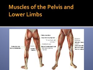
Muscles of Lower Extremity:

Hip Jt Muscle Vectors:

Thigh muscles in cross section:

Physiological crosssectional area (PCSA) of hip jt muscles Why are lateral rotators & gluteii muscles so large?

Common Injuries of the Hip • Fractures – Usually of femoral neck, a serious injury usually occurring in elderly with osteoporosis • Contusions – Usually in anterior aspect of thigh, during contact sports • Strains – Usually to hamstring during sprinting or overstriding

Knee Joint • Ligaments and cartilage – medial and lateral collateral ligaments – anterior and posterior cruciate ligaments – medial and lateral meniscus • Muscles and movements • Extensors • quadriceps femoris (rectus femoris, vastus lateralis, vastus medialis, vastus intermedius) – Flexors • hamstrings (semitendinosus, semimembranosus, biceps femoris)

Knee Joint Structure: 25% of Alpine skiing injuries are ligament injuries Peripatellar pain (runner’s knee) caused by imbalance of stress on patella

Lower Extremity Misalignment: Q angle is larger in females due to Wider hip structure, increasing potential for PFPS (Patellofemoral pain syndrome)

Quadriceps Tendon and Patella Force Lines Compressive force at PFJ is ½ body wt during normal walking, and over 3 times bw during stair climbing Comp force increases as knee flexion Angle increases

Cruciate Ligaments and Shear Stress

Loads on Knee • Forces at tibiofemoral Joint – Shear stress is greater during open kinetic chain exercises such as knee extensions and knee flexions – Compressive stress is greater during closed kinetic chain exercises such as squats and weight bearing exercises. • Forces at Patellofemoral Joint – With a squat, reaction force is 7. 6 times BW on this joint. • Beneficial to rehab of cruciate ligament or patellofemoral surgery

Thigh muscles in cross section:

PCSA of Muscles Crossing Knee

Common Injuries of the Knee and Lower Leg • • Patellofemoral Pain Syndrome Shin Splints Anterior cruciate ligament (ACL) injuries Posterior cruciate ligament (PCL) injuries Medial collateral ligament (MCL) injuries Meniscus Injuries Iliotibial Band Friction Syndrome Breaststroker’s Knee

Foot and Ankle joint structure • Bones and arches of foot • Tibia, fibula, calcaneus, talus, other tarsals, metatarsals, phalanges – Longitudinal arch, transverse arch – plantar fascia • Movements of ankle - talocrural joint • Movements of foot - subtalar, intertarsal, intermetatarsal, interphalangeal

Bones of Shank and Foot:

Ankle Joint Muscles and Movements • Anterior compartment - All dorsiflex – Tibialis anterior (also inverts) – Extensor digitorum longus (also everts) • Posterior compartment - All plantar flex – Tibialis posterior (also inverts), gastrocnemius (also flexes knee), & soleus • Lateral compartment - All plantar flex & evert – Peroneus longus & brevis • Foot pronation and supination

Ankle and Foot Muscles:

Percent PCSA of Muscles Crossing Ankle

Subtalar Axis:

Foot Pronation and Tibial Torsion:

Rearfoot Movement During Running:

Plantar Fascium • What is the plantar fascium? - attaches to calcaneus posteriorly and to the first row of phalanges anteriorly • What is its function? – passive intertarsal stabilization

Arches of the Foot:

Plantar Fascium: Plantar fascitis is 4 th most common cause of pain among runners (1 st – knee pain, 2 nd – shin splints, 3 rd- achilles tendonitis)

Plantar Fascitis – 4 th leading cause of pain in runners • What causes plantar fascitis(inflamation of plantar fascium)? – anatomic anomalies • • microtears in fascium and bone spurs inadequate flexibility of plantar flexors inadequate strength of plantar flexors functional pronation (eversion and abduction) – overuse • • overweight poorly designed and poorly fitted shoes running and jumping on hard surfaces sudden increase in stress • Treatment – remove the cause(s) – Therapeutic treatment to promote body’s natural healing • NSAIDS • Intermittent ice and heat • Ultrasound, diathermy, massage

Patellar Chrondomalacia (a. k. a. Runner’s Knee) – leading cause of pain in runners) • Primary cause is imbalance in forces on patella – Increased Q angle – Pronated feet • Tissues affected – Degrading of articular cartilage of patella & femoral condyles – Fluid collection, causing joint stiffness • Symptoms – Pain around patella with no particular injury causing it – Worse going upstairs and downstairs, or after sitting awhile – Feels like knee needs to be stretched • Prevention/treatment – Surgery is seldom beneficial – Wet test – walk with wet feet on floor and determine if you have a hypermobile foot. If so, purchase shoes and/or orthotics to decrease degree of foot pronation – Exercises to increase strength/endurance of vastus medialis

Runner’s knee, cont’d Wet test: Safe exercise to develop vasti muscles Do not use knee sleeves! Do not bend knee more than 20 -30 degrees while doing extensions with resistance!

Websites for Muscles, Movements, & Problems of Lower Extremity • Steadman-Hawkins Sports Medicine Clinic – Shoulder joint impingement syndrome – Carpal tunnel syndrome – Patellofemoral pain syndrome – Lumbar disc herniation Homework on lower extremity: (Due Wed, Oct 20) Introductory problems, p 263: 9, 10 Additional problems, p 263 -264: 6
 Lower extremity muscles
 Femoral pulse
 D2 flexion
 Left lower extremity
 Popliteal pulse location
 Dorsalis pedis pulse artery
 Salah muscle
 Lower extremity appendicular skeleton
 Eye muscles movement
 Thumb flexion muscles
 Definition of axial movement
 Diploic veins
 Anterior leg muscles
 Superficial vein of upper limb
 Mummy restraint meaning
 Upper extremity pnf patterns
 Closed kinetic chain definition
 Psoas insertion and origin
 Orrin franko md
 Hospital wristband colors
 Biomechanics of upper extremity
 Chapter 8 lifting and moving patients
 Yokochi
 Muscle movements types and names
 Attitudes and movements
 Locomotor movements dance
 Two types of physical
 Joseph stalin political movement and beliefs
 Japanese militarists aggressive actions
 Camera angle
 14.4 gases: mixtures and movements answers
 Temporary and permanent movements
 Bound and free movements
 Bound and free movements
 Camera types of shots
 Relaxed passive movement adalah

