Upper Extremity Strains and Sprains ORRIN FRANKO EAST


 Usually involves individuals with")


 Physical therapy")


")


 Pathology 30 – 50 years old Repetitive micro-trauma Chronic tear")
 Mechanism of Injury Overuse syndrome caused by repeated forceful wrist")
 Clinical Signs and Symptoms Increased pain around lateral epicondyle Tenderness")

 Pathology 30 - 50 years old Repetitive micro trauma to")
 Mechanisms of injury Throwing a baseball Racquetball or tennis Swimming")
 Clinical signs and symptoms Increased pain over medial epicondyle Tenderness")


 Caused by forced abduction of")


 Background 2% lifetime risk of developing At least 2 times")
 2% lifetime risk of developing Symptoms Painful triggering, often worse")
 Pathoanatomy")
 Pathoanatomy")
 Treatment Corticosteroid Injection 86% effective at 3 months 50% effective")


 When to Refer Patient does not want to undergo injection")
- Slides: 86
Upper Extremity Strains and Sprains ORRIN FRANKO EAST BAY HAND MEDICAL CENTER
www. EBHMC. com
Cell: 858 -337 -7149 www. ebhmc. com/referral
Table of Contents Shoulder Wrist AC dislocation De. Quervain tendonitis Shoulder dislocation Sprains Biceps tendonitis Elbow Fingers Lateral epicondylitis PIP joint dislocation/subluxation/sprain Medial epicondylitis Mallet finger / Central slip avulsion Thumb UCL rupture Trigger Finger
General Knowledge Sprains A sprain is a stretch or tear of a ligament. Ligaments connects one bone to another. Ligaments have poor blood supply (that’s why they are white!) so healing can take 3 -12 months
Grades Sprain Classification Grade 1: some stretching and some damage to the fibers Grade 2: A partial tearing with some subluxation Grade 3: Complete tears and dislocations
Signs and symptoms Pain Swelling Bruising Inflammation TREATMENT Grade 1 RICE Physical Therapy Grade 2 Bracing Grade 3 Surgery to repair the torn ligaments.
Strains A direct injury to muscles or tendons. Tendons connect muscles to bones Like ligaments, they have poor blood supply They are often under “high stress” and require modified activity or protection to heal
Symptoms Ø Pain Ø Muscle spasm Ø Muscle weakness Ø Swelling Ø Inflammation Ø Cramping TREATMENT ØRICE ØSpecific exercises to regain mobility ØSurgery
Strains
“Shoulder Separation” = AC Dislocation Def: A sprain of the acromioclavicular ligament Mechanis: shoulder Cyclists fall on the outstretched arm tip of the
Anterior View Posterior View
• Sx: Point tenderness over AC joint - inability or pain with abduction - in some cases, gross deformity • Tx: Immobilization with a sling - ice - NSAIDS - if grade III or above, refer to a surgeon - if grade I protective padding
Rotator Cuff Strain 3 degrees Most involve supraspinatus Tears usually at insertion on humerus
Rotator cuff strain
Mechanism Dynamic rotation of arm at high velocity (overhead throwing) Usually involves individuals with a history of impingement or instability
Signs & symptoms Pain w/ muscle contraction Tenderness over greater tuberosity Loss of strength Complete tear produces pain, loss of function, swelling and POT
Treatment RICE Decrease level of activity Exercises to strengthen rotator cuff
Biceps tendon rupture
Mechanism Direct blow Severe contraction of biceps
Signs & symptoms Pain and bruising through arm Deformity of biceps—balling up of muscle belly Pain with elbow flexion or supination
Treatment Ice Immobilization (short term) Physical therapy
Tendonitis Rotator cuff Biceps Common among athletes performing overhead motions due to overuse or muscle weakness
Mechanism Repetitive overhead motion causing inflammation of tendon
Signs & symptoms Tenderness to palpation Swelling Crepitus Pain with motion
Treatment Rest Ice Heat NSAIDS Stretching Strengthening Activity modification (reduction in pitching for adolescents)
Impingement syndrome Involves compression of supraspinatus tendon, subacromial bursa, long head of biceps tendon (all are under the coracoacromial arch)
Impingement
Impingement
Mechanism Repetitive overhead motions
Signs & symptoms Diffuse pain around the acromion process when arm is in overhead position External rotators are weak “+” impingement test Empty can test may increase pain Pinching sensation
Treatment RICE Restore normal biomechanics to shoulder Strengthen RC muscles and muscles that produce movement of scapula Stretch posterior and inferior joint capsule
Elbow
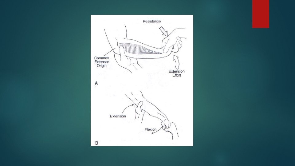
Lateral Epicondylitis (tennis elbow) Pathology 30 – 50 years old Repetitive micro-trauma Chronic tear in the origin of the extensor carpi radialis brevis
Lateral Epicondylitis (tennis elbow) Mechanism of Injury Overuse syndrome caused by repeated forceful wrist and finger movements Tennis players (rarely) Prolonged and rapid activities
Lateral Epicondylitis (tennis elbow) Clinical Signs and Symptoms Increased pain around lateral epicondyle Tenderness in palpation Tests AROM; PROM Resisted tests Lidocaine
Treatment of Tennis Elbow Time Steroid shot Time Therapy Time …and give it more time
Medial Epicondylitis (golfer’s elbow) Pathology 30 - 50 years old Repetitive micro trauma to common flexor tendon
Medial Epicondylitis (golfer’s elbow) Mechanisms of injury Throwing a baseball Racquetball or tennis Swimming backstroke Hitting a golf ball
Medial Epicondylitis (golfer’s elbow) Clinical signs and symptoms Increased pain over medial epicondyle Tenderness on palpation Tests AROM; PROM Resisted tests Lidocaine
Medial Overload Syndrome in Throwers Pathology Lateral joint line- compressive forces Shear forces posteriorly in olecranon fossa Tensile forces along medial joint line
Medial Overload Syndrome in Throwers Clinical signs and symptoms Persistent medial elbow soreness Arm fatigue is the 1 st indicator of impending injury Medial tenderness Elbow pain
Medial Overload Syndrome in Throwers: Treatment Pre throwing stretches Adequate gentle warm up with gradual increase to higher velocity throws Using safe pitch counts for adolescents Post throwing stretching ICE after throwing Surgical Intervention
Wrist
Wrist Sprain Mxn: Abnormal forced movement of the wrist Falling on hyperextended or hyperflexed wrist Violent torsion
Wrist Sprain
S/S: Pain Point tenderness Swelling Difficulty moving wrist—limited ROM
TX: RICE for mild/ moderate Physician referral to rule out fx for severe Splint if necessary Exercises for strengthening and ROM Tape for support
Treatment for wrist injuries
Wrist Tendonitis Mechanism: repetitive motion at wrist—usually in flexion/extension Seen more often in athletes involved ins sports with repetitive acceleration and deceleration i. e. weight lifters, rowers
TX: Ice Heat Analgesics Modify activity NSAIDS Splint Strengthening and ROM exercises
De. Quervain’s Tenosynovitis Tendonitis specifically to the 1 st Dorsal Compartment + Finklestein test 90% response to a single steroid injection Splinting temporarily may help NSAIDs can help Remaining require surgery for pain relief
De. Quervain’s Tenosynovitis
PIP Joint Dislocations / Subluxations Dorsal more common Simple dorsal dislocation: reduce, buddy tape Fracture-dislocation Splint in stable position Volar dislocation: Open reduction required in most cases
Reduction Techniques Digital block: 5 cc 1% lidocaine Volar at the digital crease – right in the middle One shot – works every time.
Reduction Techniques Re-create deformity, traction
Mallet Finger / Central Slip
MALLET FINGER
MALLET FINGER ANATOMY Dorsal avulsion Extensor digitorum tendon tear MECHANISM: Forced flexion of extended digit TREATMENT: No fracture: DIP extended for 6 -8 weeks FRACTURE: if <30% joint surface, splint x 4 weeks If >30% Might need ORIF Less than full passive extension? ? ? COMPLICATIONS: Pressure necrosis from splint Permanent extensor lag
Mallet Finger Presentation Pain at dorsal DIP joint Inability to actively extend the joint Characteristic flexion deformity On exam, very important to isolate the DIP joint to ensure extension from DIP and not the central slip If can’t passively extend consider bony entrapment All of these need x-rays
CENTRAL SLIP AVULSION ANATOMY Extensor digitorum communis tendon disruption Lateral bands migrate in volar direction MECHANISM: Volar-directed force on middle phalanx against semi-flexed finger attempting to extend
CENTRAL SLIP AVULSION EXAM: Pain, swelling over dorsal PIP in 15 -30 degrees flexion May have limited extension (better at 0 degrees than 30 degrees) TREATMENT Surgery if >30% joint surface involved with avulsion fx PIP splint in full extension 4 -5 weeks Protect 6 -8 weeks for sports *allow DIP to flex- relocates lateral bands COMPLICATIONS: Boutonierre deformity
Central Slip Extensor Tendon Injury. Boutonnière deformity PIP joint is forcibly flexed while actively extended Volar dislocation of the PIP joint Examine with PIP joint in 15 -30 degrees of flexion, can’t active extend but can passively extend Tenderness over dorsal aspect of the middle phalanx
Central Slip Extensor Tendon Injury Treatment A delay in proper treatment will cause boutonniere deformity Deformity can develop over several weeks or occasionally acutely Splint PIP in extension for 6 weeks Can still play sports
Central Slip Extensor Tendon Injury Avulsion fracture involving more than 30 percent of the joint Inability to achieve full passive extension
Volar Plate Injury Hyperextension, such as dorsal dislocation PIP is usually affected Collateral damage is often present The loss of joint stability can cause hyperextension deformity
VOLAR PLATE RUPTURE EXAM FINDINGS: Tender volar PIP Bruising, swelling MECHANISM: Hyperextension injury Ruptures distally from attachment at middle phalanx
VOLAR PLATE RUPTURE TREATMENT: Early mobilization Extension block splint Buddy tape Surgery if >30% joint involved COMPLICATIONS: Swan neck deformity: extensor tendons pull PIP into hyperextension, DIP flexion Swan Neck Deformity
Volar Plate Injury- Treatment Progressive splinting starting at 30 degrees flexion Followed by buddy taping If less severe, can buddy tape immediately Can play sports if splinted
GAMEKEEPER’S THUMB MECHANISM Hyperabduction of thumb EXAM: Weak, painful pinch Pain over ulnar thumb XRAYS BEFORE STRESS
GAMEKEEPER’S THUMB SIGNS Pain over ulnar thumb Stress testing positive Testing in Extension and 40 degrees of FLEXION of MCP
Ulnar Collateral Ligament Injury of the Thumb (Skier’s Thumb) Caused by forced abduction of the 1 st MCP joint Left untreated the joint will be unstable with weak grip strength
Skier’s Thumb- Diagnosis Difficulty opposing pinky to thumb Swelling and black and blue over thenar eminence Can’t hold an OK sign Consider digital block and to facilitate ligament testing
Stener Lesion
Skier’s Thumb Grading/Treatment Grade 1 Pain without instability with stress Splinting 1 -2 weeks Grade 2 Pain with mild instability: gapping <20 degrees Casting 3 -6 weeks Grade 3 Stenner’s Lesion Instability: gapping > 20 degrees or > 35 degrees compared to unaffect thumb Early surgical intervention within 2 -3 weeks
Skier’s Thumb Treatment
Stenosing Tenosynovitis (Trigger Finger) Background 2% lifetime risk of developing At least 2 times as common in women Not an inflammatory condition! Ring finger most commonly affected digit Multiple trigger digits more common in patients with: Diabetes Thyroid problems
Stenosing Tenosynovitis (Trigger Finger) 2% lifetime risk of developing Symptoms Painful triggering, often worse in the AM Patient often describes the popping as being their PIP Pain over the A 1 pulley May describe that the finger “no longer bends”
Stenosing Tenosynovitis (Trigger Finger) Pathoanatomy
Stenosing Tenosynovitis (Trigger Finger) Pathoanatomy
Stenosing Tenosynovitis (Trigger Finger) Treatment Corticosteroid Injection 86% effective at 3 months 50% effective at 12 months May take up to 6 weeks to be effective! Open surgical Release of A 1 pulley Can be done wide awake under local anesthesia only Oral Medications Tend not to be effective Splinting May be helpful for symptoms but not long term treatment
Trigger Finger Injection Technique 1. 5 cc total in 3 cc syringe (25 g x 5/8” needle) 0. 5 cc celestone 0. 5 cc 1% lidocaine 0. 5 cc 0. 5% marcaine Warn patient that finger tip may be “numb and tingly” for 4 -6 hours
Trigger Finger Injection Technique • Inject halfway between distal palmar crease and palmar digital flexion crease (except thumb) • This is right over A 1 pulley • Insert needle and inject a little to anesthetize the skin • Then advance needle and wait for “give” as you have gentle pressure on the syringe plunger • Can also do this in reverse as you withdraw • Be aware of trajectory of flexor tendons! • Studies show no difference whether or not injxn in sheath Palmar Digital Flexion Crease X X Thenar Crease X X X Distal Palmar Flexion Crease
Stenosing Tenosynovitis (Trigger Finger) When to Refer Patient does not want to undergo injection Patient has recurrent symptoms after 1 or 2 injections Locked trigger finger (*)