The Biomechanics of the Human Upper Extremity Prepared

The Biomechanics of the Human Upper Extremity Prepared by Yassr Y. Kahtan Based upon TK Koesterer, Ph. D. , ATCHumboldt State University Basic Biomechanics Susan J. Hall 6 th Edition

Objectives • Explain how anatomical structure affects movement capabilities on upper extremity articulations. • Identify factors influencing the relative mobility and stability of upper extremity movements. • Identify muscles that are active during specific upper extremity movements. • Describe the biomechanical contributions to common injuries of the upper extremity.

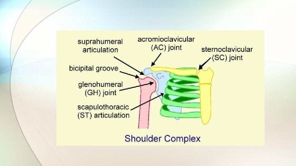
Structure of the Shoulder • Most complex joint in body. • Separate articulations: • Sternoclavicular Joint • Acromioclavicular Joint • Coracoclavicular Joint • Glenohumeral Joint • Scapulothoracic Joint • Also: Bursae

Sternoclavicular Joint • Provides major axis of rotation for movement of clavicle and scapula • Freely permitted frontal and transverse plane motion. • Allows some forward and backward sagittal plane rotation. • Rotation

Acromioclavicular Joint • Irregular diarthrodial joint between the acromion process of the scapula and the distal clavicle Øallows limited motions in all three planes. • Rotation occurs during arm elevation. • Close-packed position with humerus abducted to 90 degrees.

Coracoclavicular Joint • A syndesmosis with coracoid process of scapulabound to the inferior clavicle by the coracoclavicular ligament. • Permits little movement.

Glenohumeral Joint • Most freely moving joint in human body • Glenoid Labrum composed of: ØJoint capsule ØTendon of long head of biceps brachii ØGlenohumeral ligaments • Rotator Cuff Muscles • Most stable in close-packed position, when the humerus is abducted and laterally rotated.

Scapulothoracic Joint • Region between the anterior scapula and thoracic wall. • Functions of muscles attaching to scapula: ØContract to stabilize shoulder region ØFacilitate movements of the upper extremity through appropriate positioning of the glenohumeral joint.

Bursae • Small fibrous sacs that secrete synovial fluid internally to lessen friction between soft tissues around joints. • Shoulder contains: ØSubcoracoid bursa ØSubscapularis bursa ØSubacromial bursa

Movements of the Shoulder Complex • Humerus movement usually involves some movement at all three shoulder joints ØPositioning further facilitated by motions of spine ØScapulohumeral Rhythm

Movements of the Shoulder Complex • Muscles of the Scapula • Muscles of the Glenohumeral Joint ØFlexion ØExtension ØAbduction ØAdduction • Medial and Lateral Rotation of the Humerus • Horizontal Adduction and Abduction Glenohumeral Joint at the
 stabilize the scapula when shoulder complex")
Muscles of the Scapula • Function: Ø 1) stabilize the scapula when shoulder complex is loaded Ø 2) move and position the scapula to facilitate movement at glenohumeral joint • Are: ØLevator scapula, rhomboids, serratus anterior, pectoralis minor, subclavius, and four parts to trapezius.

Muscles of Glenohumeral Joint • Many muscles involved, some contribute more than others. • Large ROM can complicate tension development with orientation of humerus. • Tension development in one shoulder muscle is frequently accompanied by development of tension in an antagonist to prevent dislocation of the humeral head.

Flexion at Glenohumeral Joint • Prime flexors: ØAnterior deltoid ØPectoralis major: clavicular portion • Assistant flexors: ØCoracobrachialis ØBiceps brachii: short head

Extension at Glenohumeral Joint • Gravitational force is primary mover when shoulder extension isn’t resisted. ØControl by eccentric contraction of flexors • With resistance there is contraction of muscles posterior to the glenohumeral joint • Assisted by: ØPosterior deltoid ØBiceps brachii: long head

Abduction at Glenohumeral Joint • Major abductors of humerus: ØSupraspinatus • Initiates abduction • Active for first 110 degrees of abduction ØMiddle deltoid • Active degrees of abduction • Superior dislocating component neutralized infraspinatus, subscapularis, and teres minor. by

Adduction of Glenohumeral Joint • Primary adductors: ØLatissimus dorsi ØTeres major ØSternocostal pectoralis • Minor assistance: ØBiceps brachii: short head ØTriceps brachii: long head ØAbove 90 degrees- coracobrachialis and subscapularis

Medial and Lateral Rotation of Humerus • Due to action of: ØSubscapularis • Has greatest mechanical advantage for medial rotation ØTeres major • Assisted by: ØPrimarily: pectoralis major ØAlso: anterior deltoid, latissimus dorsi and short head of biceps brachii

Horizontal Adduction and Abduction at the Glenohumeral Joint • Anterior to joint: ØPectoralis major (both heads), anterior deltoid, coracobrachialis ØAssisted by short head of biceps brachi • Posterior to joint: ØMiddle and posterior deltoid, infraspinatus, teres minor ØAssisted by teres major, latissimus dorsi

Loads on the Shoulder • Arm segment moment arm: ØPerpendicular distance between weight vector and shoulder. • With elbow flexion, upper arm and forearm/hand segments must be analyzed separately. • Large torques from extended moment arms countered by shoulder muscles. ØLoad reduced by half with maximal elbow flexion

Structure of the Elbow • Humeroulnar Joint • Humeroradial Joint • Proximal Radioulnar Joint

Segments at the Elbow • Flexion and Extension ØMuscles crossing anterior side of elbow are the flexors: • Brachialis, biceps brachii, brachioradialis ØMuscles crossing posterior side of elbow are the extensors: • Triceps, anconeus muscle

Segments at the Elbow • Pronation and Supination ØInvolves rotation of radius around ulna ØArticulations: • Proximal and distal radioulnar joints (both pivot joints) • Middle radioulnar joint (syndesmosis) • Pronator quadratus • Supinator

Loads on the Elbow • Large loads generate by muscles that cross elbow during forceful pitching/throwing ØAlso in weight lifting, gymnastics • Extensor moment arm shorter flexor moment arm ØTricep attachment to ulna closer to elbow joint center than those of the brachialis on ulna an biceps on radius • Moment arm also varies with position of elbow

Structure of the Wrist • Radiocarpal joint ØReinforced by: volar radiocarpal, dorsal radiocarpal, radial collateral and ulnar collateral ligaments • Retinacula ØForm protective passageways for tendons, nerves and blood vessel to pass through

Movements of the Wrist • Sagittal and frontal plane movements • Rotary motion • Flexion • Extension and Hyperextension • Radial Deviation • Ulnar Deviation
 • Metacarpophalangeal (MP) • Interphalangeal (IP)")
Joint Structure of the Hand • Carpometacarpal (CM) • Metacarpophalangeal (MP) • Interphalangeal (IP)

Movements of the Hand • CM Joints allow large ROM because similar to ball and socket joint • Digits 2 -4 constrained by ligaments • MP joints allow flexion, extension, abduction, adduction and circumduction for digits 2 -5 • IP joints allow flexion and extension • Extrinsic Muscles • Intrinsic Muscles

Summary • Shoulder is the most complex joint in the human body. • Movements of the shoulder girdle contribute to optimal positioning of the glenohumeral joint for different humeral movements. • Humeroulnar articulation controls flexion and extension at the elbow • Pronation and supination of forearm occur at proximal and distal radioulnar joints.

Thank you for Listening
- Slides: 31