Muscle Movements Types and Names Pages 196 203


- Slides: 24

Muscle Movements, Types, and Names Pages 196 -203

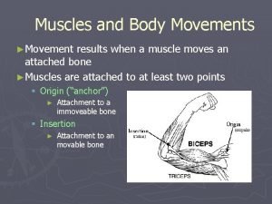
Muscles and Body Movements Movement is attained as a result of a muscle moving an attached bone Muscles are attached to at least two points 1. Origin: attaches to an immovable/less movable bone 2. Insertion: attaches to a movable bone © 2015 Pearson Education, Inc.

Figure 6. 12 Muscle attachments (origin and insertion). Muscle contracting Origin Brachialis Tendon Insertion

Table 6. 2 The Five Golden Rules of Skeletal Muscle Activity.

Types of Body Movements Flexion Decreases the angle of the joint Brings two bones closer together Typical of bending hinge joints (e. g. , knee and elbow) or ball-and-socket joints (e. g. , the hip) Extension Opposite of flexion Increases angle between two bones Typical of straightening the elbow or knee Extension beyond 180° is hyperextension © 2015 Pearson Education, Inc.

Figure 6. 13 a Body movements. Flexion Hyperextension Extension Flexion Extension (a) Flexion, extension, and hyperextension of the shoulder and knee

Figure 6. 13 b Body movements. Hyperextension Extension Flexion (b) Flexion, extension, and hyperextension

Types of Body Movements Rotation Movement of a bone around its longitudinal axis Common in ball-and-socket joints Example: moving the atlas around the dens of axis (i. e. , shaking your head “no”) © 2015 Pearson Education, Inc.

Figure 6. 13 c Body movements. Rotation Lateral rotation Medial rotation (c) Rotation

Types of Body Movements Abduction Movement of a limb away from the midline Adduction Opposite of abduction Movement of a limb toward the midline © 2015 Pearson Education, Inc.

Figure 6. 13 d Body movements. Abduction Adduction Circumduction (d) Abduction, adduction, and circumduction

Types of Body Movements Circumduction Combination of flexion, extension, abduction, and adduction Common in ball-and-socket joints Proximal end of bone is stationary, and distal end moves in a circle © 2015 Pearson Education, Inc.

Figure 6. 13 d Body movements. Abduction Adduction Circumduction (d) Abduction, adduction, and circumduction

Special Movements Dorsiflexion Lifting the foot so that the superior surface approaches the shin (toward the dorsum) Plantar flexion Depressing the foot (pointing the toes) “Planting” the foot toward the sole © 2015 Pearson Education, Inc.

Figure 6. 13 e Body movements. Dorsiflexion Plantar flexion (e) Dorsiflexion and plantar flexion

Special Movements Inversion Turning sole of foot medially Eversion Turning sole of foot laterally © 2015 Pearson Education, Inc.

Figure 6. 13 f Body movements. Inversion (f) Inversion and eversion Eversion

Special Movements Supination Forearm rotates laterally so palm faces anteriorly Radius and ulna are parallel Pronation Forearm rotates medially so palm faces posteriorly Radius and ulna cross each other like an X © 2015 Pearson Education, Inc.

Figure 6. 13 g Body movements. Pronation (radius rotates over ulna) Supination (radius and ulna are parallel) S P (g) Supination (S) and pronation (P)

Special Movements Opposition Moving the thumb to touch the tips of other fingers on the same hand © 2015 Pearson Education, Inc.

Figure 6. 13 h Body movements. Opposition (h) Opposition

Types of Muscles Prime mover— muscle with the major responsibility for a certain movement Synergist— muscle that aids a prime mover in a movement and helps prevent rotation Fixator— stabilizes the origin of a prime mover Antagonist— muscle that opposes or reverses a prime mover It is the actions of all muscles involved that provide smooth, coordinated and precise movement. © 2015 Pearson Education, Inc.

Naming Skeletal Muscles Skeletal muscles can be named according to: direction of muscle fibers: rectus (straight) relative size of the muscle: maximus (largest) location of the muscle: temporalis (temporal bone) number of origins triceps (three heads) location of origin and insertion: sterno (on the sternum) shape of the muscle: deltoid (triangular) action: flexor/extensor (flex/extend a bone) © 2015 Pearson Education, Inc.

Figure 6. 15 Relationship of fascicle arrangement to muscle structure. (a) (b) (a) Circular (orbicularis oris) (e) (c) (b) Converent (pectoralis major) (d) (e) Multipennate (deltoid) (f) (g) (c) Fusiform (biceps brachii) (d) Parallel (sartorius) (f) Bipennate (rectus femoris) (g) Unipennate (extensor digitorum longus)
 Muscle movements, types, and names
 Printed pages vs web pages
 Locomotor movements example
 Muscle movements
 Muscle names
 Biceps muscle names
 Characteristics of skeletal muscles
 Toe dancers muscle a two bellied muscle of the calf
 196 883-dimensional monster
 Perbedaan galat mutlak dan galat relatif
 Akar kuadrat 3969
 196 hangi sayının karesi
 Nilai binar dari nilai bilangan decimal 196 . ?
 Homework 196
 Odmocnina 196
 196
196 What is the decimal value of 11012?
 Luas salah satu kubus 196 cm volume kubus tersebut adalah
 Cube and square 1 to 20
 Bisp 197
 3 cloud types
 Complementary supplementary and vertical angles
 Types of ordinary body movements
 Hyperextension
 Alternative social movement
