The Diagnosis of Renal Colic Kelsey Innes PGY2

- Slides: 49
The Diagnosis of Renal Colic Kelsey Innes, PGY-2 FRCPC Emergency Medicine February 25 th 2015
Objectives • Pertinent Hx, Px • Labs • Imaging • CT • U/S • Evidence-based strategies for diagnosis
Triage • 38 yo male with severe flank pain
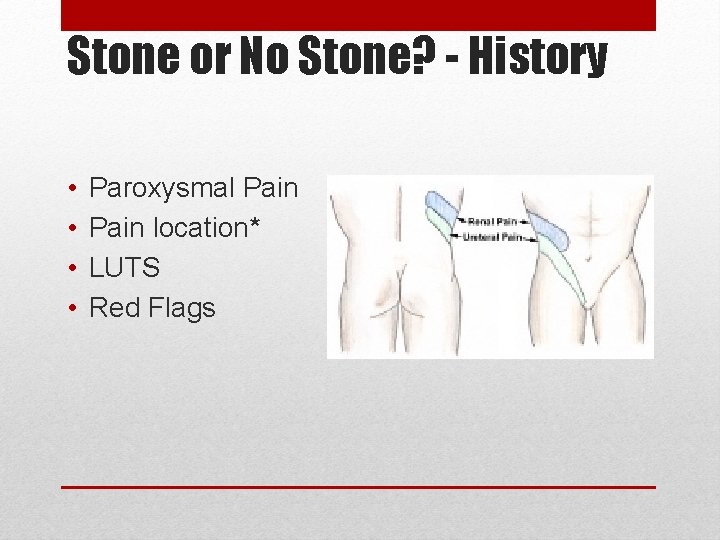
Stone or No Stone? - History • • Paroxysmal Pain location* LUTS Red Flags
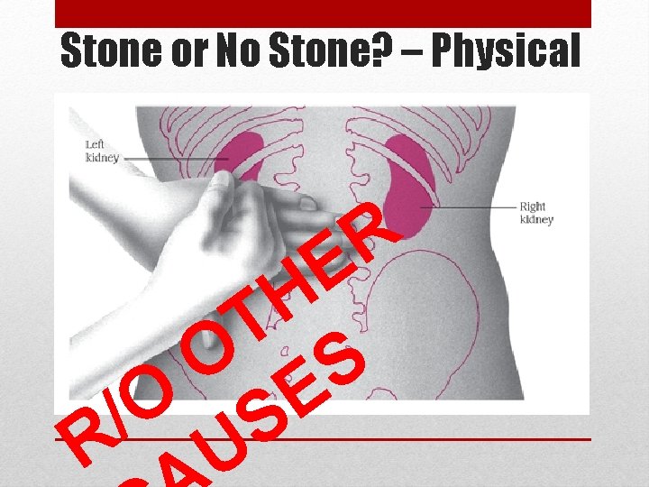
Stone or No Stone? – Physical R E H T O S E O / S R U
Stone or No Stone? - Investigations
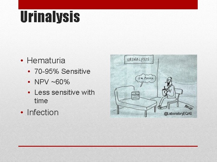
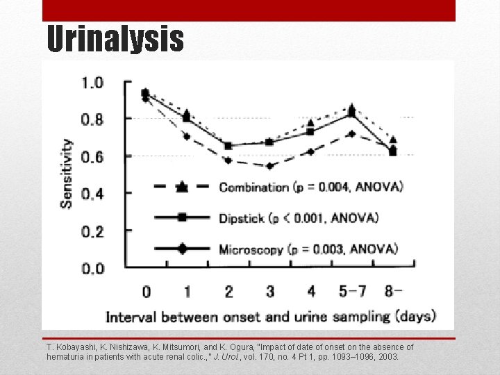
Urinalysis • Hematuria • 70 -95% Sensitive • NPV ~60% • Less sensitive with time • Infection
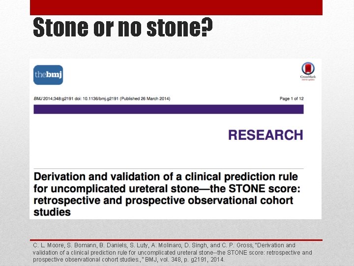
Stone or no stone? C. L. Moore, S. Bomann, B. Daniels, S. Luty, A. Molinaro, D. Singh, and C. P. Gross, “Derivation and validation of a clinical prediction rule for uncomplicated ureteral stone--the STONE score: retrospective and prospective observational cohort studies. , ” BMJ, vol. 348, p. g 2191, 2014.
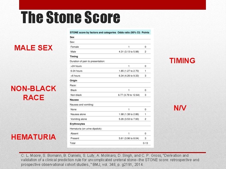
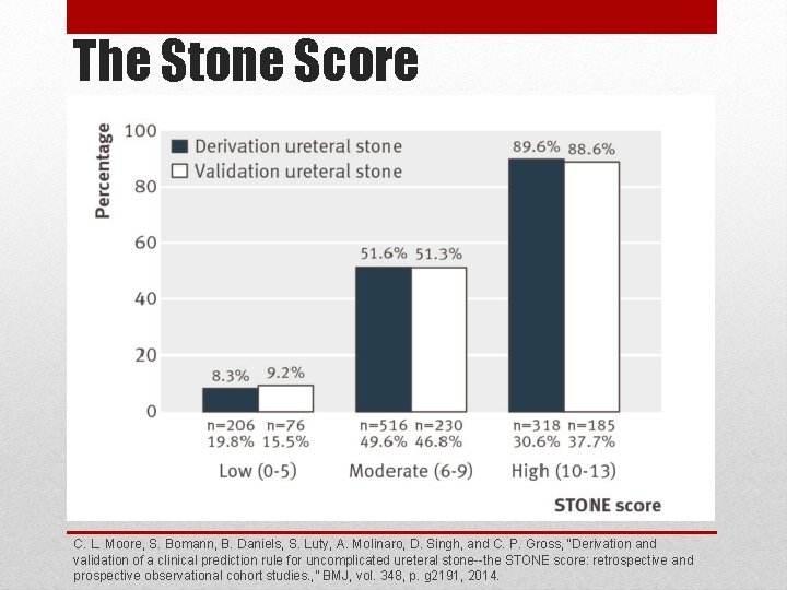
The Stone Score MALE SEX TIMING NON-BLACK RACE N/V HEMATURIA C. L. Moore, S. Bomann, B. Daniels, S. Luty, A. Molinaro, D. Singh, and C. P. Gross, “Derivation and validation of a clinical prediction rule for uncomplicated ureteral stone--the STONE score: retrospective and prospective observational cohort studies. , ” BMJ, vol. 348, p. g 2191, 2014.
The Stone Score C. L. Moore, S. Bomann, B. Daniels, S. Luty, A. Molinaro, D. Singh, and C. P. Gross, “Derivation and validation of a clinical prediction rule for uncomplicated ureteral stone--the STONE score: retrospective and prospective observational cohort studies. , ” BMJ, vol. 348, p. g 2191, 2014.
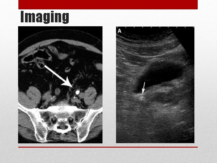
Imaging
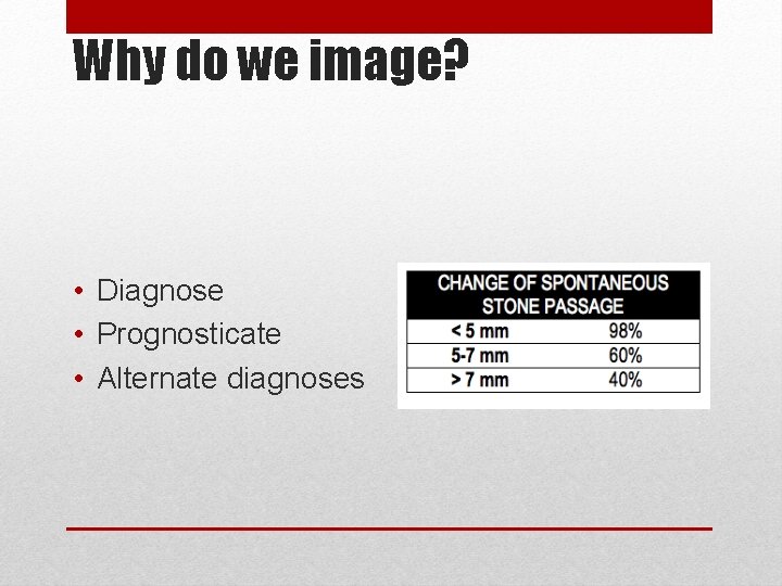
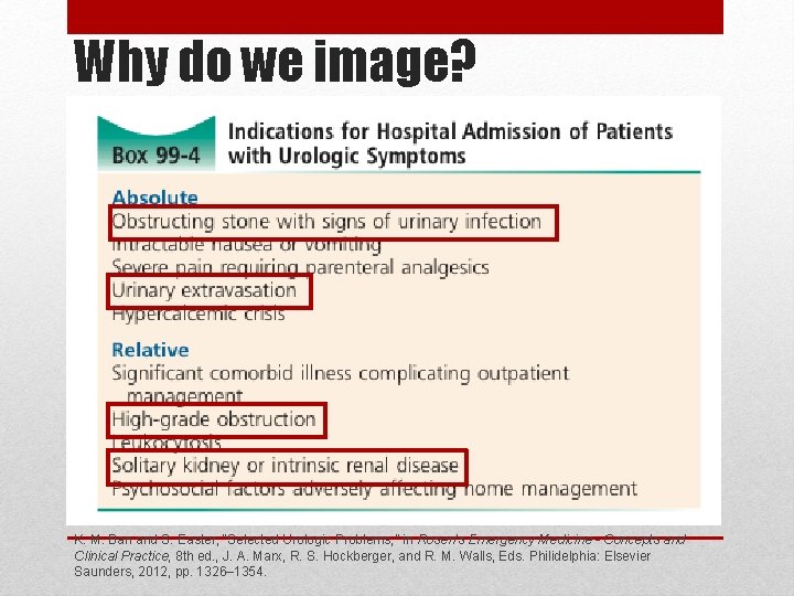
Why do we image? • Diagnose • Prognosticate • Alternate diagnoses
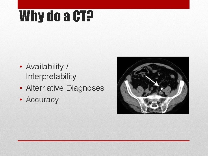
Why do a CT? • Availability / Interpretability • Alternative Diagnoses • Accuracy
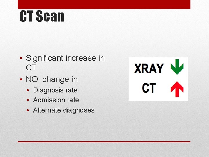
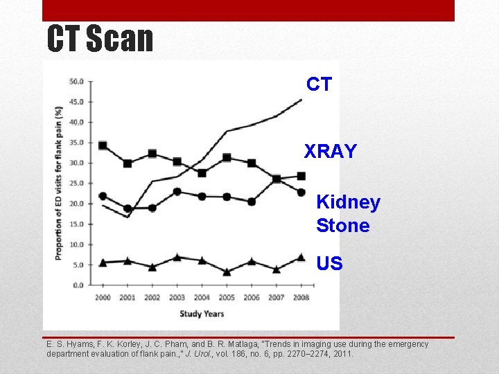
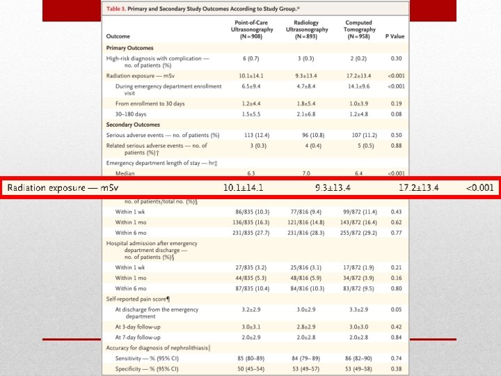
CT Scan • Significant increase in CT • NO change in • Diagnosis rate • Admission rate • Alternate diagnoses
CT Scan CT XRAY Kidney Stone US E. S. Hyams, F. K. Korley, J. C. Pham, and B. R. Matlaga, “Trends in imaging use during the emergency department evaluation of flank pain. , ” J. Urol. , vol. 186, no. 6, pp. 2270– 2274, 2011.
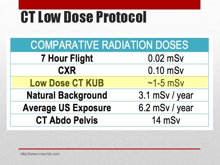
CT Low Dose Protocol http: //www. xrayrisk. com
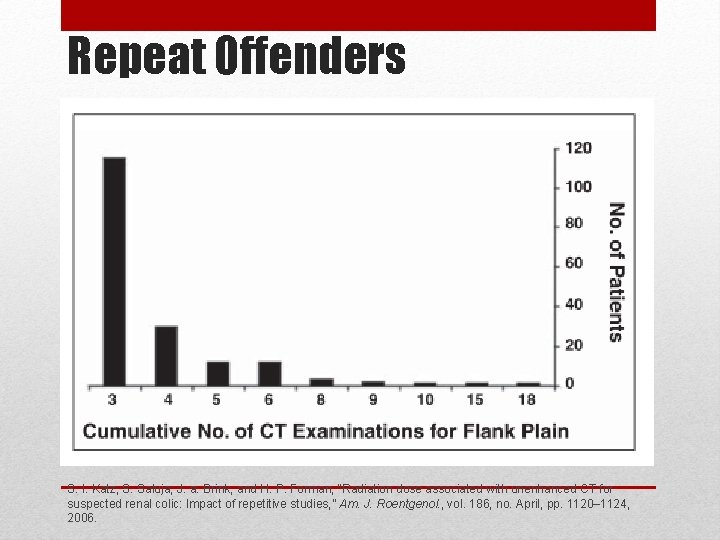
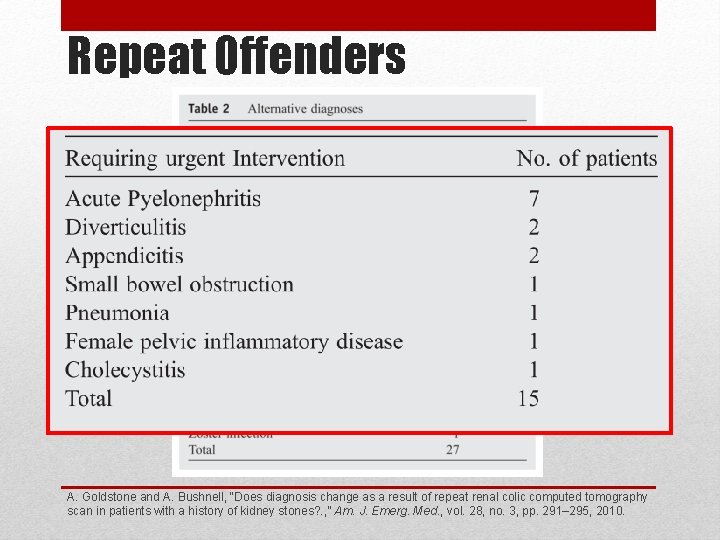
Repeat Offenders • “Repetitive disease” • Imaging begets imaging • Increased risk for serial CT • Small proportion requiring “urgent intervention”
Repeat Offenders S. I. Katz, S. Saluja, J. a. Brink, and H. P. Forman, “Radiation dose associated with unenhanced CT for suspected renal colic: Impact of repetitive studies, ” Am. J. Roentgenol. , vol. 186, no. April, pp. 1120– 1124, 2006.
Repeat Offenders A. Goldstone and A. Bushnell, “Does diagnosis change as a result of repeat renal colic computed tomography scan in patients with a history of kidney stones? . , ” Am. J. Emerg. Med. , vol. 28, no. 3, pp. 291– 295, 2010.
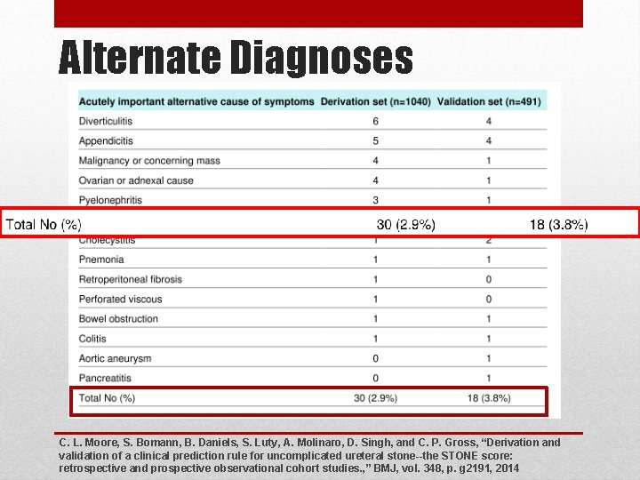
Alternate Diagnoses C. L. Moore, S. Bomann, B. Daniels, S. Luty, A. Molinaro, D. Singh, and C. P. Gross, “Derivation and validation of a clinical prediction rule for uncomplicated ureteral stone--the STONE score: retrospective and prospective observational cohort studies. , ” BMJ, vol. 348, p. g 2191, 2014
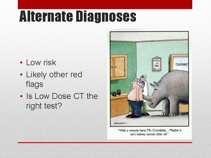
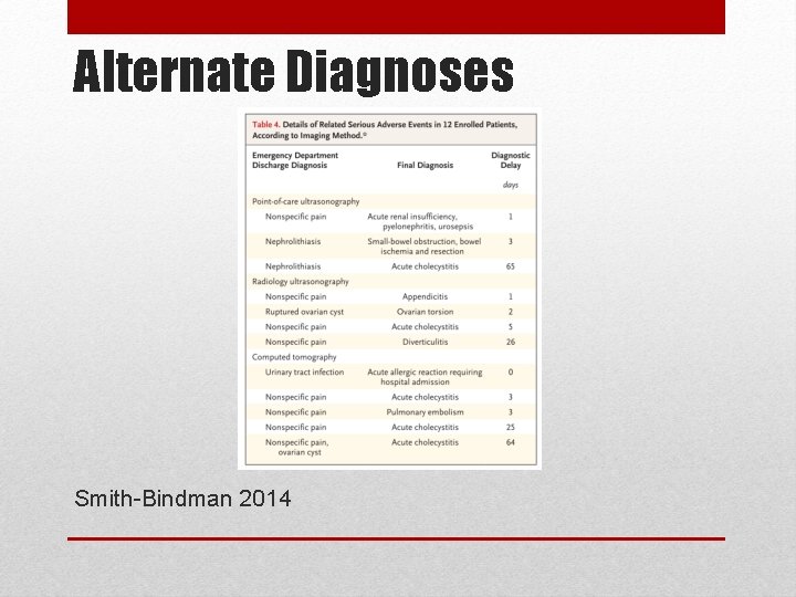
Alternate Diagnoses • Low risk • Likely other red flags • Is Low Dose CT the right test?
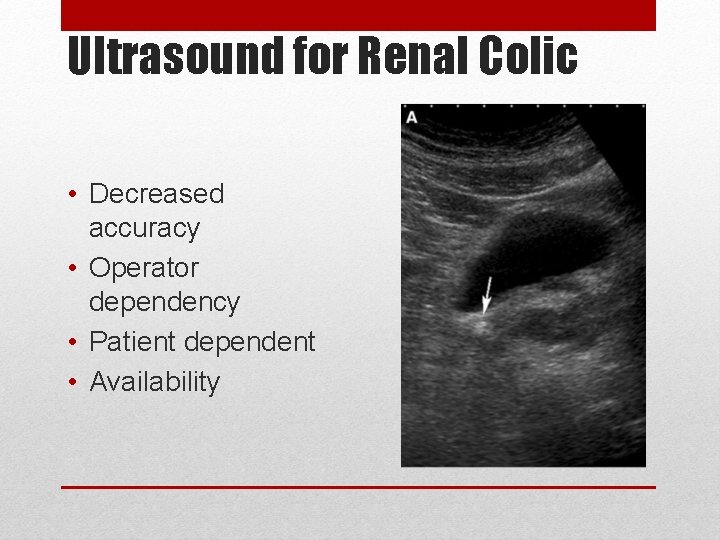
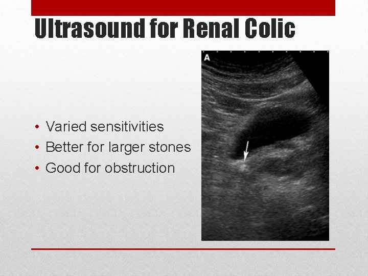
Ultrasound for Renal Colic • Decreased accuracy • Operator dependency • Patient dependent • Availability
Ultrasound for Renal Colic • Varied sensitivities • Better for larger stones • Good for obstruction
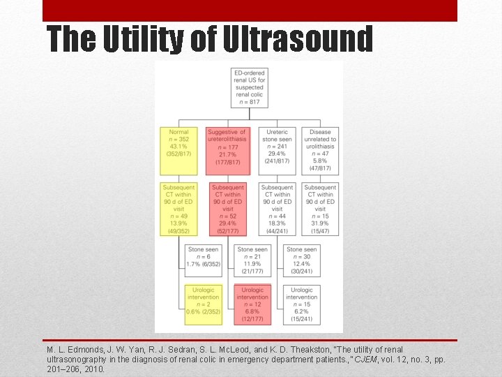
Utility of Ultrasound
The Utility of Ultrasound M. L. Edmonds, J. W. Yan, R. J. Sedran, S. L. Mc. Leod, and K. D. Theakston, “The utility of renal ultrasonography in the diagnosis of renal colic in emergency department patients. , ” CJEM, vol. 12, no. 3, pp. 201– 206, 2010.
Ultrasound vs. CT
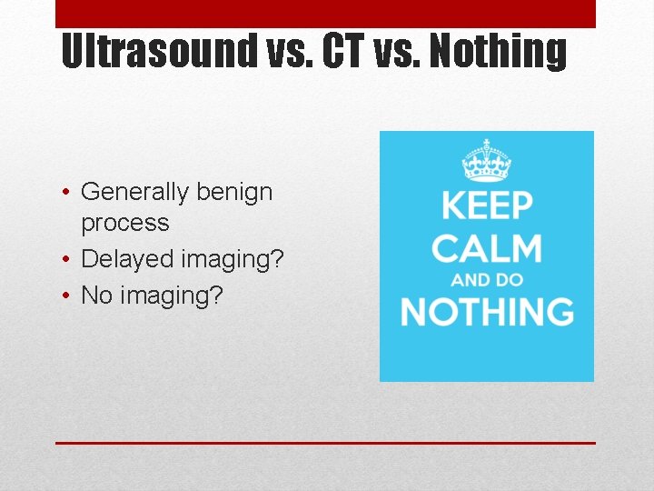
Ultrasound vs. CT vs. Nothing • Generally benign process • Delayed imaging? • No imaging?
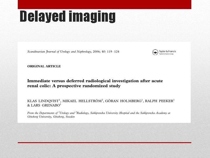
Delayed imaging
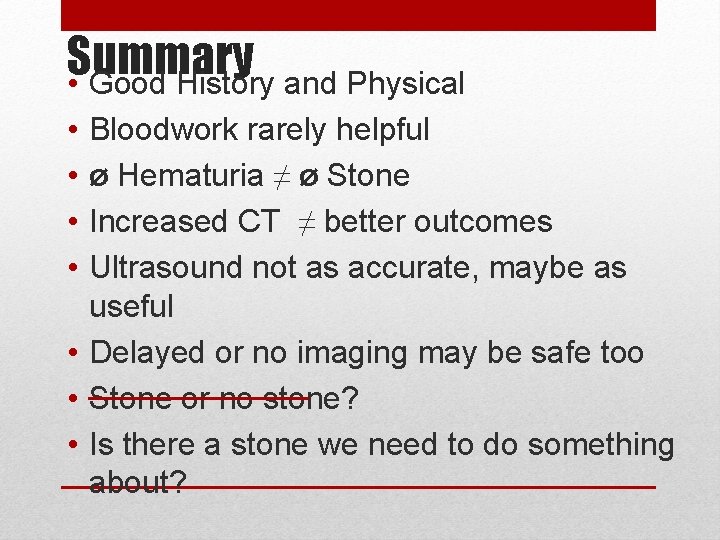
Summary • Good History and Physical • • Bloodwork rarely helpful ø Hematuria ≠ ø Stone Increased CT ≠ better outcomes Ultrasound not as accurate, maybe as useful • Delayed or no imaging may be safe too • Stone or no stone? • Is there a stone we need to do something about?
Thank You! • • • Dr. Jon Braunstein Dr. Mario Francispragasam Dr. David Hodges (Radiology) Dr. Benjamin Bay (Urology) Dr. Nick Bilby (Radiology)
Questions / Comments
References 1. 2. 3. 4. 5. 6. 7. 8. R. Smith-Bindman, C. Aubin, J. Bailitz, R. N. Bengiamin, C. A. J. Camargo, J. Corbo, A. J. Dean, R. B. Goldstein, R. T. Griffey, G. D. Jay, T. L. Kang, D. R. Kriesel, O. J. Ma, M. Mallin, W. Manson, J. Melnikow, D. L. Miglioretti, S. K. Miller, L. D. Mills, J. R. Miner, M. Moghadassi, V. E. Noble, G. M. Press, M. L. Stoller, V. E. Valencia, J. Wang, R. C. Wang, and S. R. Cummings, “Ultrasonography versus Computed Tomography for Suspected Nephrolithiasis, ” N. Engl. J. Med. , vol. 371, no. 12, pp. 1100 – 1110, 2014. C. L. Moore, S. Bomann, B. Daniels, S. Luty, A. Molinaro, D. Singh, and C. P. Gross, “Derivation and validation of a clinical prediction rule for uncomplicated ureteral stone--the STONE score: retrospective and prospective observational cohort studies. , ” BMJ, vol. 348, p. g 2191, 2014. M. Rafi, A. Shetty, and N. Gunja, “Accuracy of computed tomography of the kidneys, ureters and bladder interpretation by emergency physicians. , ” Emerg. Med. Australas. , vol. 25, no. 5, pp. 422– 426, 2013. G. Vallone, G. Napolitano, P. Fonio, G. Antinolfi, A. Romeo, L. Macarini, E. A. Genovese, and L. Brunese, “US detection of renal and ureteral calculi in patients with suspected renal colic. , ” Crit. Ultrasound J. , vol. 5 Suppl 1, p. S 3, 2013. J. H. Moak, M. S. Lyons, and C. J. Lindsell, “Bedside renal ultrasound in the evaluation of suspected ureterolithiasis. , ” Am. J. Emerg. Med. , vol. 30, no. 1, pp. 218– 21, Jan. 2012. E. S. Hyams, F. K. Korley, J. C. Pham, and B. R. Matlaga, “Trends in imaging use during the emergency department evaluation of flank pain. , ” J. Urol. , vol. 186, no. 6, pp. 2270– 2274, 2011. C. Westphalen, R. Y. Hsia, J. H. Maselli, R. Wang, and R. Gonzales, “Radiological imaging of patients with suspected urinary tract stones: national trends, diagnoses, and predictors. , ” Acad. Emerg. Med. , vol. 18, no. 7, pp. 699– 707, 2011. M. L. Edmonds, J. W. Yan, R. J. Sedran, S. L. Mc. Leod, and K. D. Theakston, “The utility of renal ultrasonography in the diagnosis of renal colic in emergency department patients. , ” CJEM, vol. 12, no. 3, pp. 201– 206, 2010.
References 9. 10. 11. 12. 13. 14. 15. A. Goldstone and A. Bushnell, “Does diagnosis change as a result of repeat renal colic computed tomography scan in patients with a history of kidney stones? . , ” Am. J. Emerg. Med. , vol. 28, no. 3, pp. 291– 295, 2010. J. S. Luchs, D. S. Katz, M. J. Lane, B. C. Mellinger, J. H. Lumerman, C. A. Stillman, E. M. Meiner, and S. Perlmutter, “Utility of hematuria testing in patients with suspected renal colic: correlation with unenhanced helical CT results. , ” J. Urol. , vol. 170, no. 1, pp. 334– 335, 2003. T. Kobayashi, K. Nishizawa, K. Mitsumori, and K. Ogura, “Impact of date of onset on the absence of hematuria in patients with acute renal colic. , ” J. Urol. , vol. 170, no. 4 Pt 1, pp. 1093– 1096, 2003. A. Farmakis, K. Doumas, and M. Lykourinas, “The presence of microscopic hematuria detected by urine dipstick test in the evaluation of patients with renal colic. , ” Urol. Res. , vol. 32, no. 4, pp. 294– 297, 2004. K. M. Ban and S. Easter, “Selected Urologic Problems, ” in Rosen’s Emergency Medicine - Concepts and Clinical Practice, 8 th ed. , J. A. Marx, R. S. Hockberger, and R. M. Walls, Eds. Philidelphia: Elsevier Saunders, 2012, pp. 1326– 1354. F. M. Abrahamian, A. Krishnadasan, W. R. Mower, G. J. Moran, and D. a. Talan, “Association of pyuria and clinical characteristics with the presence of urinary tract infection among patients with acute nephrolithiasis, ” Ann. Emerg. Med. , vol. 62, no. 5, pp. 526– 533, 2013. S. I. Katz, S. Saluja, J. a. Brink, and H. P. Forman, “Radiation dose associated with unenhanced CT for suspected renal colic: Impact of repetitive studies, ” Am. J. Roentgenol. , vol. 186, no. April, pp. 1120– 1124, 2006.
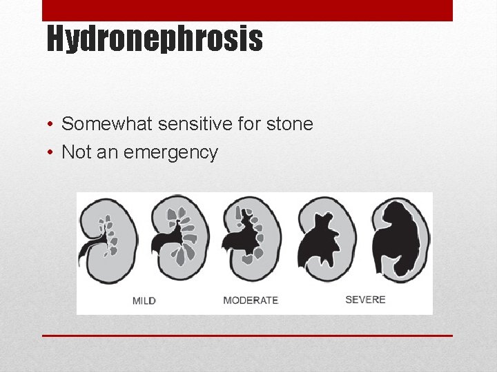
Hydronephrosis • Somewhat sensitive for stone • Not an emergency
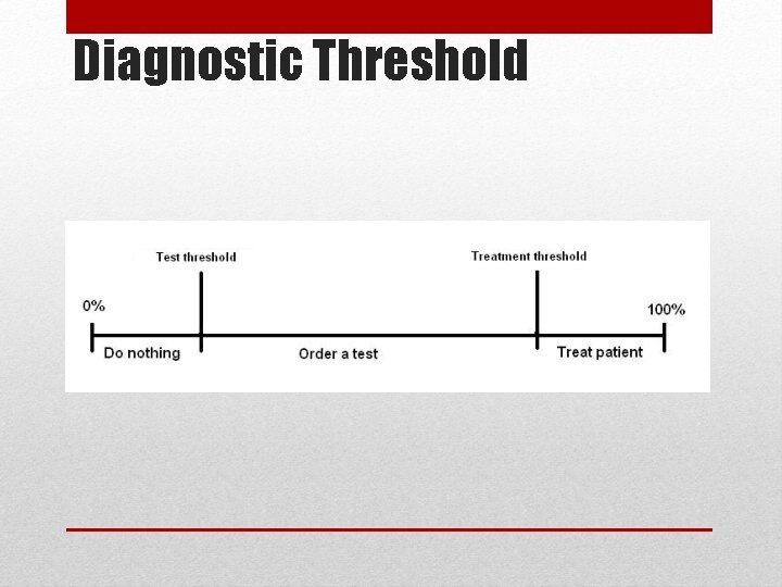
Diagnostic Threshold
Stone or No Stone?
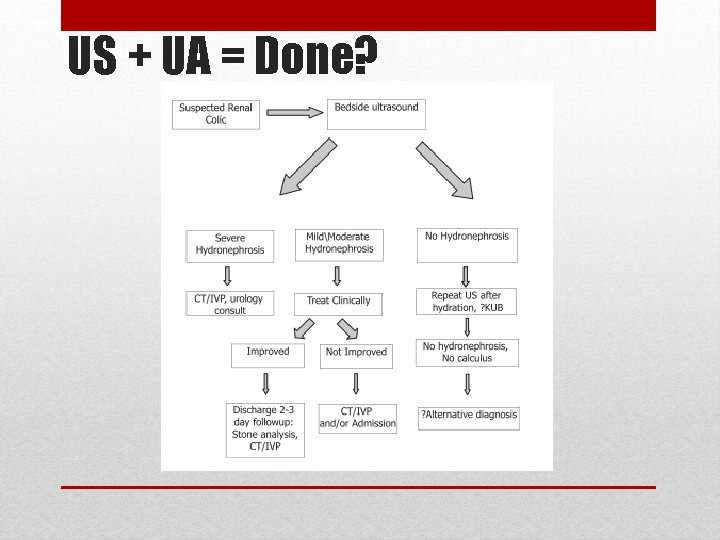
US + UA = Done?
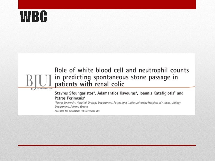
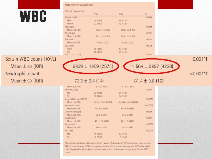
WBC
WBC
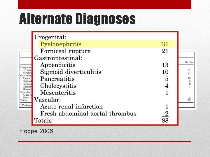
Alternate Diagnoses Hoppe 2006
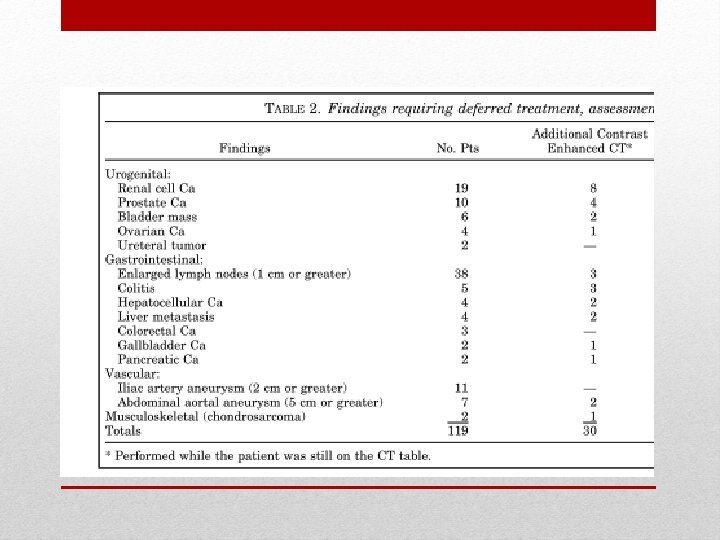
Alternate Diagnoses Smith-Bindman 2014
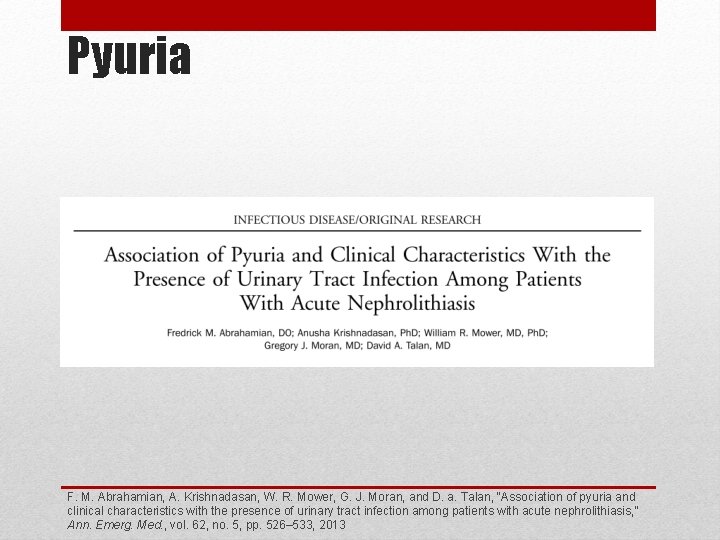
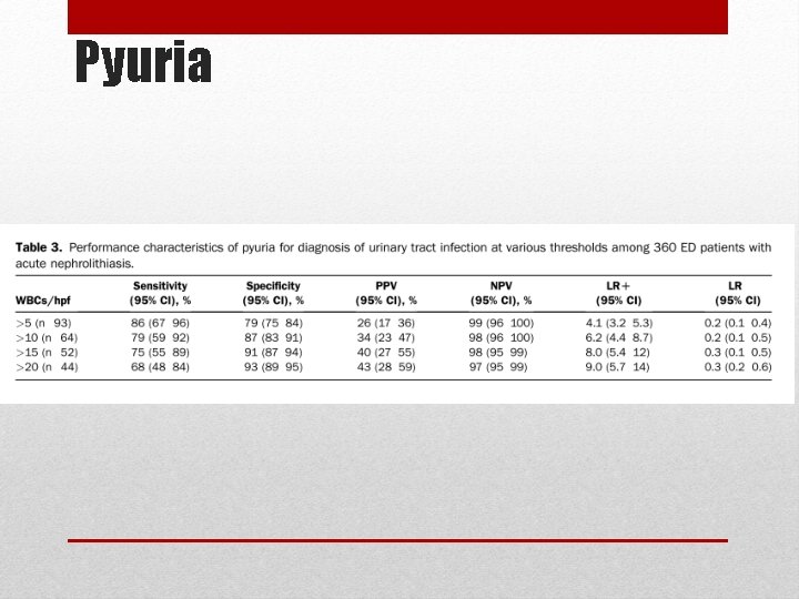
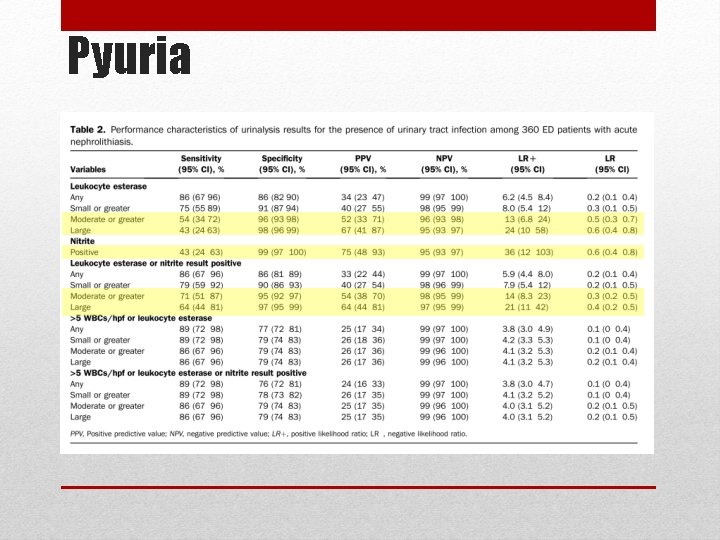
Pyuria F. M. Abrahamian, A. Krishnadasan, W. R. Mower, G. J. Moran, and D. a. Talan, “Association of pyuria and clinical characteristics with the presence of urinary tract infection among patients with acute nephrolithiasis, ” Ann. Emerg. Med. , vol. 62, no. 5, pp. 526– 533, 2013
Pyuria
Pyuria
Urinalysis T. Kobayashi, K. Nishizawa, K. Mitsumori, and K. Ogura, “Impact of date of onset on the absence of hematuria in patients with acute renal colic. , ” J. Urol. , vol. 170, no. 4 Pt 1, pp. 1093– 1096, 2003.
Why do we image? K. M. Ban and S. Easter, “Selected Urologic Problems, ” in Rosen’s Emergency Medicine - Concepts and Clinical Practice, 8 th ed. , J. A. Marx, R. S. Hockberger, and R. M. Walls, Eds. Philidelphia: Elsevier Saunders, 2012, pp. 1326– 1354.
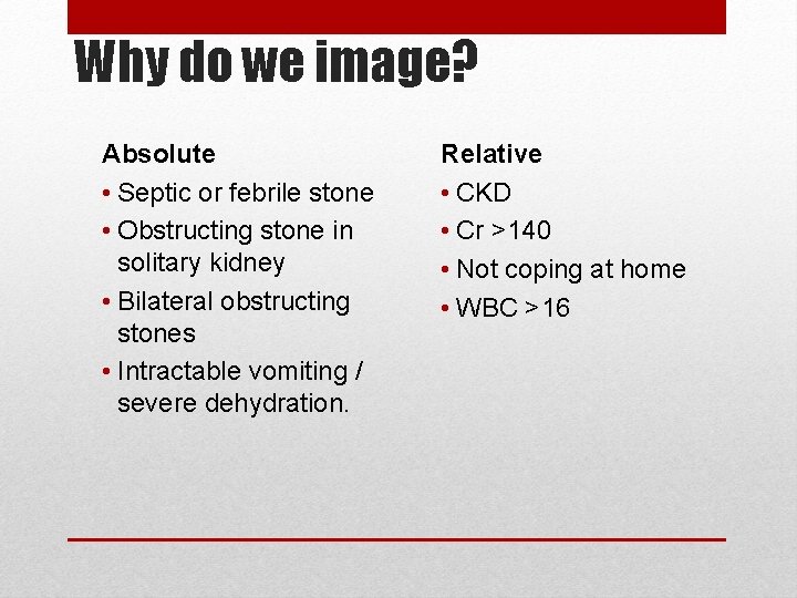
Why do we image? Absolute • Septic or febrile stone • Obstructing stone in solitary kidney • Bilateral obstructing stones • Intractable vomiting / severe dehydration. Relative • CKD • Cr >140 • Not coping at home • WBC >16