RNA YUSRON SUGIARTO STP MSc 1 Central Dogma

- Slides: 49
RNA YUSRON SUGIARTO, STP, MSc 1
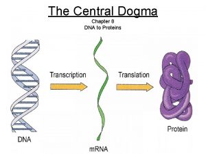
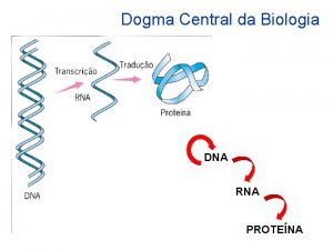
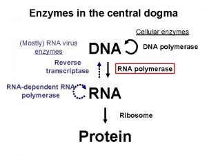
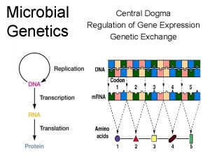
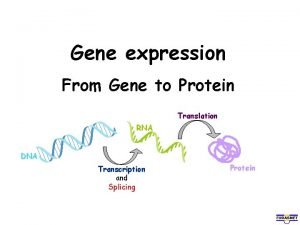
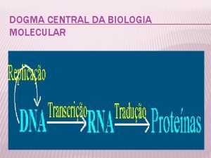
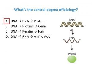
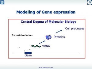
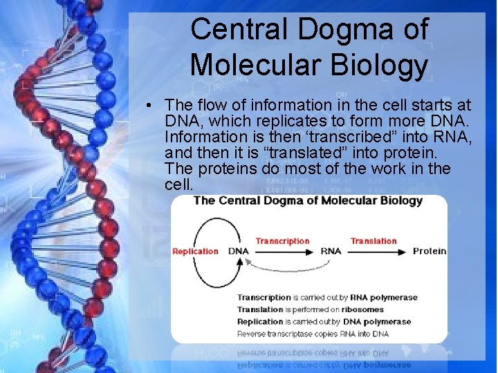
Central Dogma of Molecular Biology • The flow of information in the cell starts at DNA, which replicates to form more DNA. Information is then ‘transcribed” into RNA, and then it is “translated” into protein. The proteins do most of the work in the cell.
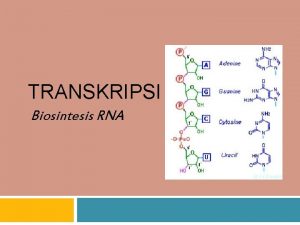
Structure of RNA A. Chain of nucleotides like DNA B. Parts of an RNA nucleotide 1. 5 carbon sugar: ribose 2. phosphate group 3. nitrogen base
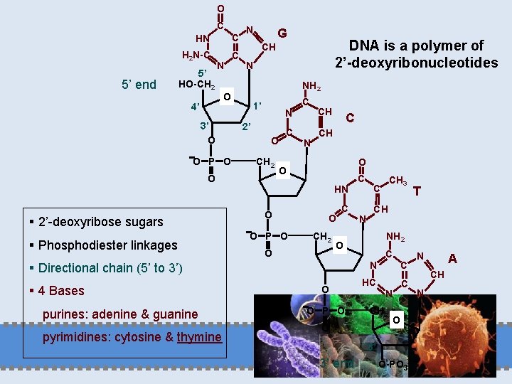
O C 5’ end HN C H 2 N-C C 5’ HO-CH 2 N N 3’ O O P O N NH 2 C 1’ N 2’ O CH 2 O § 2’-deoxyribose sugars § Phosphodiester linkages DNA is a polymer of 2’-deoxyribonucleotides CH O 4’ G C N CH C CH O O C HN O O P O O CH 2 O C purines: adenine & guanine pyrimidines: cytosine & thymine O O P O NH 2 C N C HC C N CH 2 O O 3’ 3’ end T CH O § Directional chain (5’ to 3’) § 4 Bases N CH 3 C O-PO 32 N A CH N
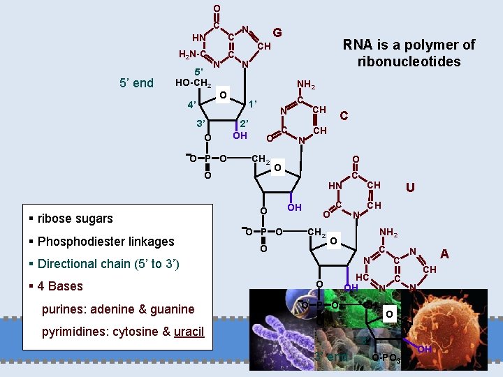
O C 5’ end HN C H 2 N-C C 5’ HO-CH 2 N N 3’ O O P O N NH 2 C 1’ 2’ OH N O CH 2 O § ribose sugars § Phosphodiester linkages RNA is a polymer of ribonucleotides CH O 4’ G C N CH C CH O O P O C OH HN CH C CH O CH 2 O N NH 2 O C N § Directional chain (5’ to 3’) § 4 Bases purines: adenine & guanine pyrimidines: cytosine & uracil U O C N HC C N OH N O P O CH 2 O CH O 3’ 3’ end A O-PO 32 OH
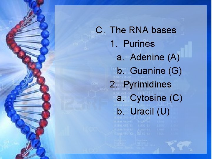
C. The RNA bases 1. Purines a. Adenine (A) b. Guanine (G) 2. Pyrimidines a. Cytosine (C) b. Uracil (U)
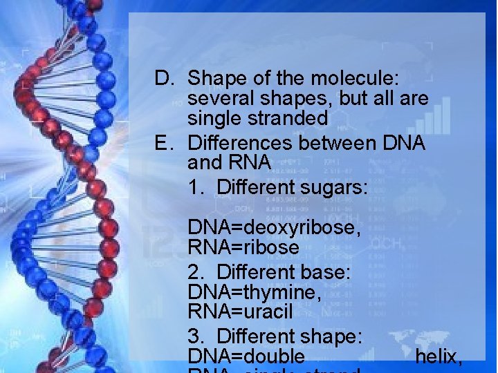
D. Shape of the molecule: several shapes, but all are single stranded E. Differences between DNA and RNA 1. Different sugars: DNA=deoxyribose, RNA=ribose 2. Different base: DNA=thymine, RNA=uracil 3. Different shape: DNA=double helix,
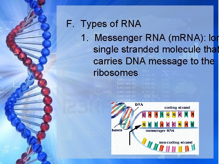
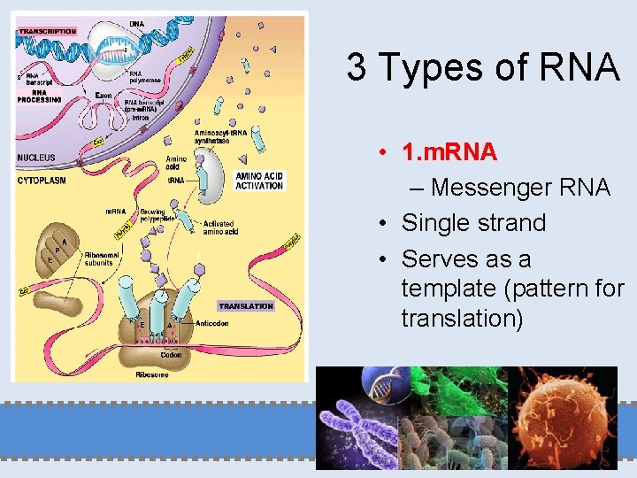
F. Types of RNA 1. Messenger RNA (m. RNA): lon single stranded molecule that carries DNA message to the ribosomes
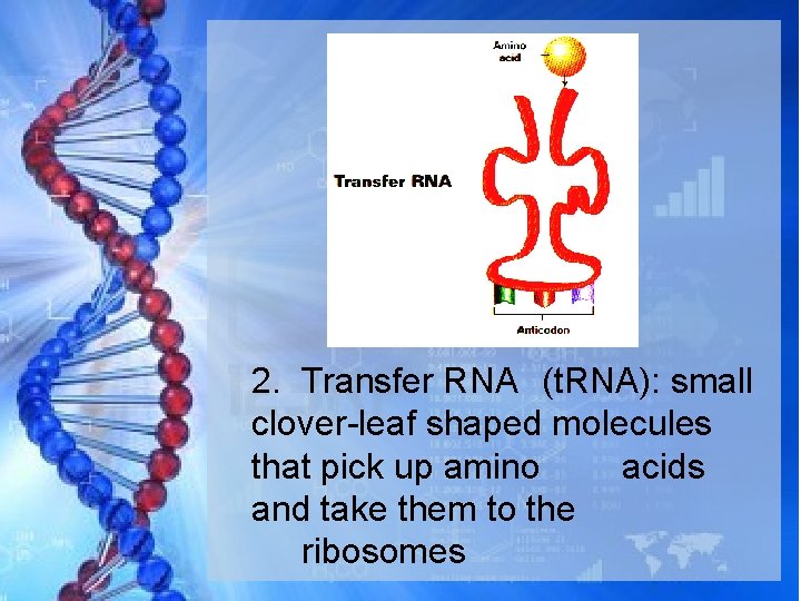
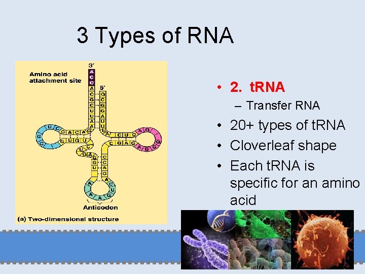
2. Transfer RNA (t. RNA): small clover-leaf shaped molecules that pick up amino acids and take them to the ribosomes
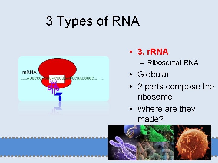
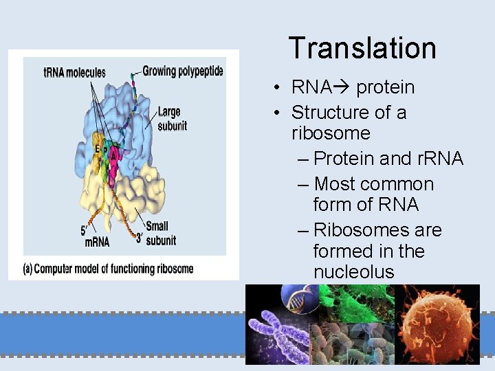
3. Ribosomal RNA (r. RNA): makes up the structure of ribosomes along with proteins
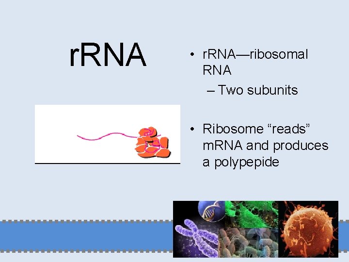
r. RNA • r. RNA—ribosomal RNA – Two subunits • Ribosome “reads” m. RNA and produces a polypepide
3 Types of RNA • 1. m. RNA – Messenger RNA • Single strand • Serves as a template (pattern for translation)
3 Types of RNA • 2. t. RNA – Transfer RNA • 20+ types of t. RNA • Cloverleaf shape • Each t. RNA is specific for an amino acid
3 Types of RNA • 3. r. RNA – Ribosomal RNA • Globular • 2 parts compose the ribosome • Where are they made?
F. The roles of RNA 1. RNA is not the genetic material and does not need to be capable of serving as a template for its own replication. 2. RNA functions as the intermediate, the m. RNA, between the gene and the proteinsynthesizing machinery. 3. RNA functions as an adaptor, the t. RNA, between the codons in the m. RNA and amino acids.
F. The roles of RNA 4. RNA also play a structural role, as in the case of the RNA components of the ribosome. 5. RNA is as a regulatory molecule, which through sequence complementarity binds to, and interferes with the translation of, certain m. RNAs. 6. Some RNAs are enzymes that catalyze essential reactions in the cell.
The phosphate groups of DNA and RNA are negatively charged 5’ HO-CH 2 N O O M+ O P O CH 2 O N O O A phosphodiester group has a p. Ka of about 1, and so will always be ionized and negatively charged under physiological conditions (p. H ~7). Nucleic acids require counterions such as Mg 2+, polyamines, histones or other proteins to balance this charge. O P O M+ CH 2 O N O O M+ O P O CH 2 O O N 3’ O-PO 32 M+
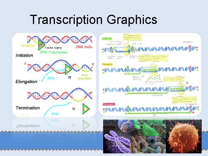
Transcription A. Transcription: the process in which DNA makes a complementary copy of m. RNA B. Steps of transcription 1. DNA untwists and bases separate 2. Only a small section of the DNA is involved and only one strand acts as the template
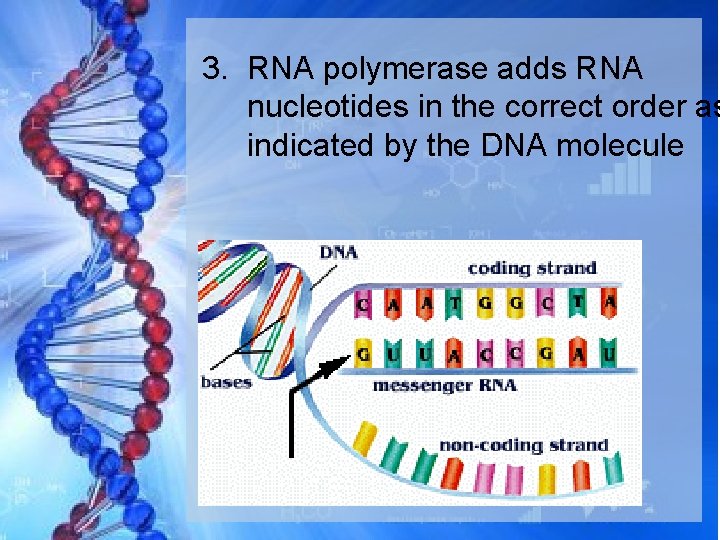
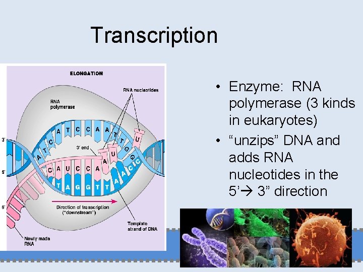
3. RNA polymerase adds RNA nucleotides in the correct order as indicated by the DNA molecule
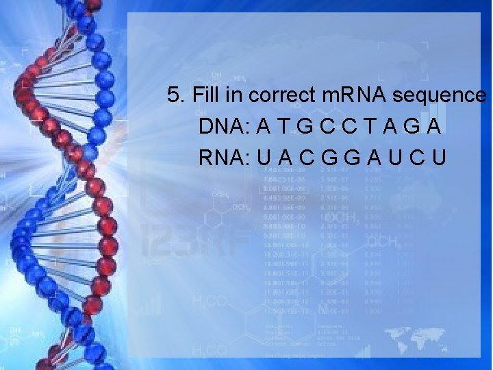
4. Base pairing rules apply a. If DNA has a T, RNA will match by adding A b. If DNA has an A, RNA will match by adding U c. If DNA has a C, RNA will match by adding G d. If DNA has a G, RNA will match by adding C
5. Fill in correct m. RNA sequence DNA: A T G C C T A G A RNA: U A C G G A U C U
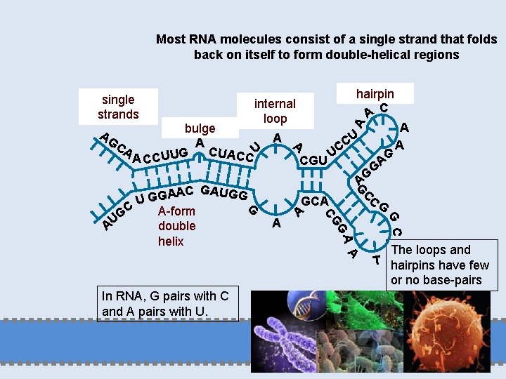
Most RNA molecules consist of a single strand that folds back on itself to form double-helical regions single strands hairpin A C A internal loop bulge U A A CA C CUACC U G U U U C AC CGU AG CC GCA A GC G A G GAUGG G In RNA, G pairs with C and A pairs with U. G AG CG AC A G G U C A-form G double AU helix G A A A The loops and T hairpins have few or no base-pairs
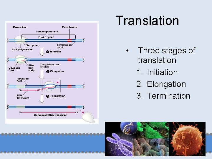
Transcription • Enzyme: RNA polymerase (3 kinds in eukaryotes) • “unzips” DNA and adds RNA nucleotides in the 5’ 3” direction
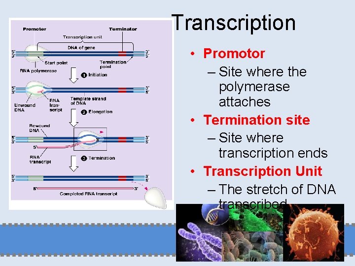
Transcription • Promotor – Site where the polymerase attaches • Termination site – Site where transcription ends • Transcription Unit – The stretch of DNA transcribed
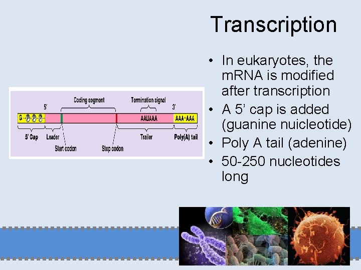
Transcription • In eukaryotes, the m. RNA is modified after transcription • A 5’ cap is added (guanine nuicleotide) • Poly A tail (adenine) • 50 -250 nucleotides long
Transcription Graphics
RNA Processing A. There are large sections of RNA molecules that are not used in making protein. These must be cut out before the RNA leaves the nucleus
B. Sections that are cut out (not used to make the protein) are called introns C. Sections that are used to make the protein are called exons (they are expressed) D. Some parts of RNA molecules may be exons when one protein is made and introns when another protein is made
The Genetic Code A. Tells the cell how to assemble a protein B. Proteins determine the structure and function of organisms C. Proteins are made of amino acids D. The bases in m. RNA (as made from DNA) determine what amino acids will be assembled into a protein
E. 20 amino acids can be assembled into thousands of proteins F. This works similar to the way letters are assembled to make words - 26 letters in English alphabet make thousands of words
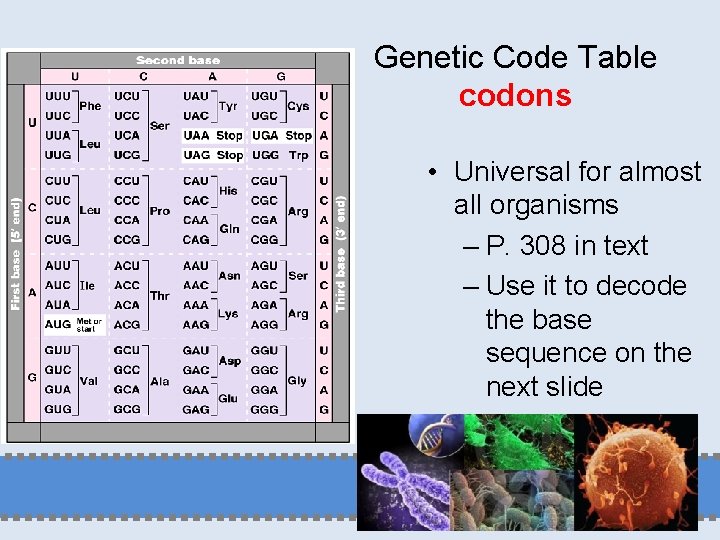
G. Codon: a sequence of 3 bases in m. RNA that codes for 1 amino acid 1. Examples GUG = valine GUA = valine GUC = valine
GAC = aspartic acid GAU = aspartic acid UCU = serine UCC = serine UCG = serine Which base could vary and still stand for the same amino acid? - 3 rd base
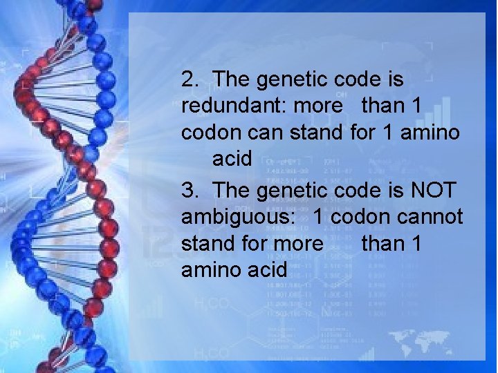
2. The genetic code is redundant: more than 1 codon can stand for 1 amino acid 3. The genetic code is NOT ambiguous: 1 codon cannot stand for more than 1 amino acid
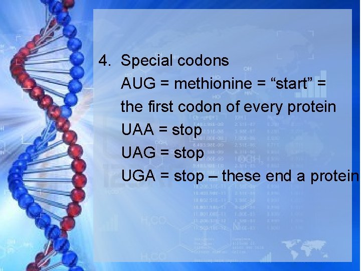
4. Special codons AUG = methionine = “start” = the first codon of every protein UAA = stop UAG = stop UGA = stop – these end a protein
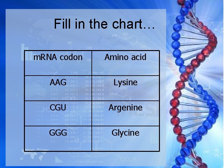
Fill in the chart… m. RNA codon Amino acid AAG Lysine CGU Argenine GGG Glycine
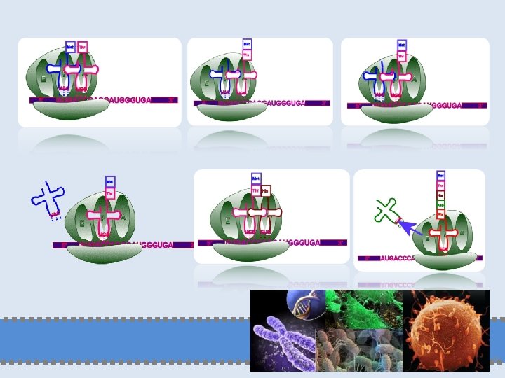
Translation A. Translation: the process in which the m. RNA message is decoded and a protein is made. B. Steps in translation 1. m. RNA made in the nucleus leaves and travels to a ribosome 2. m. RNA attaches to a ribosome
3. The ribosome reads the first codon, which is always AUG 4. A t. RNA that has a sequence of three complementary bases to m. RNA brings in the appropriate amino acid. The complementary bases on t. RNA are called an anticodon.
5. The ribosome reads the second codon and a t. RNA with a matching anticodon brings in a second amino acid 6. The ribosome joins the two amino acids with a linkage that is called a peptide bond 7. The ribosome moves down and reads the next codon
8. t. RNA molecules keep bringing in the appropriate amino acids 9. The process continues until a “stop” codon is reached 10. The polypeptide leaves the ribosome and folds to become a protein
Translation • RNA protein • Structure of a ribosome – Protein and r. RNA – Most common form of RNA – Ribosomes are formed in the nucleolus
Translation • Three stages of translation 1. Initiation 2. Elongation 3. Termination
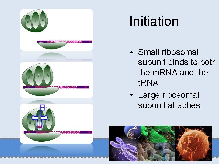
Initiation • Small ribosomal subunit binds to both the m. RNA and the t. RNA • Large ribosomal subunit attaches
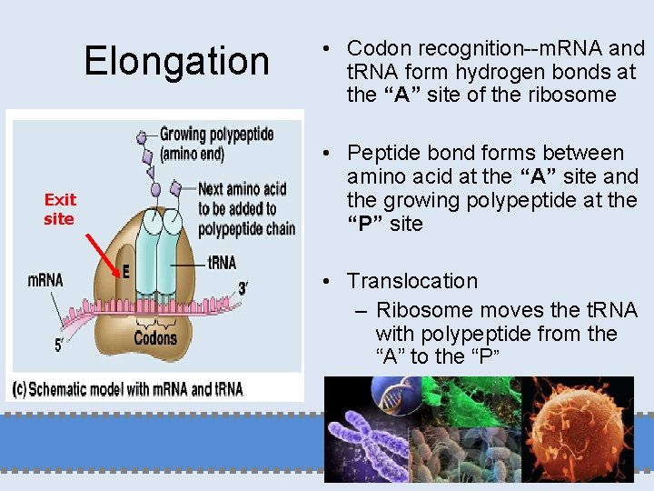
Elongation Exit site • Codon recognition--m. RNA and t. RNA form hydrogen bonds at the “A” site of the ribosome • Peptide bond forms between amino acid at the “A” site and the growing polypeptide at the “P” site • Translocation – Ribosome moves the t. RNA with polypeptide from the “A” to the “P”
45
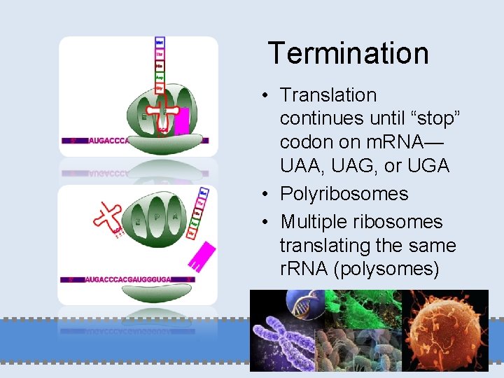
Termination • Translation continues until “stop” codon on m. RNA— UAA, UAG, or UGA • Polyribosomes • Multiple ribosomes translating the same r. RNA (polysomes)
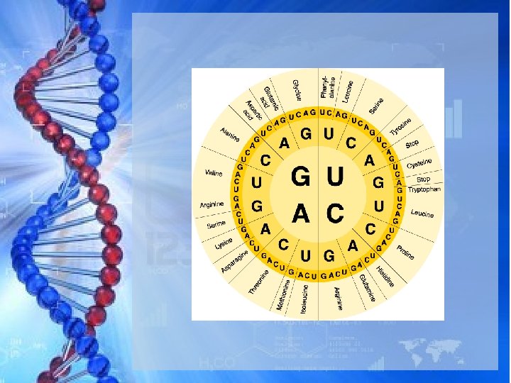
Genetic Code Table codons • Universal for almost all organisms – P. 308 in text – Use it to decode the base sequence on the next slide
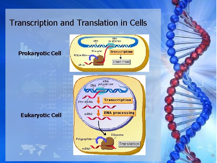
Transcription and Translation in Cells Prokaryotic Cell Eukaryotic Cell 48
THANK YOU YUSRON SUGIARTO, STP, MSc 49