Restriction enzymes Restriction endonucleases Restriction enzymes Are enzymes


- Slides: 32

Restriction enzymes Restriction endonucleases

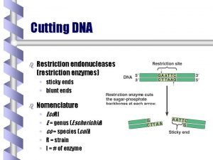
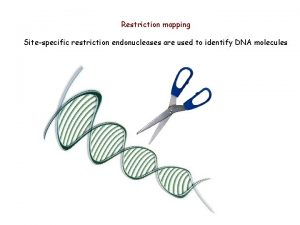
Restriction enzymes Ø Are enzymes that cut a DNA molecule at a particular place. Ø The enzyme "scans" a DNA molecule, looking for a particular sequence, usually of 4 -6 nucleotides. Ø These sequences are palindromic in that the complimentary DNA strand has the same sequence only in the reverse direction, so both strands of DNA are cut at the same location. (GAATTA – ATTAAG) Ø Once it finds this recognition sequence, it stops and cuts the strands. This is known as enzyme digestion.

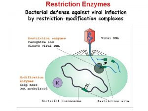
§ Restriction enzymes apparently evolved as a primitive immune system in bacteria. § The term "restriction" derives from the phenomenon in which bacterial viruses are restricted from replicating in certain strains of bacteria by enzymes that cleave the viral DNA, but leave the bacterial DNA untouched. § Methylation of DNA at the recognition sequence typically protects the microbe from cleaving its own DNA.

Nomenclature § Restriction enzymes are named based on the organism in which they were discovered. § For example, the enzyme Hin d III was isolated from Haemophilus influenzae, strain Rd. § The first three letters of the name are italicized because they abbreviate the genus and species names of the organism.

Nomenclature • The fourth letter (if present) refers to the bacterial strain designation. • The Roman numerals are used to identify specific enzymes from bacteria that contain multiple restriction enzymes. • Typically, the Roman numeral indicates the order in which restriction enzymes were discovered in a particular strain.

Nomenclature • Bam. H I: Bacillus amyloliquefaciens, strain H, enzyme I • Hind III: Haemophilus influenzae strain d, enzyme III • Sma I: Serratia marcesens, enzyme I • Kpn I: Klebsiella pneumonite, enzyme

Classification • There are three classes of restriction enzymes, labeled types I, II, and III. Ø Type I restriction systems § Consist of a single enzyme that performs both modification (methylation) and restriction activities. § These enzymes recognize specific DNA sequences, but cleave the DNA strandomly, at least 1, 000 base pairs (bp) away from the recognition site.

Classification Ø Type III restriction systems § Have separate enzymes for restriction and methylation, but these enzymes share a common subunit. § These enzymes recognize specific DNA sequences, but cleave DNA at random sequences approximately 25 bp from the recognition sequence.

Classification Ø Type II enzymes § Consist of single, separate proteins for restriction and modification. § Type II restriction enzymes cleave the DNA sequence at the same site at which they recognize it. § Neither type I nor type III restriction systems have found much application in recombinant DNA techniques.

Types of cuts of Restriction Enzymes • If they cut both strands at the center of the recognition sequence, it will generate "blunt ends" with no nucleotide overhangs. • If they cut both strands closer to one end of the recognition sequence, it will generates "sticky" or "cohesive" ends with nucleotide overhangs.

Types of cuts of Restriction Enzymes

Types of cuts of Restriction Enzymes

Use of Restriction Enzymes in Biotechnology • DNA fragments or genomes map • Verify the identity of a specific DNA fragment • Identifying individuals or strains of a particular species – Restriction fragment length polymorphism (RFLP) – Pulsed field gel electrophoresis • Generation of recombinant DNA molecules


Unit Definition One unit is the enzyme activity that completely cleaves 1 µg of substrate DNA suspended in 50 µl of the recommended reaction buffer in 60 minutes at the appropriate temperature under optimal assay conditions as stated for each restriction endonuclease.

Restriction enzyme digestion of DNA Ø Typical protocol – 1µl 10 x Buffer (A, B, C, D Salt conc, Mg conc, p. H) – 6µl H 2 O – 2µl DNA (kit extracted, salt interfere with enzyme, funny gel) – 1µl Enzyme • Do not use more enzyme than 10% of the final reaction volume • the enzyme storage buffer contains antifreeze (glycerol) to allow it to survive at – 20°C. The glycerol will inhibit the digestion if present in sufficient quantities. – Incubate at 37°C for one hour

Special conditions Ø Double digests – Digestion of DNA with two (or more) enzymes – Make sure to use the buffer that will be most compatible with all the enzymes.

Special conditions Ø A few enzymes require special conditions Ø Some require BSA (bovine serum albumin) added into the mixture. Ø Some require weak detergents (e. g. triton-X-100) to reduce surface tension. Ø Some require to be incubated at temperatures other than 37°C (e. g. 50°C)

Star activity Ø This is when the enzyme cuts at sites other than its cognate element. Ø e. g. Eco. RI is supposed to only cut GAATTC but, under extreme conditions, it might possibly cut CAATTC also.

Problems with enzyme activity Ø Problems with enzyme activity can occur under the following conditions: – High glycerol concentration – Enzyme-to-DNA ratio is too high – p. H is too high – Organic solvents, particularly ethanol, interfere with your DNA

Phosphorylation & Dephosphorylation • Digested DNA typically possesses a 5’ phosphate group that is required for ligation. • It is recommended to dephosphorylate (removal of the 5' phosphate) the linearized vector to prevent self-ligation phosphate) & facilitate ligation of recombinant DNA molecules. & • Dephosphorylation can be accomplished using any of a number of phosphatases, including Shrimp Alkaline Phosphatase (r. SAP) , Calf Intestinal Alkaline Phosphatase (CIP)

Phosphorylation & Dephosphorylation Ø ligation requires the presence of a 5'-phosphate, the insert must be phosphorylated. Ø Blunt-ended PCR product normally lacks a 5'phosphate, therefore it needs to be phosphorylated by treatment with T 4 polynucleotide kinase Ø Digestion of DNA with a restriction enzyme will always produce a 5´ phosphate

DNA Ligation Ø The final step in the construction of a recombinant plasmid is connecting the insert DNA (gene or fragment of interest) into a compatibly digested vector backbone. This reaction, called ligation & is performed by the T 4 DNA ligase enzyme Ø After ligation, the insert DNA is physically attached to the backbone and the complete plasmid can be transformed into bacterial cells for propagation.

Ligation Protocol Ø It is important to determine the amount of cut insert and vector to use for the ligation reaction. Ø The volume of vector DNA and insert DNA used in the ligation will vary depending on the size of each and their concentration. Ø for most standard cloning (where the insert is smaller than the vector) A 3 insert : 1 vector ratio will work just fine.

Ligation reaction optimization Ø http: //www. insilico. uni-duesseldorf. de/Lig_Input. html Ø Just enter the concentration, lengths of the insert and vector, and what the ratio you want, and it will tell you exactly how much of each to use.

Ligation reaction • Since ATP can be damaged by repeated freeze-thaw cycles, it is advisable to make aliquots of the buffer. • The ligation reaction itself has two basic steps. – Firstly the DNA ends have to collide by chance and stay together long enough for the ligase to join them (low temperatures. ) – The second step is the enzymatic reaction (T 4 ligase). • Normally 1 hr at 16°C is fine but since but overnight at 4°C can give even more efficiency.

Transformation Ø Introduction of exogenous genetic material (vector) into a bacterial cell. (uptake of DNA by bacterial cells) Ø The host cells must be specially prepared to take up the foreign DNA. Ø Selectable markers can be for antibiotic resistance, color changes, or any other characteristic which can distinguish transformed hosts from untransformed hosts.

Transformation Ø Most types of cells cannot take up DNA efficiently unless they have been exposed to special chemical or electrical treatments to make them competent. Ø The standard method for making the bacteria permeable to DNA involves treatment with calcium ions (Ca. Cl 2 ). Ø Brief exposure of cells to an electric field also allows the bacteria to take up DNA and this process is called as electroporation

Transformation Ø ln Ca. Cl 2 method, the competency can be obtained by creating pores in bacterial cells by suspending them in a solution containing high concentration of calcium. • The plasmid DNA is mixed with chilled cells and incubated on ice to allow the plasmid to come into close contact with the cells. Ø The plasmid-cell mixture then is briefly heated to 42°C, allowing the DNA to enter the cell through the disrupted membrane Ø The heated mixture is then placed back on ice to retain the plasmids inside the bacteria


Transformation Ø For electroporation, the competent cells also sit on ice with the plasmid DNA. Ø However, the plasmid-cell mixture is exposed to an electrical current, opening pores in the cell membrane so that the plasmid can enter the cell. Ø Both methods allow efficient recovery of transformed cells using antibiotic selection for the plasmid of interest.

Thank You For your attention Dr. Hatem Toughan Fish Medicine Faculty of Veterinary Medicine Assiut University
 Types of restriction endonucleases
 Mikael ferm
 Restriction enzymes
 Selective breeding definition biology
 Restriction enzymes
 Molecular scissors
 Restriction enzymes
 Restriction enzyme
 Polyacrimide
 Enzyme
 Carte de restriction linéaire
 Symmetric growth restriction
 Illustration of the steps in restriction digestion and pcr
 Rflp vs rapd
 John gurdon
 Molecular scissors
 Restriction codes in texas
 Molecular scissors wikipedia
 Restriction mapping
 What is restriction digestion
 How to draw a restriction map
 Symmetrical and asymmetrical iugr
 Intermediate accounting chapter 16
 Example 2
 Dna fragment length
 Sticky ends
 Cla 1 restriction enzyme
 Cronies calorie restriction
 Restriction mapping
 Restriction collocation
 Restriction enzyme
 Endonuclease restriction
 Symmetrical growth restriction
