RENAL STONES DR OHANG CHAUDHARI Etiology Idiopathic calcium

 Hypercalcaemic disorders Renal tubular syndromes Uric acid")


 DRUGS: acetazolemide, allopurinol, thiazide")
 also known as mulberry stones, brown in color having")


- Slides: 52
RENAL STONES DR. OHANG CHAUDHARI
Etiology • • • Idiopathic (calcium stone) Hypercalcaemic disorders Renal tubular syndromes Uric acid lithiasis Enzyme disorders Secondary urolithiasis
Idiopathic • Unexplained hypercalciuria with normal serum calcium level • 70% of renal stone patients have this type • 1 ) renal type- more common in children 2) absorptive type- more common in adult
HYPERCALCAEMIC DISORDERS • Parathyroid adenoma, chief cell hyperplasia • Overproduction of parathormone • Increased reabsorption of calcium from intestine, and renal tubules, &bone resorption • ( primary hyperparathyroidism)
Prolonged immobilisation • Lead to bone resorption • Hypercalcaemia, hypercalciuria • As immobility induces osteoclast mediated bone resorption
Milk alkali syndrome • Increased consumption of calcium, vitamine –D lead to hypercalcaemia, alkalosis, • Alkalosis compromise renal excretion of calcium • Other sarcoidosis
RENAL TUBULAR SYNDROME • Renal tubular acidosis • Cystinuria • Uric acid lithiasis
RTA • RTA type 1 associated with renal stone • Due to hypercalciuria & low urinary citrate excretion • Stones composed of calcium phosphate
cystinuria • Autosomal recessive disorder • Defect in amino acid transport in GIT & renal tubules. . cystine, lysine, arginine, ornithine • Increased excretion of such amino acid in urine • Cystine stones – multiple, very hard, radio opaque • Family members should be screened for it
Uric acid lithiasis • 5 -10% of renal stones • Uric acid is end product of purine metabolism • Who take exessive purine content food will excrete more uric acid in urine • Uric acid remains undissociated and insoluble at PH <5. 5 • Low urine volume contribute to uric acid stone formation
Uric acid stone • Inflammatory bowel disease, ileostomy lead to less urine formation • Uric acid stones are hard , smooth, multiple faceted • Pure uric acid stones are RADIOLUCENT but they look radio-opaque due to little bit presence of calcium
ENZYME DISORDERS • PRIMARY HYPEROXALURIA • XANTHINURIA • 2, 8 -DIHYDROADENURIA
PRIMARY HYPEROXALURIA • • • Autosomal recessive disorder Increased production of OXALATE Results in nephrocalcinosis Calcium oxalate stone Common in children
SECONDARY UROLITHIASIS • SECONDARY HYPEROXALURIA • DIETARY EXCESS- spinach, tea, coco, chocolate, peper increases urinary oxalate • INFECTION- urease producing organismproteus, pseudomonas, staphylococus,
infection • Bacteria breakdown urea to produce ammonia and co 2 • Urine becomes alkaline, , promote formation of STRUVITE STONE ( magnessium ammonium phoshphate) • Grown to form STAGHORN CALCULI
OTHER OBSTRUCTION STASIS MEDULARY SPONGE KIDNEY URINARY DIVERSION-(infection acidosis stasis) DRUGS: acetazolemide, allopurinol, thiazide diuretics • Less water intake • Sedentary life style • • •
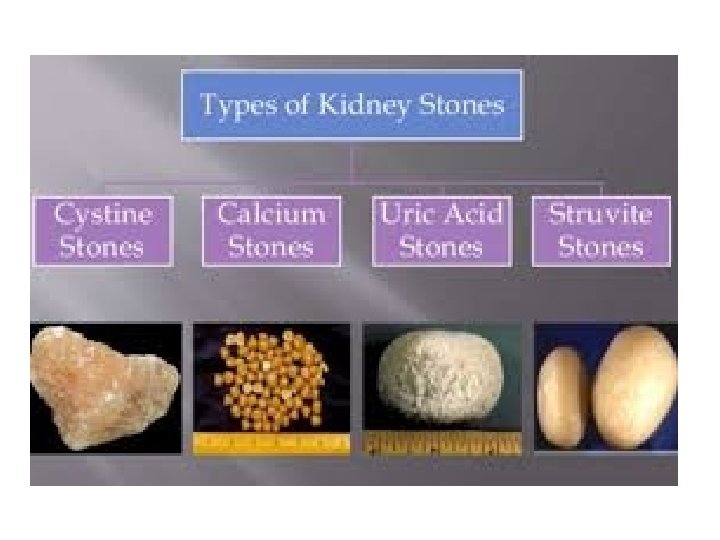
TYPES • OXALATE STONES- (75%) also known as mulberry stones, brown in color having sharp projections • PHOSPHATE STONES (15%)-in infected urine, smooth & white in color, in alkaline urine filled in major calyces , taking shape of it. ( STAGHORN CALCULUS), large size, radio opaque • URIC ACID STONES –smooth, hard, yellow, multiple, radilolucent • CYSTINE STONES- cystinuria patient, very hard stone, attains large size
TYPES • XANTHINE STONE • STRUVITE STONE • INDIGO STONE
Calcium Oxalate stone
Stag horn stone
Stag horn
Struvite stone
CLINICAL FEATURES • PAIN- pain at renal angle, and in lumbar region - radiating to groin and testis -pain worsen on movement • VOMITING- due to pylorospasm • Hematuria • Pyuria • Fever • Burning micturition
Clinical sign • Tenderness in renal angle • Mass in hypochondrium or lumbar due to hydronephrosis which moves with respirations that is bimanually palpable ballotable soft smooth • May present with renal failure due to out flow obstruction
investigations • • • CBC RFT ESR S. CALCIUM BLOOD UREA, URIC ACID PTH LEVEL
INVESTIGATIONS • • • URINE – CALCIUM, CYSTINE, URIC ACID, MICRO PLAIN X RAY KUB IVP USG CECT RENAL SCAN
MANAGEMENT • CONSERVATIVE • SURGICAL
CONSERVATIVE • • If stone size < 8 mm Plenty of liquid orally Intravenous furosemide Anti spasmodic drugs
surgical • • If size > 8 mm PCNL ESWL OPEN SURGERIES - PYELOLITHOTOMY -NEPHROLITHOTOMY -PARTIAL NEPHRECTOMY
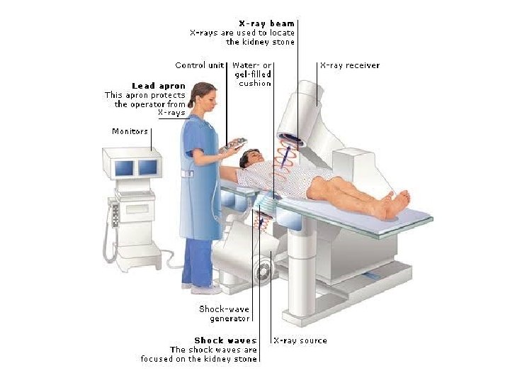
ESWL • Endoscopic shock wave lithotripsy • Piezo-electric or electromagnetic shock waves passed on stone through water bath or water cushion • Stone location confirmed by C-arm • 2 shocks per second • 1000 -4000 shocks required for each stone • Fragmented stone come out in urine
ESWL TABLE
ESWL
C ARM
WITH FLUOROSCOPY
ESWL ADVANTAGE • • • No anesthesia needed Can be done as OPD procedure , 2. 5 cm size stone can break very well Hard stones can easily eliminated Can perform multiple times as it is non invasive
COMPLICATIONS • RENAL HEMATOMA • HEMATURIA • FRAGMENTED STONE RETAINED IN URETER
CONTRAINDICATIONS • • PREGNANCY BLEEDING DISORDERS PATIENTS WITH ABDOMINAL ANEURYSM SEPSIS & RENAL FAILURE
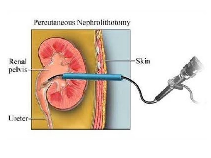
PCNL • Per cutaneous nephrolithotomy • stones - > 2. 5 cm - multiple stones - stones not responding to ESWL • Cystoscopy is done and ureteric stent is placed • Renal pelvicalyceal system identified under C-ARM guidance
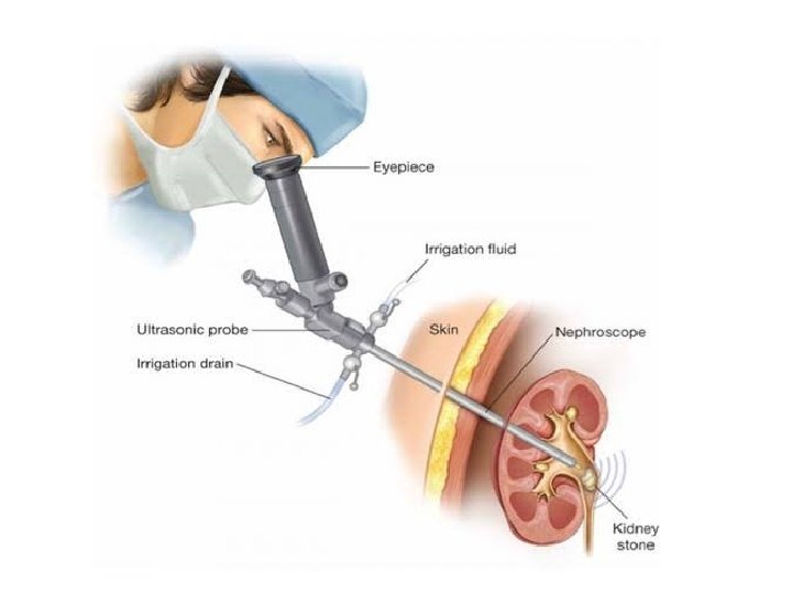
PCNL Through kidney , calyx and pelvis approached Will pass guide wire after needle puncture By using Graduated dilator track is widen NEPHROSCOPE is passed Will break stone by laser holmium, ultrasonic or electrohydraulic lithotripser • Size- 5 mm, 10 mm , 15 mm • • •
NEPHROSCOPE
COMPLICATIONS • HAEMORRHAGE • PERFORATION OF COLLECTING DUCTS • INJURY TO PLEURA, COLON
PYELOLITHOTOMY • • Stone in extra renal pelvis Posterior subcostal incision Remove 12 th RIB Open renal pelvis Remove stone Put DJ stent Put drain closure
EXTENDED PYELOLITHOTOMY • In case of intra renal pelvis • Incision on HILUM between pelvis & kidney over renal sinus
NEPHROLITHOTOMY • INCISION OVER BRODEL”S LINE • ON MOST CONVEX SURFACE OVER POSTERIOR PART • STONE REMOVED
NEPHROPYELOLITHOTOMY • INCISION OVER KIDNEY as well as on PELVIS • In case of STAGHORN STONE
PARTIAL NEPHRECTOMY • • • Multiple stones occupying in ONE POLE Usually lower Damage to that calyx Remove half kidney If not removed – can encourage further stone formation later
other • Bench surgery- kidney removed temporarily in ice pack, replaced in RIF • Coagulum pyelolithotomy • Anatrophic pyelolithotomy • Nephrectomy
THANK YOU.