Urolithiasis Renal Calculi Stones 1 Urolithiasis Renal Calculi


- Slides: 43

Urolithiasis (Renal Calculi, Stones) 1

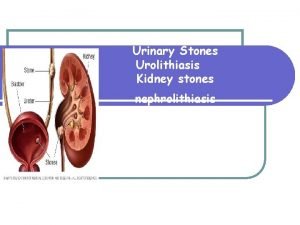
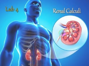
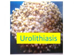
Urolithiasis (Renal Calculi, Stones) • Urolithiasis is calculus formation at any level in the urinary collecting system, but most often the calculi arise in the kidney • Symptomatic urolithiasis in Men > women • A familial tendency in stone formation was recognized. 2

Urolithiasis (Renal Calculi, Stones) • Stones are unilateral in about 80% of patients. • Common sites of formation are renal pelvis and calyces and the bladder. • average diameter, 2 to 3 mm and may be smooth or jagged. 3

4 An organic matrix of mucoprotein, making up 2. 5% of the stone by weight, is present in all calculi Although there are many causes for the initiation and propagation of stones, the most important determinant is an increased urinary concentration of the stones' constituents, such that it exceeds their solubility in urine (supersaturation). A low urine volume in some metabolically normal patients may also favor supersaturation

Three major types of stones • About 80% of renal stones are composed of either calcium oxalate or calcium oxalate mixed with calcium phosphate. • 10% are composed of magnesium ammonium phosphate. • 6%to 9% are either uric acid or cystine stones 5

6

Calcium oxalate stones 7

Calcium stones Most in this group absorb calcium from the gut in excessive amounts (absorptive hypercalciuria) and promptly excrete it in the urine, and some have a primary renal defect of calcium reabsorption(renal hypercalciuria). 8

Magnesium ammonium phosphate stones after infections by urea-splitting bacteria ( Proteus and some staphylococci), which convert urea to ammonia. The resultant alkaline urine causes the precipitation of magnesium ammonium phosphate salts. 9

Magnesium ammonium phosphate stones These form some of the largest stones, as the amounts of urea excreted normally are huge. so-called staghorn calculi occupying large portions of the renal pelvis are almost always a consequence of infection In avitaminosis A, desquamated cells from the metaplastic epithelium of the collecting system act as nidi 10

Uric acid stones • More than half of all patients with urate calculi have neither hyperuricemia nor increased urinary excretion of uric acid(Idiopathic) • patients with hyperuricemia, such as gout, leukemias. • tendency to excrete urine of p. H < 5. 5 may predispose to uric acid stones, because uric acid is insoluble in relatively acidic urine. • In contrast to the radio-opaque calcium stones, uric acid stones are radiolucent 11

Cystine stones • associated with a genetically determined defect in the renal transport of certain amino acids, including cystine. • Stones form at low urinary p. H. 12

Clinical Course Stones are of importance when they obstruct urinary flow or produce ulceration and bleeding. Stones predispose to superimposed infection, both by their obstructive nature and by the trauma they produce 13

Clinical Course smaller stones are most hazardous, because they may pass into the ureters, producing pain referred to colic as well as ureteral obstruction. Larger stones cannot enter the ureters and are more likely to remain silent within the renal pelvis. first manifest themselves is hematuria. 14

Hydronephrosis • Dilation of the renal pelvis and calyces associated with progressive atrophy of the kidney due to obstruction to the outflow of urine. 15

Morphology of hydronephrosis In sudden and complete obstruction reduction of glomerular filtration leads to mild dilation of the pelvis and calyces but sometimes to atrophy of the renal parenchyma 16

Morphology of hydronephrosis In subtotal or intermittent obstruction glomerular filtration is not suppressed, progressive dilation ensues 17

Morphology of hydronephrosis The earlier features are tubular dilation, followed by atrophy and fibrous replacement of the tubular epithelium with relative sparing of the glomeruli. in severe cases the glomeruli also become atrophic and disappear, converting the entire kidney into a thin shell of fibrous tissue 18

Morphology of hydronephrosis • With sudden and complete obstruction, there may be coagulative necrosis of the renal papillae, similar to the changes of papillary necrosis. • In uncomplicated cases the accompanying inflammatory reaction is minimal. • Superimposed pyelonephritis, however, is common 19

urinary tract Tumors • Tumors of the lower urinary tract are about twice as common as renal cell carcinomas • The most common malignant tumor of the kidney is renal cell carcinoma, followed in frequency by nephroblastoma (Wilms tumor) and by primary tumors of the calyces and pelvis. 20

Tumors of the Kidney Benign Oncocytoma Papillary adenoma in 40% of adults, have no clinical significance Malignant RCC Wilm, s tumor Medulary fibroma Angiomyolipoma 21

Oncocytoma a benign tumor about 10% of renal tumors Intercalated cells of collecting ducts associated with loss of chromosomes 1, 14, and Y-that distinguish them from other renal neoplasms. 22

Oncocytoma A central stellate scar provides a characteristic appearance on imaging studies. they are removed by nephrectomy, both to prevent such complications as spontaneous hemorrhage and to make a definitive diagnosis. 23

Histologically characterized by a excess of mitochondria, providing the basis for their tan color and their finely granular eosinophilic cytoplasm 24

KIDNEY MALIGNANT TUMORS 25

MALIGNANT TUMORS Renal cell carcinomas • 2% to 3% of all visceral cancers • 85% of renal cancers in adults • In the sixth and seventh decades of life, showing a male /female ratio of 2 to : 1 • All these tumors arise from tubular epithelium and are therefore renal adenocarcinomas. • in any portion of the kidney, more commonly, particularly the upper poles • located predominantly in the cortex 26

Epidemiology of Renal cell carcinomas Cigarette smoking obesity Hypertension cadmium acquired cystic disease (30 -fold) Most renal cancer is sporadic, but unusual forms of autosomal-dominant familial cancers occur, usually in younger individuals. 27

Classification of Renal Cell Carcinoma CLEAR CELL CARCINOMA PAPILLSRY RENAL CELL CARCINOMA CHROMOPHOB CARCINOMA 28

Clear cell carcinoma The most common type 65% of renal cell cancers They can be familial, associated with VHL disease, or in most cases (95%) sporadic. In 98% of these tumors, loss of sequences on the short arm of chromosome 3. chromosomal translocation (3; 6, 3; 8, 3; 11) resulting in loss of chromosome 3 29

recent deep sequencing of clear cell carcinoma genomes has revealed frequent loss-of-function mutations in SETD 2, JARID 1 C, and UTX, all of which encode proteins that regulate histone methylation, suggesting that changes in the" epigenome" have a central role in the genesis of this subtype of renal carcinoma. 30

Clear cell carcinoma (spherical masses 3 to 15 cm in diameter) May arise anywhere in the cortex. The margins of the tumor are well defined. Sometimes with small satellite nodules The cut surface is yellow to orange to gray-white, with prominent areas of cystic softening or of hemorrhage, either fresh or old 31

Clear cell carcinoma As the tumor enlarges it may fungate through the walls of the collecting system, extending through the calyces and pelvis as far as the ureter. Even more, the tumor invades the renal vein and inferior vena cava and even into the right side of the heart grows as a solid column within this vessel direct invasion into the perinephric fat and adrenal gland may be seen 32

• Depending on the amounts of lipid and glycogen the tumor cells may appear vacuolated (lipidladen), or solid. • At the other extreme are granular cells resembling the tubular epithelium, which have small, round, regular nuclei enclosed within granular pink cytoplasm 33

• The nuclei are usually small and round. • Some tumors are highly anaplastic, with numerous mitotic figures and markedly enlarged hyperchromatic, pleomorphic nuclei. • The cells may form abortive tubules or cluster in cords or disorganized masses. • The stroma is usually scant but highly vascularize 34

Papillary carcinoma • 10% to 15% of renal cancers • characterized by a papillary growth pattern and also occurs in both familial and sporadic forms • Frequently multifocal and bilateral and appear as early-stage tumors 35

Papillary carcinoma • These tumors are not associated with 3 p deletions. • The culprit in most cases of hereditary PRCC is the MET proto-oncogene, located on chromosomal subband 7 q 31. (The MET gene is a tyrosine kinase receptor for the hepatocyte growth factor) 36

• may show gross evidence of necrosis, hemorrhage, and cystic degeneration, but they are less vibrantly orange-yellow because of their lower lipid content. • various degrees of papilla formation with fibrovascular cores. • The cells may have clear or, more commonly pink cytoplasm. 37

Papillary carcinomas are the most common type of renal cancer in patients who develop dialysisassociated cystic disease 38

Chromophobe renal carcinoma • 5% of renal cell cancers • On cytogenetic examination, these tumors exhibit multiple chromosome losses including chromosomes 1, 2, 6, 10, 13, 17, and 21 • Thus, they show extreme hypodiploidy. Because of multiple losses, the" critical hit" has not been determined 39

Chromophobe renal carcinoma • They arise from intercalated cells of collecting ducts. • Their name derives from the observation that the tumor cells stain more dark (they are less clear) than cells in clear cell carcinomas. 40

grossly tan-brown. The cells have clear, flocculent cytoplasm with very prominent, distinct cell membranes. The nuclei are surrounded by halos of clear cytoplasm. Ultrastructurally, large numbers of characteristic macrovesicles are seen. 41

Clinical Course The three classic diagnostic features of renal cell carcinoma are : costovertebral pain Palpable mass Hematuria, but these are seen in only 10% of cases. The most reliable of the three is hematuria, ( intermittent/ microscopic) in more than 50% 42

A number of paraneoplastic syndromes associated to RCC : • • • Polycytemia Hypercalcemia Hypertension Feminization or masculinization Cushing syndrome 43
 Res extra commercium
 Ira pré renal renal e pós renal
 Struvite vs calcium oxalate
 Calculi sumeri
 Calcium phosphate calculi
 Vasa recta vs peritubular capillaries
 Collective noun for cotton
 The lottery shirley jackson worksheet
 Stepping stones portsmouth ohio
 Built of living stones
 Nephrology
 Stepping stones to glory analysis
 Triple p stepping stones
 Rolling stone origin
 Odowa stones
 Sufi proverb in the desert there is no sign
 Magnesium ammonium phosphate stones
 Hoarse shoe kidney
 Cart before the horse
 Magnesium ammonium phosphate stones
 Large monuments created from huge stone slabs
 Parable of stones by gemino h. abad
 Date and title
 Stick and stones poem
 Difference between rock and stone
 Sticks and stones origin
 Rolling stone sympathy for the devil chords
 The rolling stones history
 Summation formulas
 What does kidney stones look like
 Crossing jordon
 Weihnachtskugel rolling stones
 Stones bones and petroglyphs reading street
 Sticks and stones origin
 Stepping stones to glory cartoon analysis
 Chronic hyperplastic pulpitis
 Emily wants to buy turquoise stones
 Renal
Renal Insuficiencia renal prerrenal y postrenal
 Palidez terrosa renal
 Nefrone iuxtamidollare
 Loading dose formula in pharmacology
 Renal osteodystrophy
 Girft renal


