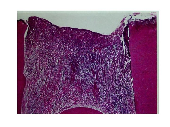
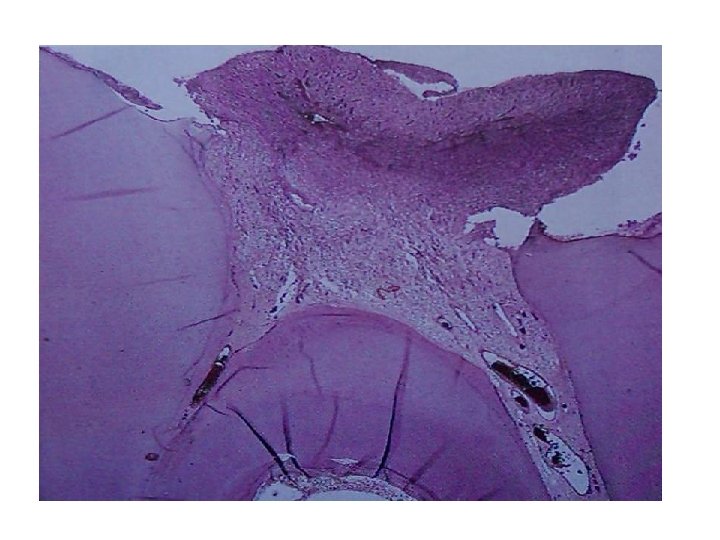
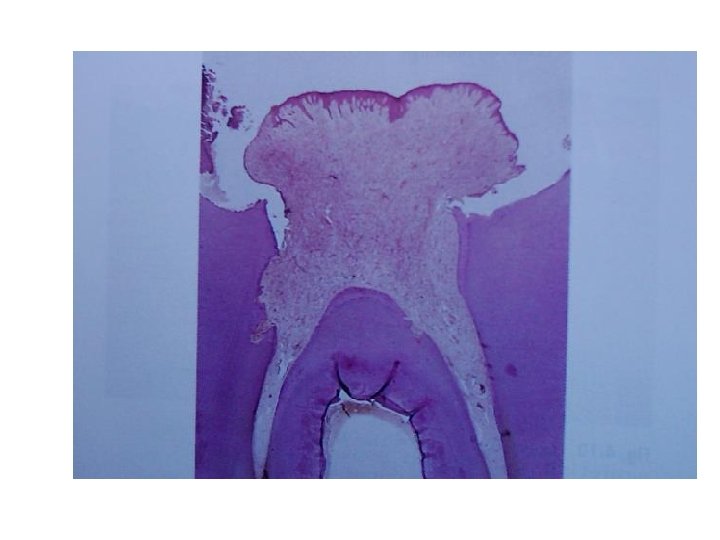
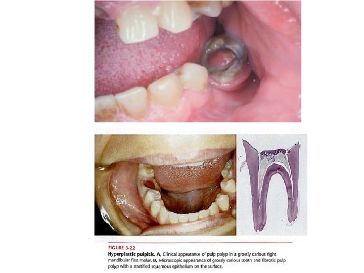
Histopathological features of chronic hyperplastic pulpitis This demonstrates


 are calcified bodies with an organic matrix.")


- Slides: 22
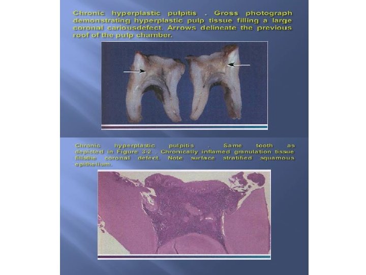
Histopathological features of chronic hyperplastic pulpitis �This demonstrates a cap of sub acutely inflamed granulation tissue that fills the entire space of the original pulp chamber The surface of the polyp may or may not be covered with stratified squamous epithelium, which migrates from the adjacent gingiva or arise from sloughed epithelium within the oral fluids. �The deeper pulp tissue within the canals typically demonstrate fibrosis and chronic inflammation. .
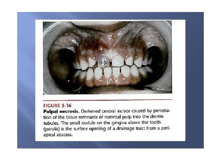
Pulp Necrosis �May follow either pulpitis or a traumatic injury to the apical blood vessels. �A coagulative type of necrosis is seen after ischemia, trauma and the patient usually has no symptoms. �If the necrosis follows pulpitis may lead to liquefactive type of necrosis : - Which may become infected by bacteria from caries. - This type is usually associated with foul odour when opened with endodontic treatment.
Diagnosis of pulp pain �The diagnostic procedure that are commonly used to assess the status of a symptomatic tooth and pulp are as follows: 1 -History and nature of pain. 2 -Visual clinical examination. 3 -Reaction to thermal changes. 4 -Reaction to electric stimulation. 5 -Reaction to tooth percussion. 6 -Radiographic examination. 7 -Palpation of the surrounding area.
Vitality test Cold test: Ethyl chloride Electrical pulp test
� Numerous disorders have been reported to mimic pulpalgia, e. g. migrain, headache, and myofacial pain. �If these conditions are not considered then the results would be: Sequential extractions or endodontic treatment which is all not needed and inappropriate.
Treatment and prognosis �Reversible pulpitis -- Removal and elimination of the cause, on occasion analgesics are required. -- Prognosis is good if action taken early. -- Pulp testing is essential periodically to ensure that irreversible damage has not occurred. �Irreversible pulpitis Acute, chronic hyperplastic are treated by endodontic treatment or extraction.
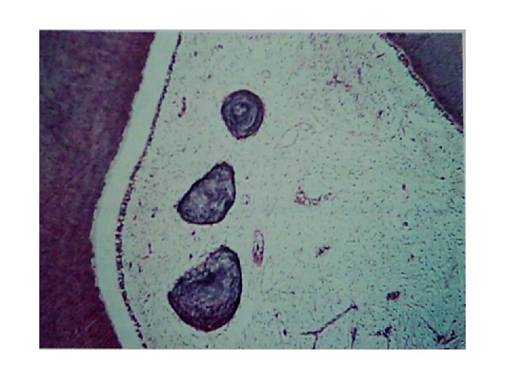
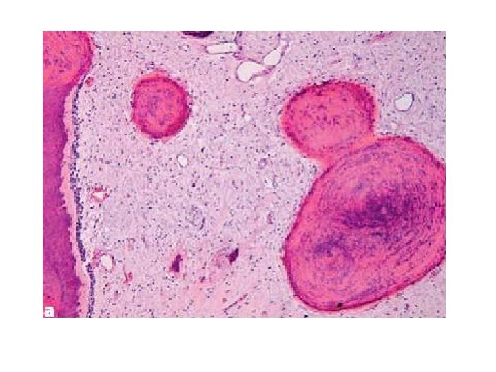
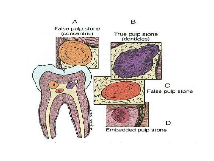
Pulp calcification • Pulp stones (or denticles) are calcified bodies with an organic matrix. • Occur most frequently in the coronal pulp. • True pulp stones contain tubules (scanty and irregular). may have an outer layer of predentine and adjacent odontoblasts.
False pulp stones are composed of concentric layers of calcified material with no tubular structure. �According to their location in the pulp, stones may be described as free, adherent, or interstitial. �pulp stones increases in number and size with age and are apparently more numerous after operative procedures on the tooth. �when large they may be recognized on radiographs. �They do not cause symptoms. Although neuralgic pain has sometimes been attributed to their presence.
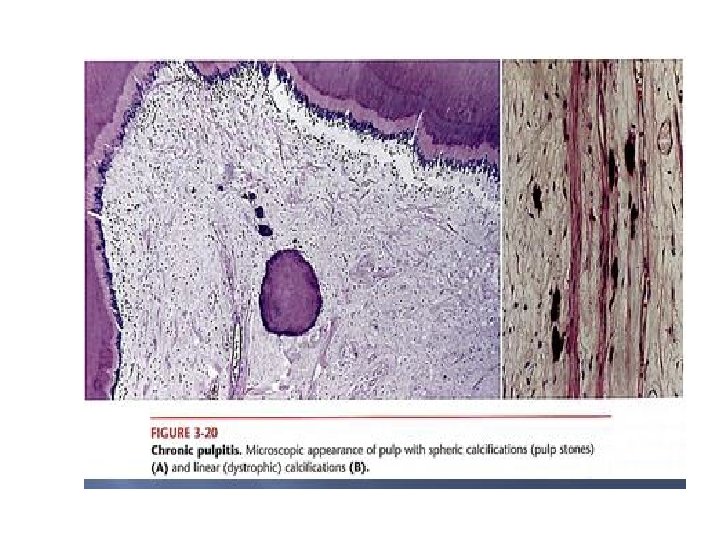
Diffuse linear calcifications • Consist entirely of fine, fibrillar, and irregular calcifications that develop in the pulp chambers and canals. • This material often is deposited in a linear fashion along the course of a blood vessel or nerve.
Dystrophic calcifications Granules of amorphous calcific material which may be scattered along collagen fibers or aggregated into larger masses. �They are most commonly found in the root canals. �Dystrophic calcifications and pulp stones may obstruct endodontic therapy. �Pulp calcification may follow traumatic injury to the apical blood vessels which is not sufficient to cause pulp necrosis. �Large quantities of irregular dentine form in the pulp chamber and root canals which become obliterated. Pulp obliteration is also seen in dentinogenesis imperfect and dentinal dysplasia.
Age changes in the pulp �The volume of the pulp gradually decreases with the age due to the continued production of secondary dentine. Decreased vascularity, reduction in cellularity and increase in collagen fiber content have been reported. These changes may impair the response of the tissue to injury and its healing potential. �It is generally accepted that the prevalence of the pulp stones and diffuse calcification increase.
Physiologic secondary dentin. Primary dentin and physiologic secondary dent in are separated by a distinct line of demarcation (arrow).