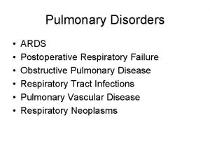
Pulmonary Diseases Disorders Assessment Pulmonary Diseases Disorders n


- Slides: 38

Pulmonary Diseases & Disorders: Assessment

Pulmonary Diseases & Disorders n Epidemiology u 28% of all EMS Chief Complaints in the US u >200, 000 deaths annually due to respiratory emergencies

Pulmonary Diseases & Disorders n Many, many pulmonary diseases u Difficult to learn all pathophysiologies u All can be categorized as affecting: F Ventilation F Diffusion (Respiration) F Perfusion u Treatment can be focused on identifying and treating source of ventilatory/respiratory impairment

Sources of Pulmonary Impairment n n Pulmonary Diseases Disorders of the Pulmonary System Non-Pulmonary Disorders/Disease Impairing Ventilation or Respiration What examples can you list for each of these?

Sources of Pulmonary Impairment n Ventilation u Upper Airway Trauma F Epiglottitis F FBAO F Inflammation of tonsils F u Lower Airway Trauma F Obstructive lung disease F Mucous accumulation F Smooth muscle spasm F Airway edema F

Sources of Pulmonary Impairment n Ventilation u Chest Wall Impairment Trauma F Hemothorax F Pneumothorax F Empyema F Pleural inflammation F Neuromuscular diseases F u Neurologic Control Brainstem dysfunction F Phrenic or spinal nerve dysfunction F

Sources of Pulmonary Impairment n Diffusion Inadequate Fi. O 2 u Diseased alveoli u asbestosis F COPD F inhalation injury F u Capillary bed disease F atherosclerosis u Interstitial space disease High pressure pulmonary edema F High permeability pulmonary edema F

Sources of Pulmonary Impairment n Perfusion u Inadequate blood u Impaired blood flow F pulmonary embolus volume or hemoblogin u Capillary wall F hypovolemia pathology F anemia F trauma

Risk Factors for Pulmonary Disease n Intrinsic Risk Factors u Genetic predisposition F asthma F COPD F carcinoma u Cardiac or Circulatory pathologies F Source for pulmonary edema F Source for pulmonary emboli u Stress

Risk Factors for Pulmonary Disease n Extrinsic Factors u Smoking prevalence of COPD & carcinomas F severity of pulmonary disease F u Environmental Factors prevalence of COPD & asthma F severity of all obstructive disorders F

Function of the Pulmonary System n Gas Exchange System u ~10, 000 liters of air are filtered, warmed and humidified daily u Oxygen diffused into blood u Carbon dioxide excreted from the body

Function of the Pulmonary System n Physiology of Ventilation u Requires neurologic initiation (brainstem) u Nerve conduction pathways between brainstem and muscles of respiration u Intact & patent Upper and Lower airways u Intact & non-collapsed alveoli

Function of the Pulmonary System n Physiology of Respiration u Simple diffusion process at the pulmonarycapillary bed u Diffusion Requirements F Intact, non-thickened alveolar walls F Minimal interstitial space & without additional fluid F Intact, non-thickened capillary walls

Function of the Pulmonary System n Physiology of Perfusion u Process of circulating blood through the capillary bed u Perfusion Requirements F Adequate blood volume F Adequate hemoglobin F Intact, non-occluded pulmonary capillaries F Functioning Left Heart

Control of Ventilation n Control ventilation in response to physiologic needs u Driven 1° by p. H of CSF F influenced largely by Pa. CO 2 u 2° drive = Pa. CO 2 u 3° drive = Pa. O 2 detected by chemoreceptors F very small population with severe COPD

Nervous System Effect on Ventilation n Medulla u Stimulation n Phrenic Nerve u Innervation n of the diaphragm Spinal Nerves at Thoracic levels u Innervation n to initiate ventilation of intercostal muscles Hering-Breuer reflex u Prevents overinflation

General Assessment n Size-Up u Environment F Airborne Hazards F Number of patients F Needs • Specialized rescue equipment • Protective equipment u Is the environment creating or exacerbating the pulmonary condition?

General Assessment n Initial Goal u Identify potentially life-threatening pulmonary conditions n Perform minimal PE & Hx u Initiate n immediate & appropriate therapies Then, continue PE & Hx u Try to determine if origin is ventilation, diffusion, perfusion or combination

General Assessment n Signs of potentially life-threatening pulmonary condition u altered mental status u absent signs of ventilation u Audible stridor or wheezing u Able to speak in short phrases only u Sustained Tachycardia u Pallor / Diaphoresis u Accessory muscle use / Retractions

Assessment: H&P n Present History (focused hx) u Chief Complaint F Dyspnea • “Subjective sensation that breathing is excessive, difficult or uncomfortable F CP F Cough, Hemoptysis u Associated F Fever, Symptoms Chills F sputum production F Fatigue

Assessment: H&P n Present History (focused hx) u Sputum Findings amount of sputum infection F Thick green or brown pneumonia or infection F Yellow or gray allergic or inflammatory response F Hemoptysis tuberculosis or carcinoma F Pink, frothy severe pulmonary edema F

Assessment: H&P n HX of Present Illness u How long has dyspnea been present? u Gradual or sudden onset? u What aggravates or alleviates? F Hx of orthopnea? u Coughing? u Productive cough? u What does sputum look/smell like? u Pain? u What does the pain feel like?

Assessment: H&P n Listen - To Pt. Breathe or Talk u Noisy Breathing is Obstructed Breathing u Not All Obstructed Breathing is Noisy u Snoring - Tongue Blocking Airway u Stridor - “Tight” Upper Airway from Partial Obstruction n Observe Breathing u Tachypnea u Bradypnea

Assessment: H&P n Observe u Body Positioning F Tripod F Legs in dependent position u Mental Status u Ventilatory Effort F Accessory muscle use / retractions F Abdominal muscle use F Chest wall expansion F Nasal flaring, pursed lips

Assessment: H&P n Physical Exam of the Chest u Increased A-P Diameter u Lung Sounds F Abnormal: stridor, wheezing, rhonchi, rales, pleural rub u Chest expansion u Symmetrical Findings u Evidence of Trauma

Assessment: H&P n Physical Exam u Cyanosis? F Late, unreliable sign of Hypoxia u Oxygenate Immediately! Especially If: Decreased LOC F Possible Shock F Possible Severe Hemorrhage F Chest Pain F Chest Trauma F Respiratory distress or dyspnea F HX of any Kind of Hypoxia F

Assessment: H&P n Physical Exam u Vital Signs F Skin Color, Temp & Moisture F Respiratory Rate • No an accurate lone indicator of respiratory status unless very slow F Respiratory Rhythm/Pattern F Pulse • Bradycardia vs Tachycardia F Blood Pressure

Assessment: H&P n Physical Exam - Circulatory assessment u Is the heart beating? u Is there major external hemorrhage? u Is the Pt. Perfusing vital organs? u Effects of hypoxia: F Early in adults - Tachycardia F Late in adults - Bradycardia F Children - Bradycardia

Assessment: H&P n n Don’t let respiratory failure distract you from assessing for circulatory failure. Vascular Access

Assessment: H&P n Physical Exam u Extremities F Peripheral Cyanosis F Clubbing F Carpopedal spasm F Peripheral edema

Assessment: H&P n Diagnostic Testing u Pulse oximetry Saturation F Inaccuracies & Disadvantages F u Peak Flow Meter Baseline measurement for obstructive lung disease F Often available from patient F u Capnometry real-time assessment of endotracheal tube placement F quantitative vs qualitative F

Assessment: H&P n Past History u Similar Episodes in Past F Patient’s description of acuity F “What happened last time you had an episode this bad? ” n Chronic Symptoms u Acute, Seasonal SOB episodes u Seasonal Allergies u Chronic cough u Recurrent flu, pulmonary infection or SOB

Assessment: H&P n Past History u Known diagnosis F Does the present H&P correlate with this past history? • CHF • Hypertension • Renal Failure u Previous intubation or hospitalization u Aggravating Factors (e. g. smoking)

Assessment: H&P n Past History u Medications F Class, Route, Frequency of Use F Pulmonary • • Sympathomimetics Corticosteroids MAST Cell Stabilizer Methylxanthines F Cardiovascular • Diuretics • Antihypertensives • Cardiac glycosides

Assessment: H&P n Disability u Restlessness, anxiety, combativeness = HYPOXIA Until Proven Otherwise u Drowsiness, lethargy = HYPERCARBIA When the patient stops fighting, he is not necessarily getting Better!!

Other Adventitious Sounds n Cough u Forced exhalation against partially closed glottis u Reflex response to mucosa irritation u Determine circumstances At work F Postural changes F Lying down F u Productive vs non-productive

Other Adventitious Sounds n Sneeze u Forced exhalation via nasal route u Clears nasal passages u Reflex response to mucosa irritation n Sigh u Slow, deep inspiration - Prolonged, audible exhalation u Reexpands areas of atelectasis

Other Adventitious Sounds n Hiccough u Hiccups, singultus u Spasm of diaphragm followed by glottic closure u No useful purpose u Benign, transient
 Milady chapter 8 skin disorders and diseases
 Chapter 21 mental health diseases and disorders
 Seborrheic keratoses
 Chapter 10 lymphatic system diseases and disorders
 Chapter 17 reproductive system diseases and disorders
 Chapter 10 nail disorders and diseases
 Define a primary skin lesion and list three types
 Chapter 15 nervous system diseases and disorders
 Cardiovascular system diseases and disorders chapter 8
 Chapter 6 musculoskeletal system
 Milady nail diseases and disorders
 Portfolio assessment matches assessment to teaching
 Stages in implementing portfolio assessment
 Define dynamic assessment
 Nursing management of pulmonary tuberculosis
 Pac pulmonary artery catheter
 Bronchial segments
 Svr calculation
 Muscles of inspiration and expiration
 Pulmonary hypertension
 Hypoventilation
 Rib anatomy anterior
 Mean pulmonary arterial pressure
 Thoracic aortic aneurysm
 Pulmonary embolism
 Function of coronary artery
 5 cyanotic congenital heart disease
 Pac pulmonary artery catheter
 Pulmonary fibrosis
 Management of hemorrhage
 Bronchopneumonia
 Bronchopulmonary segments
 Chest pediatric pulmonary board review
 Pulmonary hypoplasia with anasarca
 Cardiogenic vs noncardiogenic pulmonary edema
 Pah vs pulmonary hypertension
 Saturacion 94
 Pulmonary artery and aorta
 Right atrium
