Principles of Anatomy and Physiology Thirteenth Edition Gerard

- Slides: 45
Principles of Anatomy and Physiology Thirteenth Edition Gerard J. Tortora • Bryan H. Derrickson Chapter 1 An Introduction to the Human Body Copyright © 2012 by John Wiley & Sons, Inc.
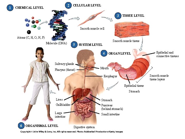
1 2 CHEMICAL LEVEL CELLULAR LEVEL 3 TISSUE LEVEL Smooth muscle cell Atoms (C, H, O, N, P) Molecule (DNA) Smooth muscle tissue 5 SYSTEM LEVEL 4 ORGAN LEVEL Epithelial and connective tissues Salivary glands Pharynx (throat) Mouth Smooth muscle tissue layers Esophagus Epithelial tissue Stomach Liver Stomach Gallbladder Pancreas (behind stomach) Large intestine Small intestine 6 ORGANISMAL LEVEL Digestive system
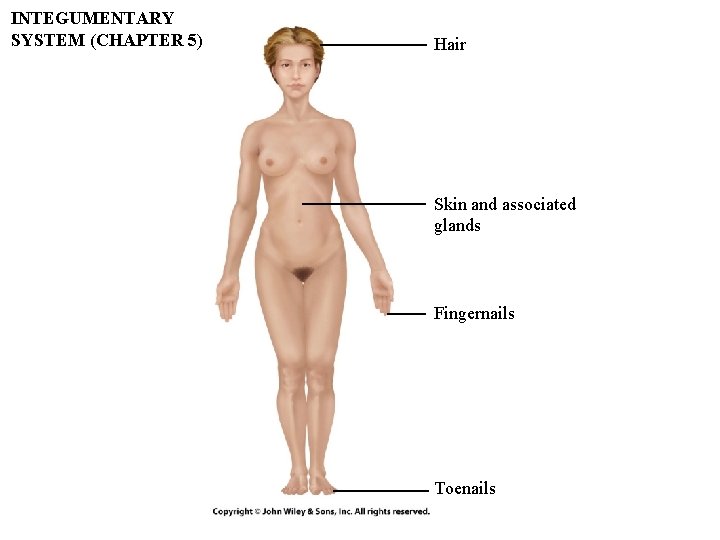
INTEGUMENTARY SYSTEM (CHAPTER 5) Hair Skin and associated glands Fingernails Toenails
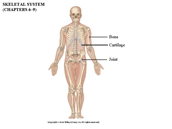
SKELETAL SYSTEM (CHAPTERS 6– 9) Bone Cartilage Joint
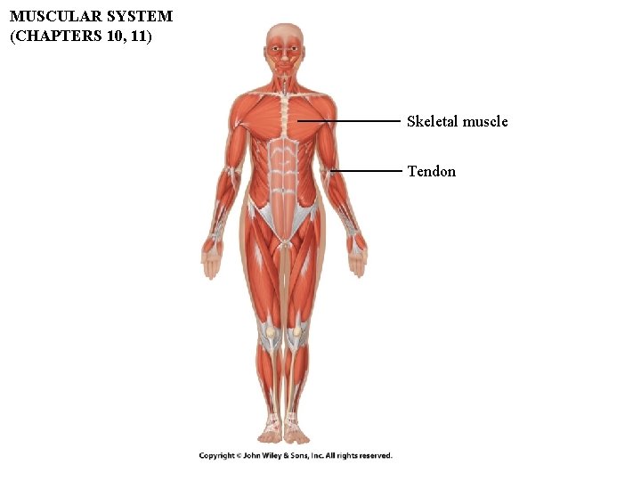
MUSCULAR SYSTEM (CHAPTERS 10, 11) Skeletal muscle Tendon
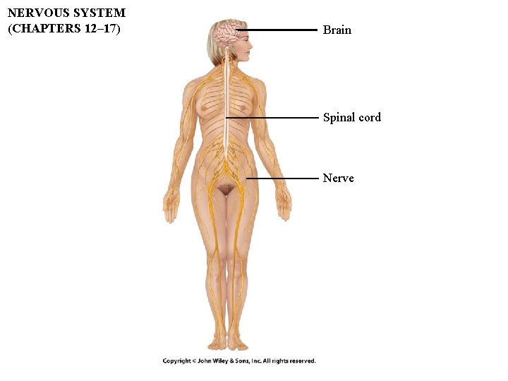
NERVOUS SYSTEM (CHAPTERS 12– 17) Brain Spinal cord Nerve
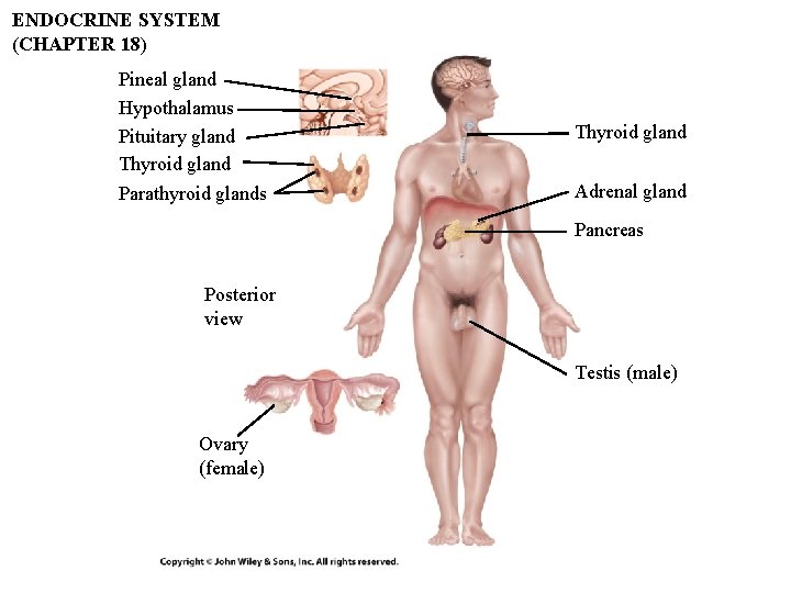
ENDOCRINE SYSTEM (CHAPTER 18) Pineal gland Hypothalamus Pituitary gland Thyroid gland Parathyroid glands Thyroid gland Adrenal gland Pancreas Posterior view Testis (male) Ovary (female)
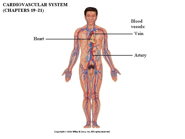
CARDIOVASCULAR SYSTEM (CHAPTERS 19– 21) Blood vessels: Vein Heart Artery
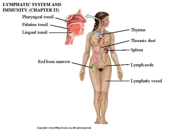
LYMPHATIC SYSTEM AND IMMUNITY (CHAPTER 22) Pharyngeal tonsil Palatine tonsil Lingual tonsil Thymus Thoracic duct Spleen Red bone marrow Lymph node Lymphatic vessel
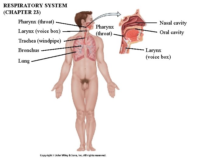
RESPIRATORY SYSTEM (CHAPTER 23) Pharynx (throat) Larynx (voice box) Pharynx (throat) Nasal cavity Oral cavity Trachea (windpipe) Bronchus Lung Larynx (voice box)
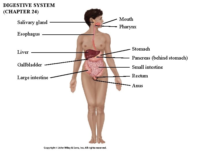
DIGESTIVE SYSTEM (CHAPTER 24) Salivary gland Mouth Pharynx Esophagus Liver Stomach Pancreas (behind stomach) Gallbladder Small intestine Large intestine Rectum Anus
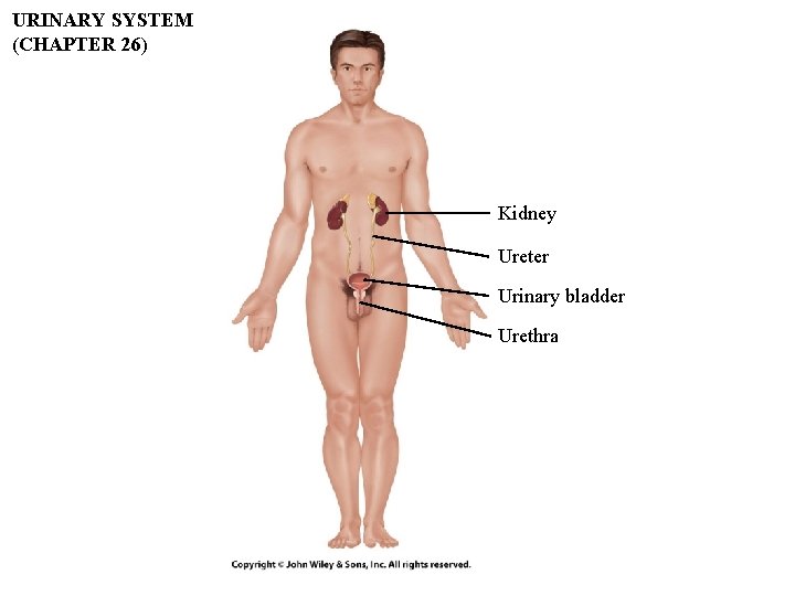
URINARY SYSTEM (CHAPTER 26) Kidney Ureter Urinary bladder Urethra
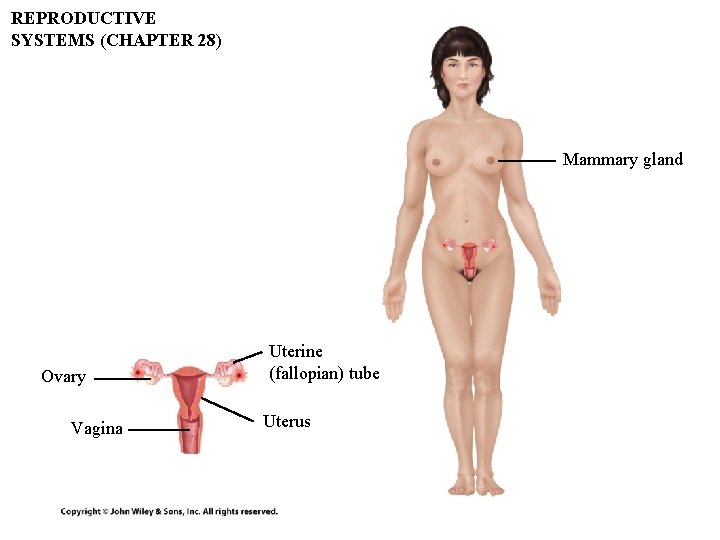
REPRODUCTIVE SYSTEMS (CHAPTER 28) Mammary gland Ovary Vagina Uterine (fallopian) tube Uterus
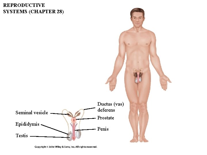
REPRODUCTIVE SYSTEMS (CHAPTER 28) Seminal vesicle Epididymis Testis Ductus (vas) deferens Prostate Penis
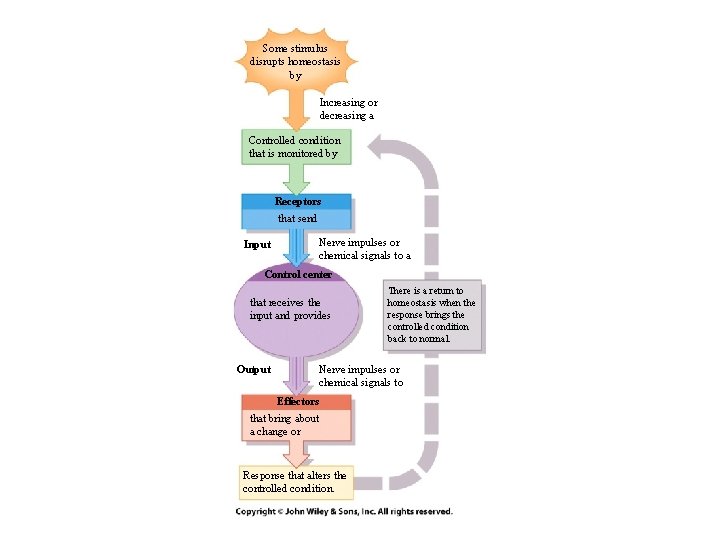
Some stimulus disrupts homeostasis by Increasing or decreasing a Controlled condition that is monitored by Receptors that send Input Nerve impulses or chemical signals to a Control center that receives the input and provides Output There is a return to homeostasis when the response brings the controlled condition back to normal. Nerve impulses or chemical signals to Effectors that bring about a change or Response that alters the controlled condition.
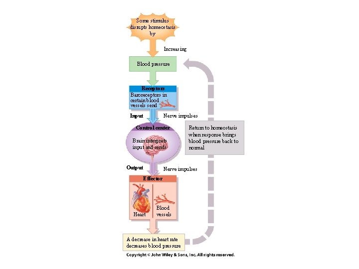
Some stimulus disrupts homeostasis by Increasing Blood pressure Receptors Baroreceptors in certain blood vessels send Nerve impulses Input Control center Brain interprets input and sends Output Return to homeostasis when response brings blood pressure back to normal Nerve impulses Effector Heart Blood vessels A decrease in heart rate decreases blood pressure
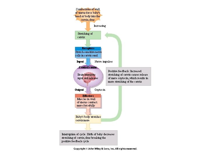
Contractions of wall of uterus force baby's head or body into the cervix, thus Increasing Stretching of cervix Receptors Stretch-sensitive nerve cells in cervix send Input Nerve impulses Control center Brain interprets input and releases Output Positive feedback: Increased stretching of cervix causes release of more oxytocin, which results in more stretching of the cervix Oxytocin Effectors Muscles in wall of uterus contract more forcefully Baby's body stretches cervix more Interruption of cycle: Birth of baby decreases stretching of cervix, thus breaking the positive feedback cycle
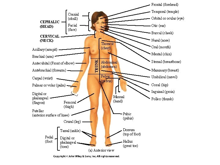
Frontal (forehead) Temporal (temple) Cranial (skull) CEPHALIC (HEAD) Orbital or ocular (eye) Facial (face) Otic (ear) Buccal (cheek) CERVICAL (NECK) Nasal (nose) Thoracic (chest) Axillary (armpit) Oral (mouth) Mental (chin) Antecubital (Front of elbow) Antebrachial (forearm) Carpal (wrist) TRUNK Brachial (arm) Abdominal (abdomen) Sternal (breastbone) Pelvic (pelvis) Umbilical (navel) Mammary (breast) Coxal (hip) Palmar or volar (palm) Digital or phalangeal (fingers) Inguinal (groin) Femoral (thigh) Manual (hand) Patellar (anterior surface of knee) Pubic (pubis) Crural (leg) Dorsum (top of foot) Tarsal (ankle) Pedal (foot Digital or phalangeal (toes) Hallux (great toe) (a) Anterior view Pollex (thumb)
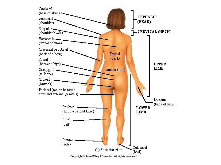
Occipital (base of skull) Acromial (shoulder) CEPHALIC (HEAD) Scapular (shoulder blade) CERVICAL (NECK) Vertebral (spinal column) Olecranal or cubital (back of elbow) Sacral (between hips) Coccygeal (tailbone) Dorsal (back) UPPER LIMB Lumbar (loin) Gluteal (buttock) Perineal (region between anus and external genitals) Popliteal (hollow behind knee) LOWER LIMB Sural (calf) Plantar (sole) (b) Posterior view Calcaneal (heel) Dorsum (back of hand)
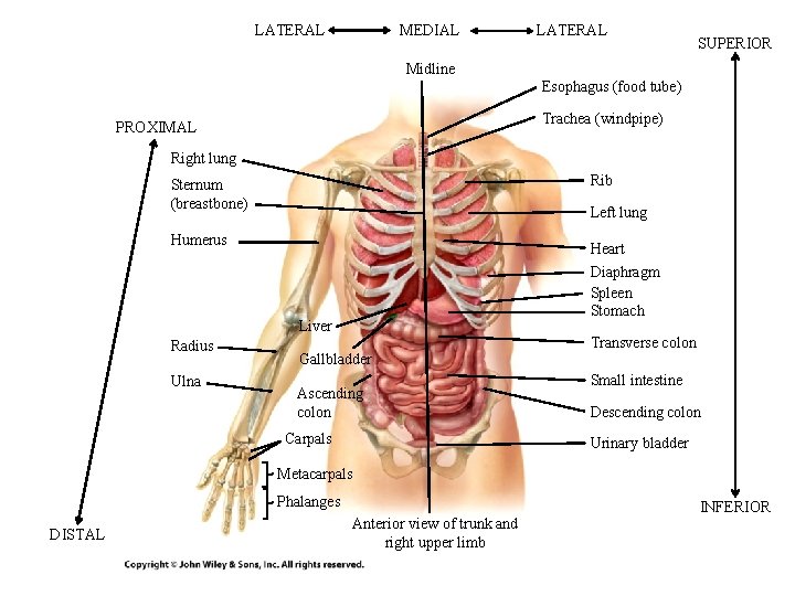
LATERAL MEDIAL LATERAL SUPERIOR Midline Esophagus (food tube) Trachea (windpipe) PROXIMAL Right lung Rib Sternum (breastbone) Left lung Humerus Heart Diaphragm Spleen Stomach Liver Radius Ulna Gallbladder Ascending colon Carpals Transverse colon Small intestine Descending colon Urinary bladder Metacarpals Phalanges DISTAL Anterior view of trunk and right upper limb INFERIOR
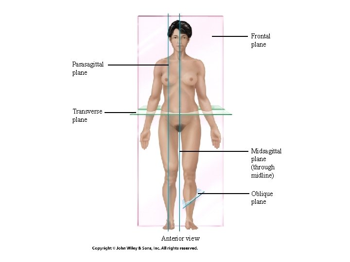
Frontal plane Parasagittal plane Transverse plane Midsagittal plane (through midline) Oblique plane Anterior view
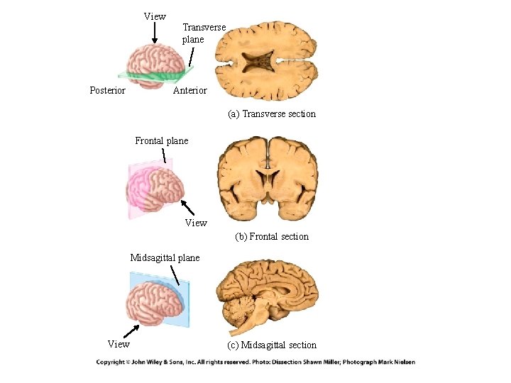
View Posterior Transverse plane Anterior (a) Transverse section Frontal plane View (b) Frontal section Midsagittal plane View (c) Midsagittal section
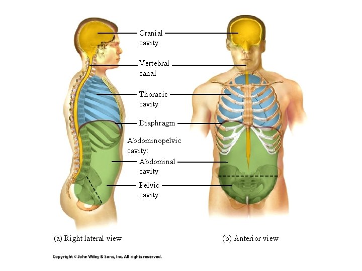
Cranial cavity Vertebral canal Thoracic cavity Diaphragm Abdominopelvic cavity: Abdominal cavity Pelvic cavity (a) Right lateral view (b) Anterior view
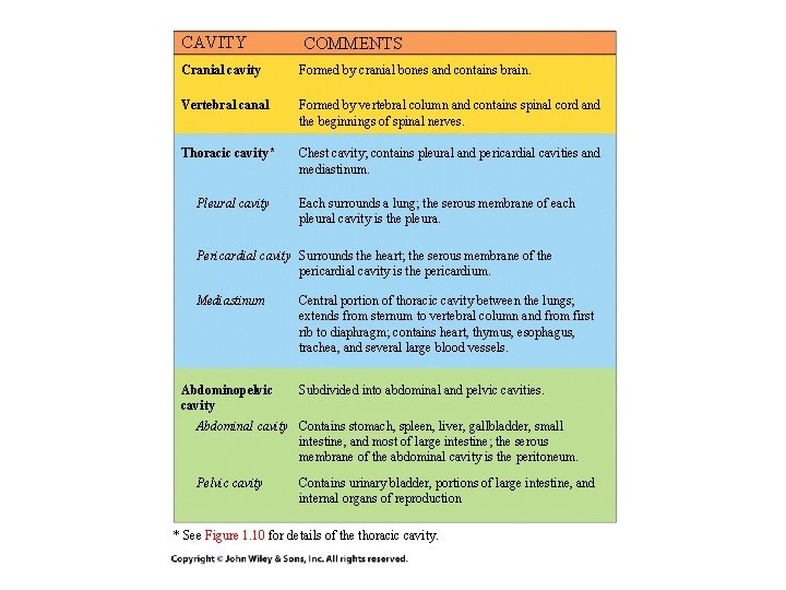
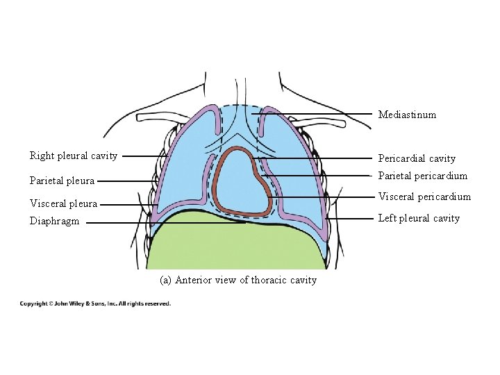
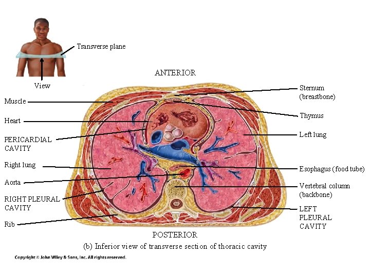
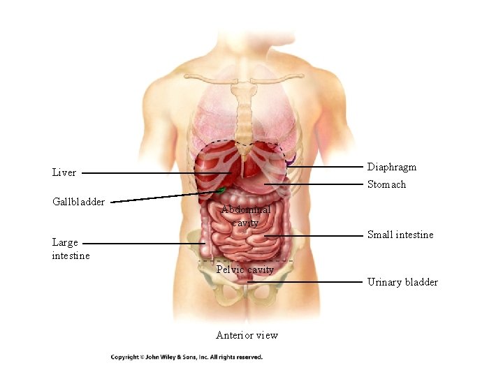
CAVITY COMMENTS Cranial cavity Formed by cranial bones and contains brain. Vertebral canal Formed by vertebral column and contains spinal cord and the beginnings of spinal nerves. Thoracic cavity* Chest cavity; contains pleural and pericardial cavities and mediastinum. Pleural cavity Each surrounds a lung; the serous membrane of each pleural cavity is the pleura. Pericardial cavity Surrounds the heart; the serous membrane of the pericardial cavity is the pericardium. Mediastinum Abdominopelvic cavity Central portion of thoracic cavity between the lungs; extends from sternum to vertebral column and from first rib to diaphragm; contains heart, thymus, esophagus, trachea, and several large blood vessels. Subdivided into abdominal and pelvic cavities. Abdominal cavity Contains stomach, spleen, liver, gallbladder, small intestine, and most of large intestine; the serous membrane of the abdominal cavity is the peritoneum. Pelvic cavity Contains urinary bladder, portions of large intestine, and internal organs of reproduction * See Figure 1. 10 for details of the thoracic cavity.
Mediastinum Right pleural cavity Pericardial cavity Parietal pleura Parietal pericardium Visceral pleura Left pleural cavity Diaphragm (a) Anterior view of thoracic cavity
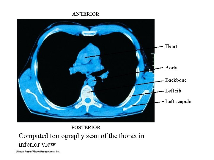
Transverse plane ANTERIOR View Sternum (breastbone) Muscle Thymus Heart Left lung PERICARDIAL CAVITY Right lung Esophagus (food tube) Aorta Vertebral column (backbone) RIGHT PLEURAL CAVITY LEFT PLEURAL CAVITY Rib POSTERIOR (b) Inferior view of transverse section of thoracic cavity
Diaphragm Liver Gallbladder Stomach Abdominal cavity Large intestine Small intestine Pelvic cavity Urinary bladder Anterior view
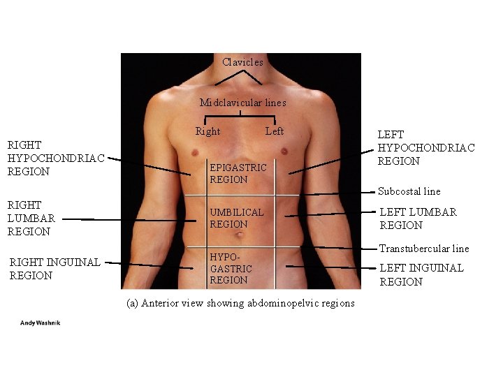
Clavicles Midclavicular lines Right RIGHT HYPOCHONDRIAC REGION Left EPIGASTRIC REGION LEFT HYPOCHONDRIAC REGION Subcostal line RIGHT LUMBAR REGION UMBILICAL REGION RIGHT INGUINAL REGION HYPOGASTRIC REGION (a) Anterior view showing abdominopelvic regions LEFT LUMBAR REGION Transtubercular line LEFT INGUINAL REGION
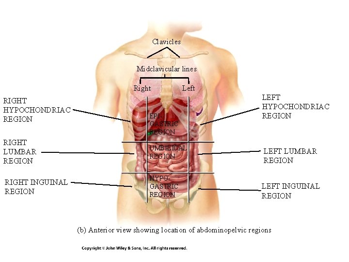
Clavicles Midclavicular lines Right RIGHT HYPOCHONDRIAC REGION Left EPIGASTRIC REGION RIGHT LUMBAR REGION UMBILICAL REGION RIGHT INGUINAL REGION HYPOGASTRIC REGION LEFT HYPOCHONDRIAC REGION LEFT LUMBAR REGION LEFT INGUINAL REGION (b) Anterior view showing location of abdominopelvic regions
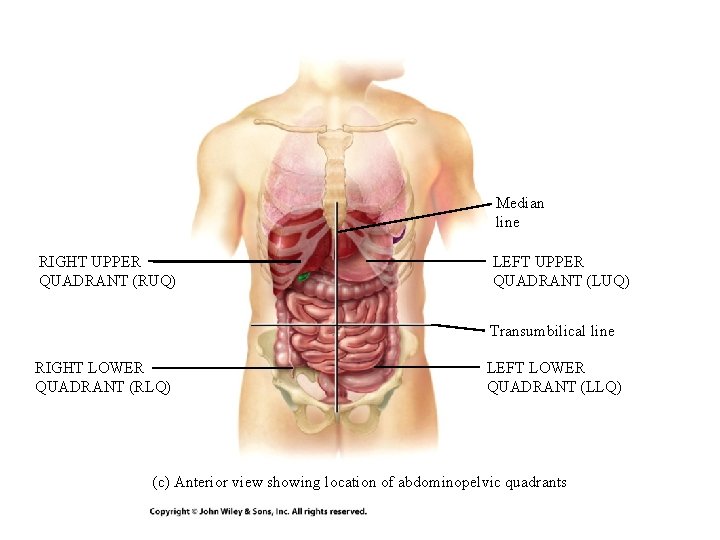
Median line RIGHT UPPER QUADRANT (RUQ) LEFT UPPER QUADRANT (LUQ) Transumbilical line RIGHT LOWER QUADRANT (RLQ) LEFT LOWER QUADRANT (LLQ) (c) Anterior view showing location of abdominopelvic quadrants
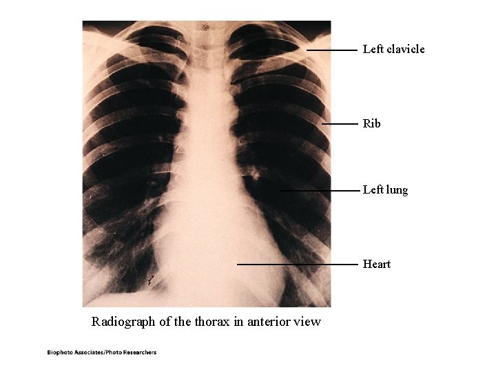
Left clavicle Rib Left lung Heart Radiograph of the thorax in anterior view
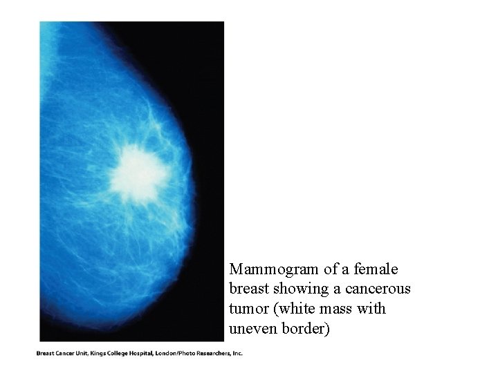
Mammogram of a female breast showing a cancerous tumor (white mass with uneven border)
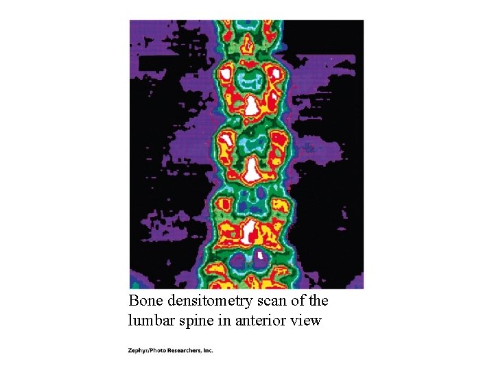
Bone densitometry scan of the lumbar spine in anterior view
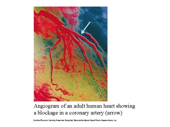
Angiogram of an adult human heart showing a blockage in a coronary artery (arrow)
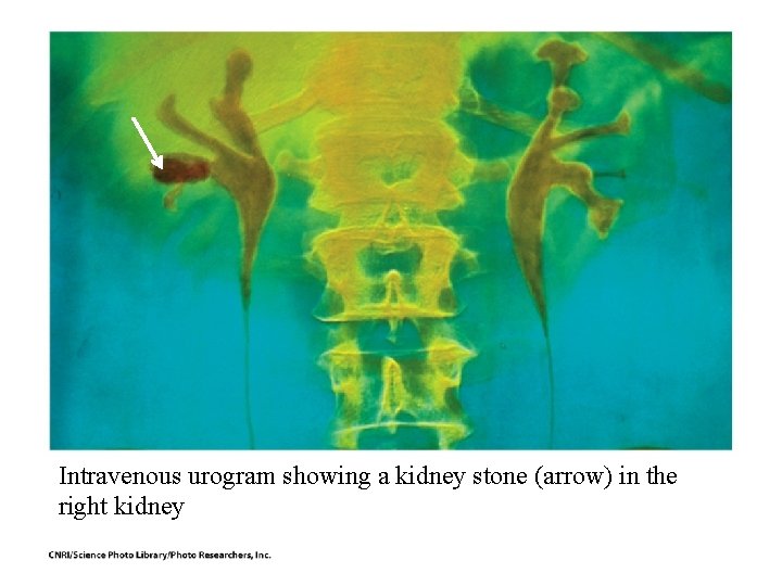
Intravenous urogram showing a kidney stone (arrow) in the right kidney
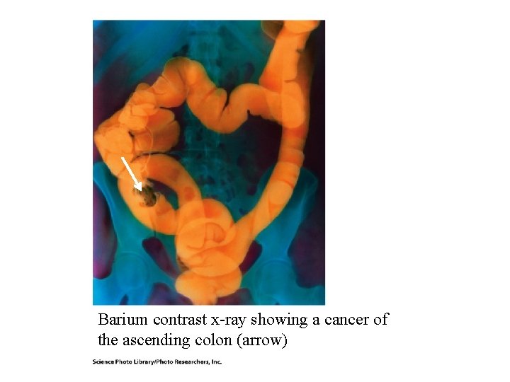
Barium contrast x-ray showing a cancer of the ascending colon (arrow)
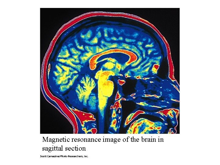
Magnetic resonance image of the brain in sagittal section
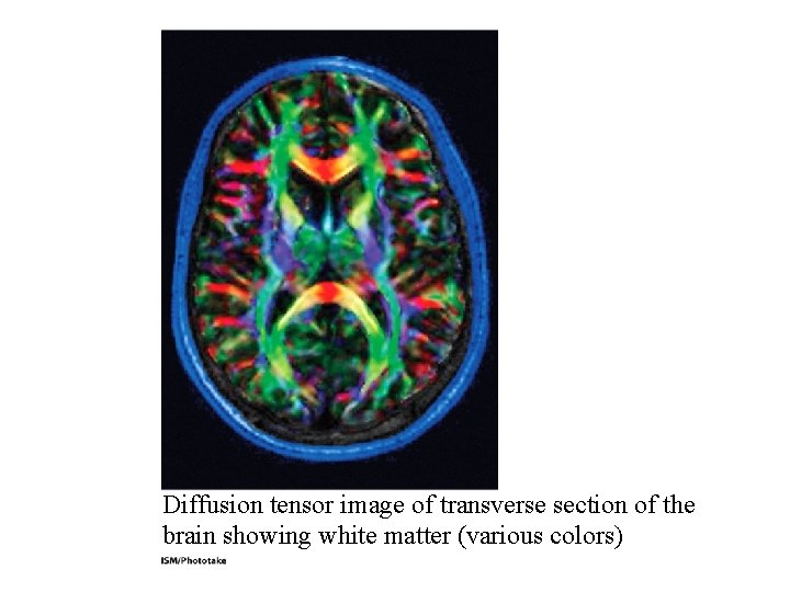
Diffusion tensor image of transverse section of the brain showing white matter (various colors)
ANTERIOR Heart Aorta Backbone Left rib Left scapula POSTERIOR Computed tomography scan of the thorax in inferior view
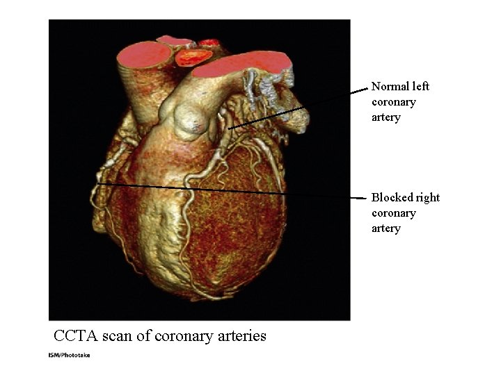
Normal left coronary artery Blocked right coronary artery CCTA scan of coronary arteries
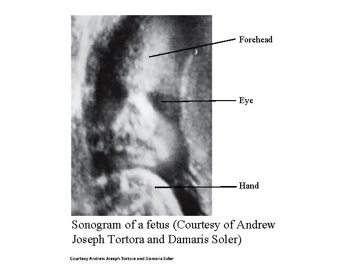
Forehead Eye Hand Sonogram of a fetus (Courtesy of Andrew Joseph Tortora and Damaris Soler)
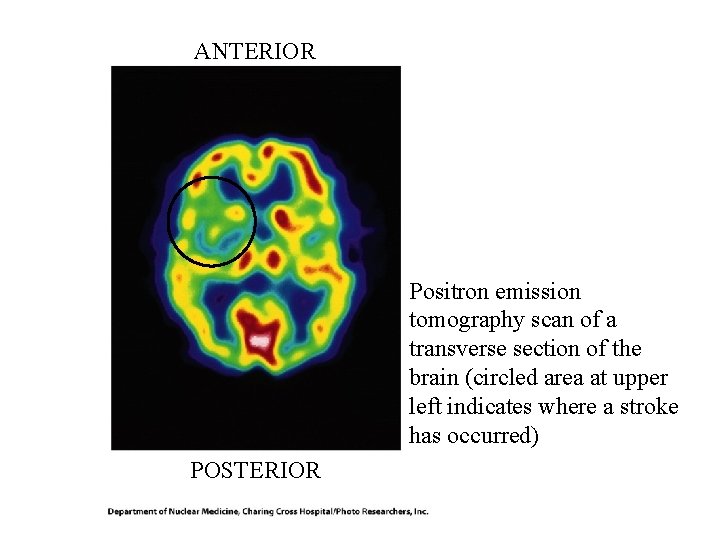
ANTERIOR Positron emission tomography scan of a transverse section of the brain (circled area at upper left indicates where a stroke has occurred) POSTERIOR
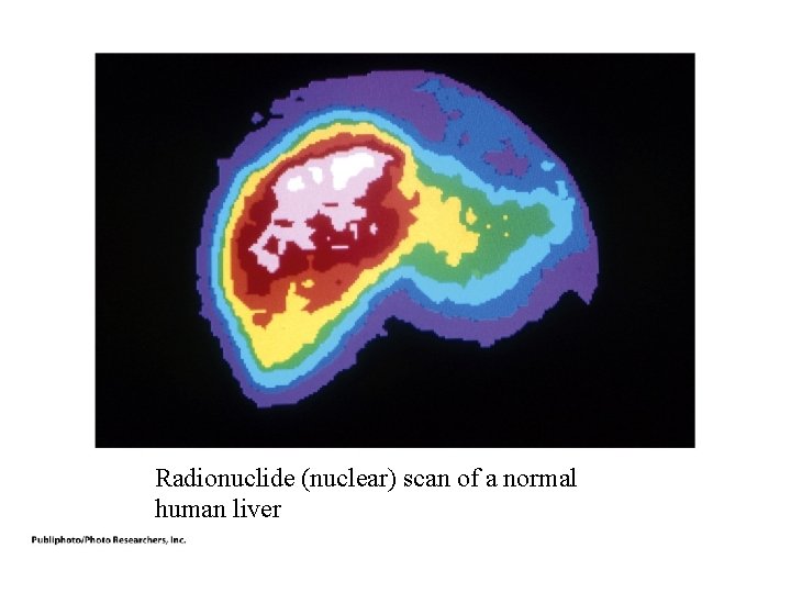
Radionuclide (nuclear) scan of a normal human liver
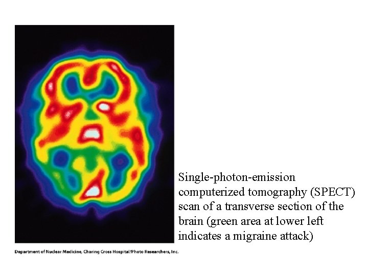
Single-photon-emission computerized tomography (SPECT) scan of a transverse section of the brain (green area at lower left indicates a migraine attack)
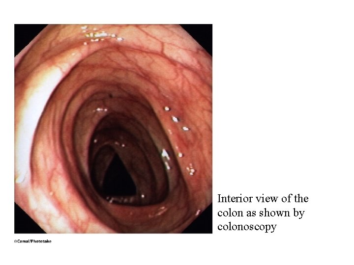
Interior view of the colon as shown by colonoscopy