Nervous System Unit 5 Nervous System Sensesp 35


 – is made up of the brain & spinal")
")

")

: center for homeostasis in the body (temperature, appetite,")


 the cranial & spinal nerves Humans have: • 12")


")
")


 Remember the nerve pathway? Sometimes it is simpler to address")


")

: • constricts the pupils of your eyes • increases")


 to its")


 or dendrite")


- Slides: 86
Nervous System
Unit 5: Nervous System & Sensesp. 35 3 a. Interpret interactions among hormones, senses nerves senses, and nerves which make possible functions the coordination of functions of the body. 3 b. Investigate the physiology of electrochemical impulses and neural integration and trace the pathway of an impulse, relating biochemical changes involved in the conduction of the impulse. 3 c. Describe how the body perceives internal and external stimuli and responds to maintain a stable internal environment, as it relates to biofeedback. (homeostasis)
I. Function of the Nervous System Control & coordination of body systems Three basic functions to accomplish this mission: A. Sensory input B. Integration C. Motor output
The three functions… visualized: Integration Evaluate the info Sensory input Information IN Send response Motor output instruction(s)
Comp Book: -p. 38 Flow Map: From Stimulus to Nervous Response
II. General Overview A. Structural Classification There are 2 major structural divisions of the nervous system: the central nervous system (CNS) the peripheral nervous system (PNS)
1. Central Nervous System (CNS) – is made up of the brain & spinal cord a. Brain – contains about 100 billion neurons Click here for intro to the brain video
The brain can be divided into 4 major regions: i. Cerebrum ii. Diencephalon (midbrain) iv. Brain stem iii. Cerebellum
i. The cerebrum governs: • intelligence • interpretation of • memory sensory info. (smell, taste, sight, hearing) • language
• motor functions (activity of muscles and glands)
ii. The diencephalon is found beneath / within the cerebrum The diencephalon contains: • Thalamus: conducts information between the spinal cord & cerebrum
• Hypothalamus (below the thalamus): center for homeostasis in the body (temperature, appetite, & water balance)
iii. Cerebellum: below the cerebrum & behind the brain stem Cerebellum
iii. Cerebellum governs: • smooth, coordinated movements • helps maintain posture, muscle tone, and equilibrium (balance) (you lose your equilibrium when you drink because the alcohol inhibits the function of the cerebellum)
iv. brain stem: connects brain to spinal cord—it regulates: • heartbeat • respiration • blood pressure
• swallowing • coughing • hiccupping • sneezing • vomiting
Neurons passing through the brain stem cross from one side of the body to the other. (In other words, if your right hand was burned, the pain signals would travel to the left side of the cerebrum to be interpreted) (Also, if you wanted to lift your left arm, the signal would be sent to your left arm from the right side of your cerebrum)
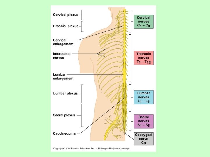
b. Spinal cord – extends from the brain stem, through the vertebrae to the 2 nd lumbar vertebra
• Notice: the spinal cord does not extend to the tip of the vertebral column
• information traveling to and from the brain must pass through the spinal cord to reach other parts of the body • often called the “information highway” of the CNS • made of 2 layers: “gray” & “white” matter
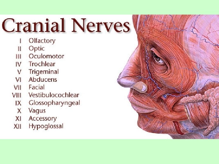
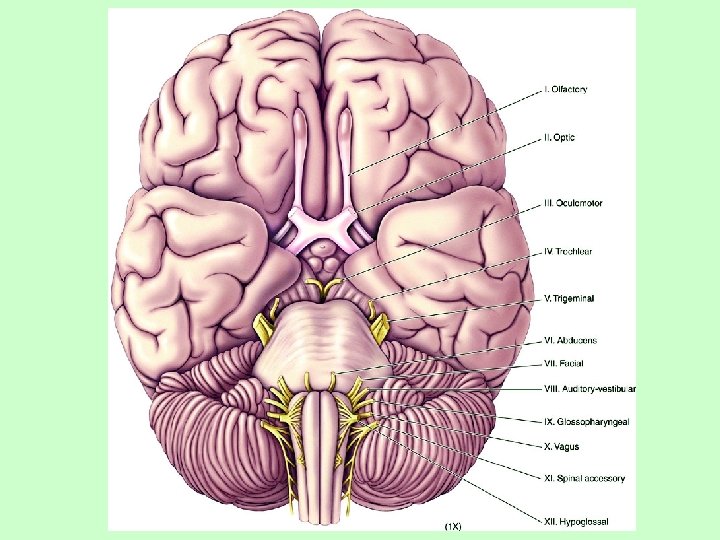
2. Peripheral Nervous System (PNS) the cranial & spinal nerves Humans have: • 12 pairs of cranial nerves ex. optic nerves, olfactory nerves, facial nerves
• 31 pairs of spinal nerves
One of the most important functions of the PNS is to perform reflex actions, which do not involve the brain A reflex is a sudden, rapid, involuntary response to a stimulus
Reflex examples: • knee jerk reflex (lower leg kicks when tendon is tapped)
• withdrawal reflex (hand withdraws if it touches something hot)
• papillary reflex (pupil constricts if a bright light is shown in your eyes)
How do reflexes work? Knee jerk reflex: the patellar tendon is tapped • this generates nerve impulses which travel along sensory neurons to the spinal cord
• the spinal cord then transmits impulses through motor neurons • the rectus femoris muscle is instructed to contract, causing extension of the lower leg • the brain is not involved in the response
• information travels over a shorter distance – producing faster reaction times • the reflex is completed before your brain is aware of what happened
• you will typically withdraw your hand from something hot before you recognize the pain of the burn
Comp Book: -set it up -back of p. 35 structural classification
II. General Overview (continued) Remember the nerve pathway? Sometimes it is simpler to address each part of the nervous system based Sensory integration motor output on the general role it plays in the nerve (brain) pathway… B. Functional Classification of PNS: categorizing parts of the nervous system based on the nerve function in the overall nerve pathway; in this way, there are 2 types of nerves: 1. Sensory 2. Motor
1. Sensory Nerves = Afferent Division: The body’s receptive nerves taking info from both the internal and external environment toward the CNS: a. Somatic sensory fibers—nerve endings & receptors in skin, skeletal muscles & joints b. visceral sensory fibers—nerve endings and receptors in abdominal & pelvic organs
Plus all your special senses… ch. 9 c. Vision from the eyes d. Hearing & balance from the ears e. Smelling from the nose (chemoreceptors) f. Taste from the tongue (chemoreceptors)
2. Motor Nerves = Efferent Division After the brain integrates the sensory input, outgoing response is sent through the efferent nerves The motor or efferent portion of the PNS is typically divided into 2 subsystems: a. Somatic Nervous System b. Autonomic Nervous System
a. Somatic Nervous System – includes the cranial & spinal nerves that regulate voluntary actions
b. Autonomic Nervous System – includes the cranial & spinal nerves that regulate involuntary actions ex. cardiac muscle smooth muscle gland activity
Keep in mind that although the ANS functions “automatically”, you can consciously control it for a short period of time When you hold your breath, you are overriding your ANS – however, you cannot override it indefinitely
The Autonomic Nervous System can be further subdivided into: i. Sympathetic Division ii. Parasympathetic Division
i. Sympathetic Division: regulates involuntary actions during periods of high stress or increased activity • increases blood pressure • heart rate • breathing rate • blood flow to muscles
• dilates the pupils of your eyes (increases amount of light; see better) • decreases digestion of food & blood flow to the skin All of these changes prepare your body for action (“fight or flight”)
ii. Parasympathetic Division: regulates involuntary actions during periods of rest or relaxation • decreases blood pressure • decreases heart & respiratory rates • decreases blood flow to muscles
ii. Parasympathetic Division (cont. ’d): • constricts the pupils of your eyes • increases digestion of food and blood flow to the skin
*** Most internal organs are regulated by both the sympathetic and parasympathetic divisions. *** • the sympathetic division dominates when you are under stress • the parasympathetic division dominates when you are relaxed
Comp Book: -p. 36 functional classification -back of p. 37 Venn: Autonomic vs. Somatic
III. The Electrochemical Impulse A. Anatomy of a neuron Click here for intro to nerve cells video
Neuron structure – 3 basic parts 1. One or more dendrites • Short, highly branched extensions • Receive impulses from receptors or other neurons • transmit impulses toward the cell body
dendrites
2. Cell body – main portion of the neuron • processes information sent by the dendrites cell body
3. One axon – • long extension that transmits impulses away from the cell body • axon tip usually branched forming axon terminals
axon
Impulses always travel in this direction: Dendrites cell body axon terminals
Many axons are surrounded by specialized cells called Schwann cells • Schwann cells produce a myelin sheath that functions to protect the axon and speed up impulse conduction myelin sheath • Similar to the coating on electrical cords
A disease that involves the destruction of the myelin sheath – multiple sclerosis
• destruction of the sheath impairs the transmission of nerve impulses
• this causes muscular weakness, loss of memory, double vision, and paralysis
Comp Book: p. 37 Frayer: neuron
III. The Electrochemical Impulse A. Anatomy of a neuron B. Physiology of a nerve impulse Nerves are the functional units of the nervous system Electrical signals called nerve impulses are transmitted by neurons
III. The Electrochemical Impulse B. Physiology of a nerve impulse 1. Resting Conditions Click here for animation about resting potential 2. Stimulus & Local Depolarization Neurons that are not conducting an impulse are called “resting” 3. Depolarization & Action Potential neurons 4. Propagation 5. Repolarization http: //www. youtube. com/watch? v=n. Iojq. RFJWb. M&feature=related (basic) 6. Initial Ionic Conditions Restored
A resting neuron has: • a high concentration of sodium ions (Na+) to its outside • a high concentration of potassium ions (K+) and negatively charged organic molecules on its inside
2. Stimulus & Local Depolarization • a resting neuron is disturbed (by pressure or other stimulus) • Na+ diffuses into the cell in one, local spot
3. Depolarization & Action Potential • If enough stimulus is present, the whole membrane’s polarity is completely reversed, generating an action potential
• this sudden reversal of voltage across the neuron membrane is called an action potential
4. Propagation Depolarization of the local patch changes the membrane beside it… And the same repeats on the patch beside that… This sends the signal down the length of the neuron.
5. Repolarization Membrane changes cause K+ to diffuse and re-establish the initial charges. 6. Restoration The sodium-potassium pump restores the resting state
The sodium/potassium pumps located within the cell membrane of the neuron maintains the high concentration of Na+ on the outside and K+ on the inside
All of these events travel down the entire length of the neuron (like the falling of dominoes) until the action potential reaches the axon terminals The neuron cannot transmit another action potential until the recovery period is completed
More Action Potential Videos http: //www. youtube. com/watch? v=R 0 Td. Xkx. BOk. E&feature=related http: //www. youtube. com/watch? v=if. D 1 YG 07 f. B 8&feature=related
Comp Book: -back of p. 36 Flow Map: Conduction of a Nerve Impulse -Frayer Model: action potential
C. After the axon terminal The action potential when it reaches the axon terminals must be transmitted to either: —another neuron — an effector (muscle or gland)
Action potentials transferring to another neuron must cross the space between the two cells.
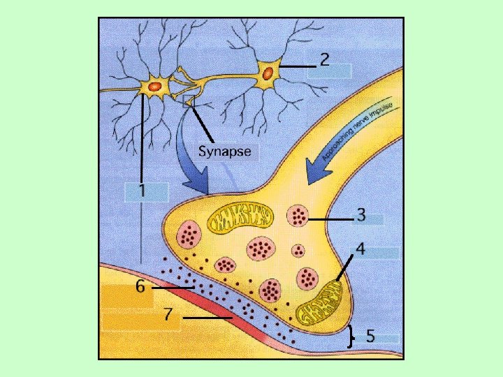
The gap between the 2 neurons or a neuron and its effector is called the synapse
Conduction of Impulses Across a Synapse: Click here for intro video about synapses An action potential is transmitted across a synapse by chemical messengers called neurotransmitters
Neurotransmitters are found in tiny sacs at the end of axon terminals
When impulses reach the axon terminal, they: 1. the tiny sacs release the neurotransmitters into the synapse 2. The neurotransmitters diffuse across the synapse
3. neurotransmitters bind to receptor proteins on the effector (muscle or gland) or dendrite of another neuron 4. the muscle, gland or neuron responds
Over 60 different neurotransmitters have been identified • the response observed depends upon which neurotransmitter was released
Some neurotransmitters are stimulators, causing: • muscles to contract, • glands to secrete, or the generation of another action potential
Other neurotransmitters are inhibitors, making: • muscles more difficult to contract, • reduced secretion of the gland, • or inhibiting the transfer of the action potential in the adjacent neuron
IV. Perception Can be internal or external Internal = nerve endings or receptors External = sensory organs
I cdnuolt blveiee taht I cluod aulaclty uesdnatnrd waht I was rdanieg. The phaonmneal pweor of the hmuan mnid. Aoccdrnig to a rscheearch at Cmabrigde Uinervtisy, it deosn't mttaer in waht oredr the ltteers in a wrod are, the olny iprmoatnt tihng is taht the frist and lsat ltteer be in the rghit pclae. The rset can be a taotl mses and you can sitll raed it wouthit a porbelm. Tihs is bcuseae the huamn mnid deos not raed ervey lteter by istlef, but the wrod as a wlohe. Amzanig huh? yaeh and I awlyas thought slpeling was ipmorantt!
Comp Book: Back of p. 38 and p. 39 Visual and Auditory Perception