neisseria infections Diagnosis of neisseria infections Diagnostical model


- Slides: 24

neisseria infections • Diagnosis of neisseria infections • Diagnostical model: • N. gonorrhoeae – swab from vagina, uretral discharge • Microscopy, cultivation, biochemical tests, detection of enzym, ATB susceptibility tests

Biology, Virulence, and Disease Gram-negative diplococci with fastidious growth requirements Growth best at 35° C to 37° C in a humid atmosphere supplemented with CO 2 Oxidase and catalase positive; acid produced from glucose oxidatively Outer surface with multiple antigens: pili protein; receptors for transferrin, lactoferrin, and hemoglobin; lipooligosaccharide; immunoglobulin protease; β -lactamase Neisseria gonorrhoeae Epidemiology Humans are the only natural hosts Carriage can be asymptomatic, particularly in women Transmission is primarily by sexual contact

Diagnosis of neisseria infections - gonococcal • Gram stain - very sensitive (90%) and specific (98%) – in purulent uretritis in men and purulent artritis. • In other cases (cervicitis, anorectal infections, pharyngitis) – sensitivity and specificity is very low. • Staining of cultures.

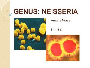
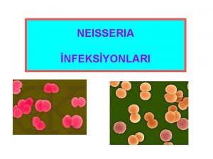
http: //www. warwickshire. gov. uk http: //medicine. plosjournals. org N. gonorrhoeae G - diplococci http: //www. ratsteachm www. medmicro. info,

http: //www. microbelibrary. org

Cultivation of Neisseria gonorrhoe • Swab from vagina or discharge – on blood agar, modified blood agar, chocolate agar + ATB – inhibition of contaminating flora • Grey colonies • after application of cytochromoxidase – become black • - biochemical tests for diff. dg. from other Neisseria

Cultivation • - Selective media Thayer Martin´s, - is a Mueller-Hinton agar with 5% chocolate sheep blood and ATB. It is used for culturing and primarily isolating pathogenic Neisseria, including N. gonorrhoeae and N. Meningitidis, as the medium inhibits the growth of most other microorganisms. - chocolate agar + ATB (vancomycin), - is a non-selective, enriched medium. . It is a variant of the blood agar plate. 10% of fresh sheep blood is heated at 80° c by the lysis of RBc. C cells. Cultivation : 5% CO 2 !drying and cold! – collected material cannot be placed in the refrigerator -

Oxidase test • The oxidase test uses Kovac’s reagent –tetramethyl-phenylenediamine dihydrochloride) to detect the presence of cytochrome c in a bacterial organism’s respiratory chain; if the oxidase reagent is catalyzed, it turns purple. Neisseria species give a positive oxidase reaction, • gram-negative oxidase-positive diplococci isolated on gonococcal selective media may be identified presumptively as N. gonorrhoeae. If the isolate is N. gonorrhoeae, • positive (purple) reaction should occur within 10 seconds.

N. gonorrhoeae cytochromoxidase +

Neisseria meningitidis • is the one with the potential to cause large epidemics. • Twelve serogroups of N. meningitidis have been identified, six of which (A, B, C, W 135, X and Y) can cause epidemics. • Geographic distribution and epidemic potential differ according to serogroup.


Mikroskopia. Gram Latex aglutinácia Ag Kultivácia PCR

Neisseria meningitidis

ATB susceptibility testing • Disc diffusion method • 6 -8 ATB discs in one plate • Zone of inhibitionof the growth in mms – comparison with standards Without zone of inhibition – resistence to tested ATB Zone of inhibition of growth sufficiently large ATB disc Growth of tested bacteria Insufficient zone of inhibition

ATB susceptibility • Neisseria gonorrhoe - PNC – penicilinase production - chromosome type of resistence - changes in cell surface, - ceftriaxon - TTC, chinolons, makrolides - azitromycin – therapy of chlamydia infection

Haemophillus infections • Microscopy – Gram staining Haemophilus influenzae • Cultivation -Haemophilus influenzae on chocolate agar - satelite growth, identification withgrow factors X, V, XV • Biochemical activity – not common • Antigen detection of H. influenzae typ b – diagnosis of bacterial infection of CNS • antigennic structure H. influenzae typ b – capsule detection (a, b, c, d, e, f type)

Microscopy • - Gram staining • Haemophilus influenzae - G - rods ( coccoid to filamentous (pleiomorphic) • - aglutination with specific antiserum - agglutination and clearing of suspension after application of specific antiserum to the colony on slide

Capsule detection • quellung reaction- aplication of specific antiserum (anti a, b, c, d, e or f) to the testing culture and staining Burri or in native smear -growing – magnification of capsule • Capsular swelling in the presence of type specific antibody is demonstrated by the halo • - aglutination with specific antiserum - agglutination and clearing of suspension after application of specific antiserum to the colony on slide

Cultivation • Haemophilus influenzae prefers a complex medium and requires preformed growth factors that are present in blood: • X factor (i. e. , hemin) and V factor (NAD or NADP). • it is usually grown on chocolate blood agar which is prepared by adding blood to an agar base at 80 o. C. • The heat releases X and V factors from the RBCs and turns the medium a chocolate brown color.

• Cross-feeding between Staphylococcus aureus and Haemophilus influenzae growing on blood agar. • Haemophilus influenzae was first streaked on to the blood agar plate • followed by a cross streak with Staphylococcus aureus. • H. influenzae is a fastidious bacterium which requires both hemin (X) and NAD(V) for growth. • There is sufficient hemin in blood for growth of Haemophilus, but the medium is insufficient in NAD. • S. aureus produces NAD in excess of its own needs and secretes it into the medium, which supports the growth of Haemophilus as satellite colonies.

Haemophillus CHOC www. medmicro. info KA (satelit)

H. Influenzae - satellite http: //phil. cdc. gov

Growth factors test faktor X, faktor V, X+V

• Antigen detection of Hib – rapid test for detection of bacterial infection of CNS • Presence of H. i. b in CSM, blood, urine can be detected by use of rapid test of latex agglutination Antibody against Hib is bound on latex particules If Hib is present in the sample, it will make a strong bound to latex and eye visible agglutination is detected. Very rapid dg - 30 minutes. Reaction of nonvital bacteria- even after previous therapy with ATB • Need of rather a big quantitiy of bacteria – correlating with visible bacteria in microscopic smear and clear clinical signs.
 What is the nursing process steps
 Medical diagnosis and nursing diagnosis difference
 Types of nursing diagnosis
 Types of nursing diagnoses
 Perbedaan diagnosis gizi dan diagnosis medis
 Neisseria meningitidis prophylaxis
 Meio de cultura para neisseria gonorrhoeae
 Meningokoklar
 Neisseria aerobic or anaerobic
 Neisseria meningitidis factores de virulencia
 Epididymitis
 Neisseria gonorrhoeae
 Caracteristicas de neisseria meningitidis
 Neisseria
 Bacteria phylum
 L
L Nitrate reduction test pseudomonas aeruginosa
 Neisseria meningitidis tratamiento
 Neisseria meningitidis
 Gonorrhea
 Neisseri
 Opportunistic infections
 Methotrexate and yeast infections
 Opportunistic infections
 Chapter 25 sexually transmitted infections and hiv/aids

