Mj s pancreas fejldse Semmelweis Egyetem OK Anatmiai


: BMP (2, 4, 7),")


 duodenum pancreas ventrale dexter")


")

 •")
- Slides: 35
Máj és pancreas fejlődése Semmelweis Egyetem, ÁOK Anatómiai, Szövet- és Fejlődéstani Intézet Dr. Kocsis Katalin 2020. 04. 22.
Előbél fejlődése
Hox gének expressziója a test anterior-posterior tengelye mentén eltérő mintázatot mutat. mellső végtag caudális határa atlas
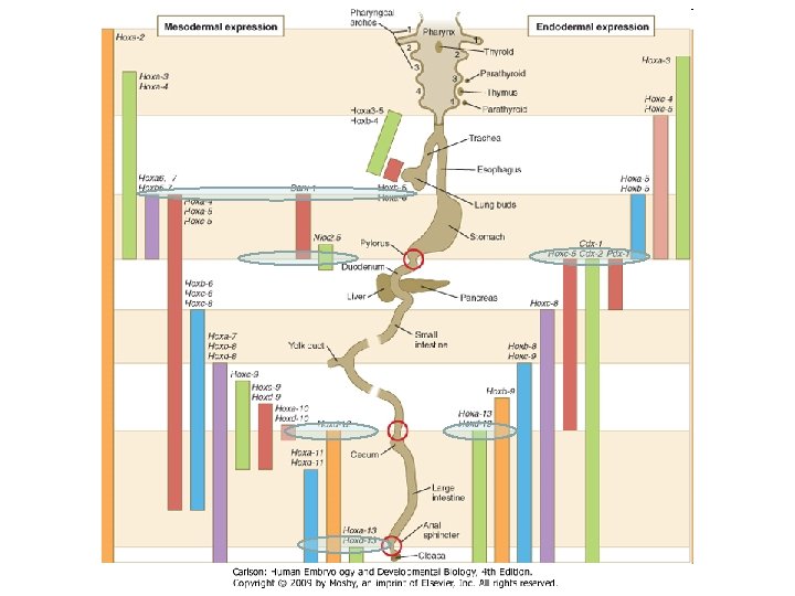
Hox gene expression boundaries in the endoderm and mesoderm during early chick gut development. Specific combinations of homeobox gene expression can be mapped to specific regions of the gastrointestinal tract, with some combinations demarcating the position of sphincters and organs. The regional expression patterns of mouse homologs are similar.
Figure 14 -5 Signals and transcription factors important in establishing regional differences in the early developing gastrointestinal tract. The top drawing represents an early mammalian embryo shortly after the initiation of embryonic folding, with areas enlarged below showing some of the signaling events involved in liver, pancreas, and hindgut specification. After initiation of cranial body folding, the endoderm of the ventral foregut is situated adjacent to the caudal cardiogenic mesoderm and septum transversum. Tissue-tissue interactions between the cardiogenic mesoderm and the endoderm, mediated by. Fgf and Bmp signals, induce hepatocytic markers within the endoderm (e. g. , Albumin and Alpha fetoprotein) while suppressing pancreatic development by upregulating Shh expression. The homeoprotein pancreatic/duodenal marker, Pdx 1, promotes pancreatic development. However, in the presence of Shh, pancreatic development is repressed. Much of the endoderm expresses Shh, but it is repressed by notochordal release of Fgfs and Activin B in the future pancreatic region. Shh expression within the hindgut endoderm induces Bmp 4 and Hoxd 13 expression within the caudal mesoderm. Shh/Bmp 4 are only capable of inducing Hoxd 13 expression in the caudal gut, possibly due to the caudal restriction of Cdx 2 expression established during gastrulation. Hoxd 13 instills a caudal identity to the hindgut. Alb, Albumin, Afp, Alpha fetoprotein; Ipf 1, insulin promoter factor 1.
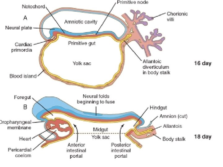
A bélcsatorna korai fejlődése aorta 20 D a. vitellina előbél a. umbilicalis utóbél entoderma szikhólyag előbél aortaív ductus omphaloentericus középbél szikhólyag utóbél 24 D szikhólyag 26 D allantois ductus omphaloentericus szikhólyag 26 D
Primitív bél régiói
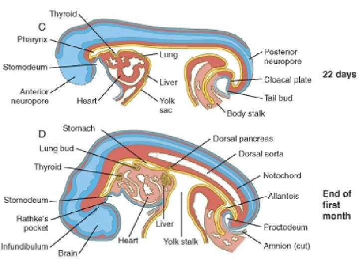
Primitív bélcső garatbél membrana buccopharyngea ductus thyreoglossus szikhólyag esophagus tüdő ductus omphaloentericus gyomor előbél szívtelep flexura duodenojejunalis testnyél középbél farokbél cecum flexura coli sinistra membrana cloacalis allantois cloaca utóbél
Máj fejlődése
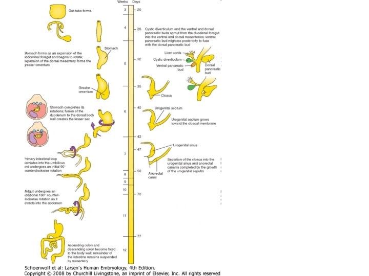
Figure 14 -9 Development of the liver, gallbladder, pancreas, and their duct systems from endodermal diverticula of the duodenum. The liver bud sprouts during the 4 th week and expands in the ventral (anterior) mesentery. The cystic diverticulum and ventral pancreatic bud also grow into the ventral mesentery, whereas the dorsal pancreatic bud grows into the dorsal mesentery. During the 5 th week, the ventral pancreatic bud migrates around the posterior side (former right side) of the duodenum to fuse with the dorsal pancreatic bud. The main duct of the ventral bud ultimately becomes the major pancreatic duct, which drains the entire pancreas.
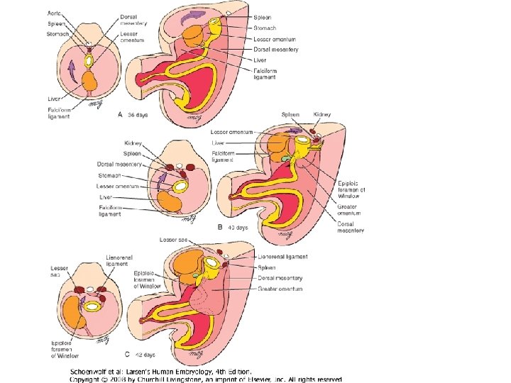
Figure 14 -14 Formation of the liver and associated membranes. As the liver bud grows into the ventral mesentery, its expanding crown makes direct contact with the developing diaphragm. The ventral mesentery that encloses the growing liver bud differentiates into the visceral peritoneum of the liver, which is reflected onto the diaphragm. This zone of reflection, which encircles the area where the liver directly contacts the diaphragm (the bare area), becomes the coronary ligament. The remnant of ventral mesentery connecting the liver with the anterior body wall becomes the falciform ligament, whereas the ventral mesentery between the liver and lesser curvature of the stomach forms the lesser omentum.
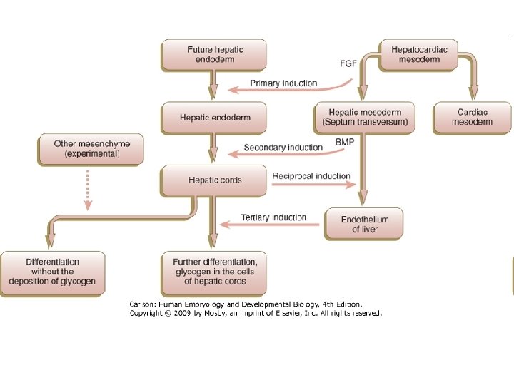
A máj fejlődésében szerepet játszó molekulák: indukció (SHH, cardiogén mesoderma): BMP (2, 4, 7), FGF (1, 2, 8), (Wnt, GATA 4, Hnf 3 (Hepatic nuclear factor -3), C/EBP (CAAT-enhancer binding proteins) Hex (hematopoietically expressed homeobox) gén kell a hepatocyta differenciálódáshoz sejtek differenciálódása, mesoderma, endothel bevándorlása: Notched / Jagged 1 (májsejt vagy ductus sejt - Alagille syndrome) Vegf, BMP, FGF érett máj funkciók (albumin, alfa fetoprotein szintézis beindítása): Hnf (hepatic nuclear factor family) C/EBP, Egf, Hgf, Tgf Tissue-tissue interactions between the cardiogenic mesoderm and the endoderm, mediated by Fgf and Bmp signals, induce hepatocytic markers within the endoderm (e. g. , Albumin and Alpha fetoprotein) while suppressing pancreatic development by upregulating Shh expression.
Sites of hematopoiesis in the human embryo. The graph highlights the relative importance of the various sites of hematopoiesis. AGM, aorta/genital ridge/mesonephros region. (Based on Carlson B: Patten's foundations of embryology, ed 6, New York, 1996, Mc. Graw-Hill).
Máj regenerációs kapacitása regeneration capacity of the liver cells Figure 18. 29. Kinetics of DNA synthesis in the four major cell types of the mammalian liver. It is possible that, since the hepatocytes respond fastest, they are secreting paracrine factors that induce DNA replication in the other cells.
Pancreas fejlődése
Figure 14 -9 Development of the liver, gallbladder, pancreas, and their duct systems from endodermal diverticula of the duodenum. The liver bud sprouts during the 4 th week and expands in the ventral (anterior) mesentery. The cystic diverticulum and ventral pancreatic bud also grow into the ventral mesentery, whereas the dorsal pancreatic bud grows into the dorsal mesentery. During the 5 th week, the ventral pancreatic bud migrates around the posterior side (former right side) of the duodenum to fuse with the dorsal pancreatic bud. The main duct of the ventral bud ultimately becomes the major pancreatic duct, which drains the entire pancreas.
A bélcsatorna mirigyeinek telepei dorsalis pancreastelep (corpus és cauda pancreatis) duodenum pancreas ventrale dexter (megmarad: caput pancreatis és proc. uncinatus) pancreas ventrale sinister (regrediál) májtelep májöböl
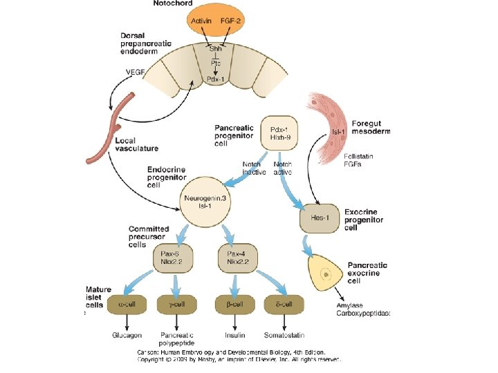
Pancreas telepek indukciója: aorta, Pdx 1 homeobox gének • Pancreas / blood vessel
Figure 14 -10 Initiation of pancreatic development in a day 10 mouse embryo. Pancreatic development begins with the formation of endodermal buds projecting into the splanchnic mesoderm near the stomach-duodenal border. Pdx 1 is expressed (seen here by immunostaining in green) in both the dorsal and ventral pancreatic bud endoderm. Glucagon-positive cells are also detected at this early stage (cells in red) within the pancreatic endodermal buds.
ventralis pancreas telep: Pdx+, Shh-, a cardiogén mesoderma gátolja a pancreas fejlődést (máj indukciójával) dorsalis pancreas telep: Shh-, ezzel indukálódik a pancreas fejlődése a bél fejlődése helyett, és a sejtek Pdx 1+-ak lesznek Pdx 1: homeoprotein
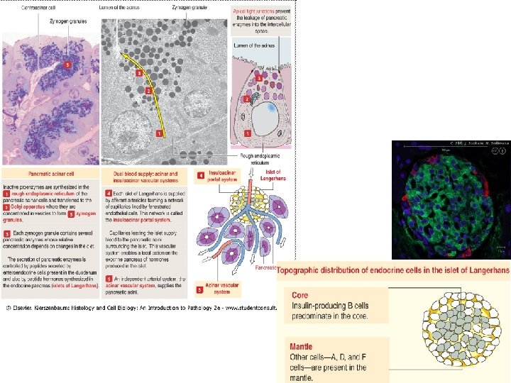
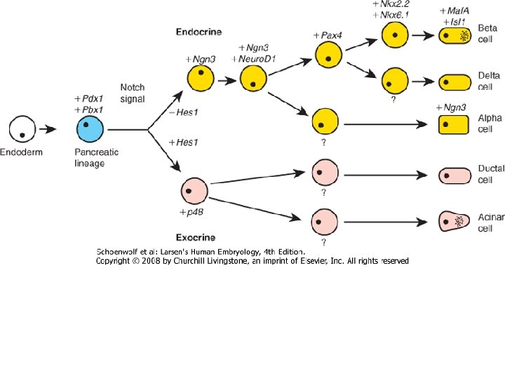
Fig. 8. Model of beta-cell development. In beta-cell precursors, Ngn 3 induces the expression of Pax 4; these cells also express Nkx 2. 2 and Nkx 6. 1, and shortly thereafter, they express Islet 1 and Pax 6. The parallel activities of Pax 4 and Nkx 2. 2 enable the program of beta-cell differentiation, and as a result, the level of Pdx 1 increases; the expression of HB 9 is induced; and the synthesis of insulin ensues. In fully mature beta cells, the activity of Pax 4 is no longer required.
Irodalom • Scott F. Gilbert: Developmental Biology (7 th edition, Sinauer Associates, 2003) • Gary Schoenwolf: Larsen's: Human Embryology (4 th edition, Elsevier, 2009) • Thomas W. Sadler: Langman's Medical Embryology (10 th edition, Lippincott Williams and Wilkins, Philadelphia, 2006) • Carlson BM. Human Embryology and Developmental Biology (Mosby, Philadelphia, 2004)