Ministry of Health of Ukraine Ukrainian Medical Dental


, wrinkles lower face (2, 3) of the chin")

 regrown hair and hairstyle make it")


 the cheeks and wrinkl")
 passes through the temporal area and")


.")


 runs along the natural crease,")


 polymer polyethylene polytetrafluoroethylene porous polytetrafluoroethylene cross-linked polymers")


- Slides: 73
Ministry of Health of Ukraine Ukrainian Medical Dental Academy Chair of surgical stomatology and maxillo-facial surgery with plastic and reconstructive surgery of head and neck Problems of plastic surgery. Pathogenetic features (involutional ptosis) of wrinkles of skin of face and neck. Low-invazive methods of their removal. Contour plastic arts of face. Basic methods of surgical removal of aesthetic defects of tissues of head and neck (blepharoplasty, rhinoplasty, facelifting). Errors and complications of plastic surgery. Candidate of sciences, reader Lokes K. P.
Lecture plan 1. 2. 3. 4. 5. Problems of plastic surgery Pathogenetic features of wrinkles of skin of face and neck. Contour plastic arts of face. Basic methods of surgical removal of aesthetic defects of tissues of head and neck. Errors and complications of plastic surgery.
Plastic surgery aimed at removing wrinkles on the face, began to develop rapidly since the beginning of the XX century. When attempts were made to improve the facial expression by injecting under the skin of wax. This method was proposed in 1890 by Gersuni. The face takes the form of masks became puffy, paraffin rolled up into a ball under the skin, bumps and ridges. Eliminating of these inequalities has been associated with difficulties, and sometimes even was impossible. Sometimes necrosis of the skin advanced, which remained after the removal of scars, and often fistulas that don’t heal.
The first interventions on the face to remove wrinkles and skin tightening were spent by Lexer, Joseph, Passot. Operations were performed under local anesthesia with numerous elipsal deletions of skin in the temporal and parotid areas, while not always was expected result. The progress that has occurred in understanding the essence of secular changes of face in many ways transformed the traditional approaches to surgical lifting techniques - from simple leather garters to the combined correction of deeper layers of the face and neck. The leading role in the modern modifications given to the correction of the superficial musculoaponeurotic system (SMAS), which can significantly improve the aesthetic result of the operation, eliminating the age-old gravitational ptosis of the soft tissues of the cheeks and nasolabial folds, neck and chin.
The following basic types of rhytidectomy: 1. Full lifting. 2. Partial lifting. 3. Mini-lifting. 4. Lifting in the forehead area.
Indications Complete lifting - presence of wrinkles and folds in the upper, middle and lower zones, the surfaces side of the face and neck. Partial lifting - removal of "crow's feet" at the outer corner of the eye and cheek wrinkles. Mini-lifting - removing of "crow's feet" at the outer corner of the eye. Lifting the forehead - the presence of wrinkles, furrows and folds in the upper part of the face.
Anesthesia As a rule, the operation is performed under general endotracheal anesthesia in combination with infiltration anesthesia (hydropreparation). Full lifting The operation consists of several stages: 1. Carrying of incision. 2. Flaking of skin-fat flap. 3. Flaking of surface musculo-fascial (musculo-aponeurotic) flap (in operations using SMAS plastics). 4. The tension of the flap within the optimal quantities of plastic deformation, given the biomechanical properties of the skin-fat grafts in each topografo-anatomical area. 5. Removal of excess skin. 6. Imposition of the internal sutures, which are absorbed in order to reduce the tension in the construction of the wound edges. 7. Imposition of superficial skin (sometimes intradermal) stitches.
Technique of incision: After careful shaving of hair in the temporal and mastoid areas and marking the line of the future incision it is started in the temporal region, dipped below continue along the front edge of the ear, bypassing the earlobe, Upward on parodit furrow and reaching the level of the external auditory canal and continue to bend it toward capitis mastoid region where it ends.
In the temporal area it is possible the conduction of two types of incisions: 1. Straight-line - space of 5 -6 mm from the line of hair growth. 2. Arcuate incision within the hairline - if "crow's feet" strongly pronounced. In parotid-masticatory area preauriculary it is possible the conduction of following incisions : - In front of tragus - to prevent the development of rough scar. - Behind of tragus - masks the scar, but can lead to the disappearance impressions in front of tragus.
In the mastoid area of the incision is at a distance of 3 -6 mm from the crease behind the ear to the level of the external auditory canal of the ear, where it smoothly at an angle 40 -60° shifted to the mastoid area. Such a move allows of the incision line formed on the rear surface of the ear thin scar is not visible when viewed from the outside. Then, incision down on continued 5 -6 cm hairline or derogate from the fibrous cover on 3 -5 mm.
Flaking of flap After the incision is necessary to perform exfoliation of skin-fat flap all over the incision. A proper selection of the plane delamination to flap has sufficient thickness and bleeding was minimal. Flaking in the temporal area should be careful not to catch the temporal branch of the facial nerve and superficial blood vessels. Hemostasis after exfoliation should be done very carefully using bipolar electrocautery or coalescent with minimal light (monopolar gifrekator) to prevent damage to a branch of the facial nerve.
Technick of detachment of skin-fat flap.
SMAS plication technique is as follows: 1. Mastectomy - removal of strips of SMAS over the parotidchewing site and linking dissects the edges: - Moving away a small strip of a width of 7 -10 mm from the top of the parotid-masticatory area and up to the middle of the cheek; - The two ends of the divided SMAS are stapled. 2. Plication of the SMAS, where the seams are tightening the SMAS without its incision: - Numerous large intermittent interrupted sutures 2 -0 or 3 -0 thread around the parotid-masticatory area, wherever the SMAS is weakened; - Surround the sutures weakened SMAS and can be fixed to a stable tissues in the upper zone in front of the tragus of the ear. The best vector of tension - the vertical direction, which improve
Numerous plicational seams by thread Maxson or Vicryl tighten the hyposthenic SMAS. These seams is better to fix the stable tissue against the ear on the side of disposition of temporal branch of the facial nerve
According to many surgeons and the results of many paired comparative studies the tightening of plicational seams can pull SMAS as well as any facelifting in the deep plane.
Removal of excess skin The first stage - the tightening of the skin vertically to improve the condition of the oval face and neck. This vector is directed upward tension provides a more natural look than the back side vector, and improves the condition of the neck. The flap is cut out and thin, so that the tension around the wound was minimal for the greatest improvement of the oval face and neck. At the points of maximum stress can impose some stitches. The first seam is applied at a tension placed on the ear.
The first tension suture over the ear
Skin-fat flap is pulled up and excised a small piece of skin to the point where it is possible to impose cosmetic sutures or staples with a cosmetic effect on the edges of the skin
The postoperative period In the postoperative period edema and bruising may develop. It is difficult to envisage the mesure of edema. Somewhat bruising can be prevented with a minimum pressure dressings. Should see patients after operation to follow the postoperative complications, such as hematomas. Damage of the facial nerve is rare, but always is possible.
Partial lifting When doing a partial lifting of the incision line is limited in the temporal and parotid-masticatory areas and ends at a point which is located midway between the trestle and the point of attachment of the ear lobe. Incision and flaking of skin is carried out in analogy to the complete lifting, after which the wound sutured by intradermally cosmetic seam.
Mini-lift The incision is carried out within the temporal scalp area. Skin-fat flap cuts out and peeled down, mobilize, and pull in the upper-rear direction. After removal of excess skin is imposed by intradermal cosmetic seam.
Complications and its prevention postoperative hematoma reasons: - Increased of blood pressure in the postoperative period, - Violation of the blood coagulation system, for example, after taking certain medications. In most cases everything passes without burdening the postoperative period and not leaving any consequences. Hematoma of small volumes (30 ml) is effectively removed by puncture. Big and growing hematomas require reoperation to identify the source of bleeding and reliable lock it. You can reduce the risk of bleeding with hematoma formation if to make sure that the patient is not taking anticoagulants, and examine the hemostatic system. Other factors that reduce the risk of hematoma - neat thorough hemostasis.
The violations of face innervation Violation of motor nerves leads to noticeable changes and sad facial expressions. This occurs in one case of 150 operations. At deep lifting the number of such complications increases manifold. At low degree of injury of the nerve trunk disorders can mimic walking alone for six months. Careful detachment with plication make the risk of accidental nerve damage low. The facial nerve will not damaged, if not doing too deep puncture in the plicational seams by nonabsorbable sutures.
Postoperative inflammation and suppuration Becouse head and neck areas are supplied very well with blood, this complication is very rare. Usually the symptoms (local pain, fever, redness of the skin) is quickly removed by antiinflammatory drugs. This problem - an inevitable consequence of damage to the hair follicles during surgery. Typically, it is most pronounced in the temporal areas in the immediate vicinity of the operational scar. The process of hair loss is usually very short-lived, and in the weeks after the operation of their growth resumes. More than others prone to this complication smokers.
The formation of pathological scars The quality of scars depends on two factors: the competence of the surgeon and the individual characteristics of the patient's skin. Instead of the normal normotrofic scar (white, thin, does not rise above the surface of the skin), which is produced in about six months after operation, can form pathological scar - hypertrophic or keloid.
Skin necrosis The risk of skin necrosis can be minimized by adequate thickness of the flap and low tension. When lifting the entire face necrosis is more reliable because the behind-the flap is thinner and often lends itself to strong tension.
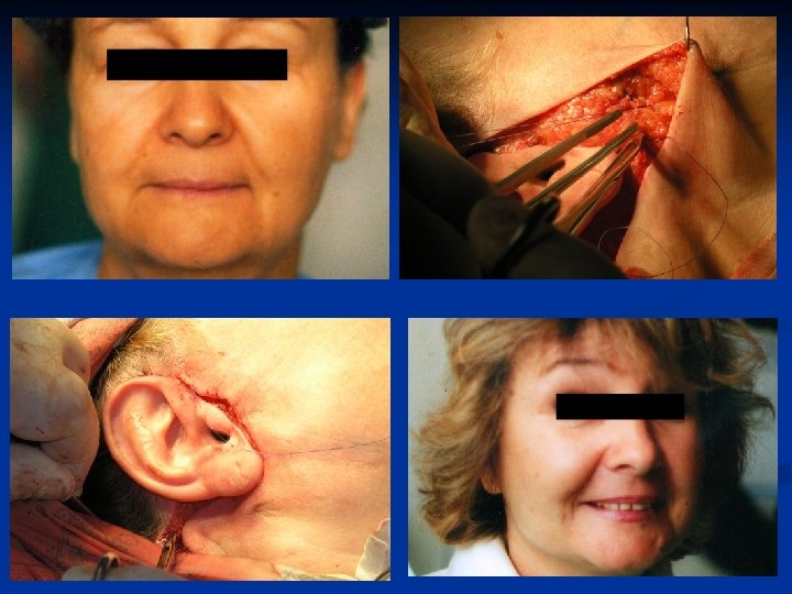
At full lifting "crow's feet" (1), wrinkles lower face (2, 3) of the chin and the neck (4) are elim
reoperative manipulation: shaving of a narrow strip of hair along the line of the futu incision, so that it could be easily hidden under other hair.
First stage: the intended incision line (black line) regrown hair and hairstyle make it invisible very soon.
Second stage: exfoliation of skin from the line of incision.
Third stage: the exfoliated skin is pulled upwards. The final result depends on the experience of the surgeon, who must determine “The golden mean" between excessive and insufficient tension.
Fourth stage: after the necessary tension, the excess part of the skin is exci
Fifth stage: the incision is closed, and soon it becomes invisible
Partial lifting eliminates "crow's feet" (1) the cheeks and wrinkl
First stage: the future incision line (black line) passes through the temporal area and front of the ea
Second stage: exfoliation of cheeks skin.
Third stage: the cheeks skin is pulled up in direction of the arrow.
Fourth stage: after pulling the excess skin is excised and the wound is sutured.
"Mini-Lifting" eliminates "Crow's feet" (A).
First step: marking fusiform skin flap with its subsequent excision.
Second step: lifting, excision of excess skin and wound closure
At men, the line of incision is shorter at the temple due to lack of large amounts of hair.
ELIMINATION OF DOUBLE CHIN First stage: incision (black line) runs along the natural crease, rendering it invisible.
Second stage: the skin and fatty tissue is striping. Excess of adipose tissue is
Third stage: the small spindly flap of skin removed to shape under-beard.
Fourth stage: the incision is closed. Scar is almost invisible.
Contour plastic INDICATIONS Injectable implant are used for filling of the face defects and to increase its some parts. In the upper third of the face are used to fill in scars ( eg on the forehead ), after acne, and in combination with botulinum toxin - for smoothing deep glabellar creases. Injection under lowered lateral third of brows raises its outer edge. Some doctors prefer the introduction of injectable implants in upper eyelid instead the blepharoplasty. Modern approach to correcting the upper third of the face based on the reduction of volume and contour of the lower eyelid and eliminates traditional fat removal. In the midface injectable implants can be used to eliminate the asymmetry due after acne scars or injuries. The most popular object of contouring plastic are nasolabial folds. The results of its correction persist especially long. In the lower part of the face above the other contours and profiles dominates the red border of lips. Sign of aging of the lower face is uneven contour of the mandible. This is
Although the first injected implant was autologous fat, the most popular implant until 2003 remained bovine collagen, which is made on the basis of Zyderm I, Zyderm II and Zyplast. These drugs are great for correction of atrophic acne scars , nasolabial folds and lips. Development of injectable implants based on hyaluronic acid allowed to make a breakthrough in contouring plastic. Hyaluronic acid is produced biotechnologically using bacteria and rooster combs. The high degree of purification of hyaluronic acid practically eliminates the risk of allergic reactions and makes it unnecessary to conduct skin tests. This versatile injectable implant with longer duration of action than collagen.
The injectable implants with polymethylmethacrylate beads recently appeared. The granules are stored permanently in the skin and lead to a proliferation of connective tissue around it, thereby providing a stable effect. These implants have been successfully used for the correction of deep wrinkles , including nasolabial folds and "marionette lines“. Hydroxylapatite (Radiesse), authorized for use as contrast agents in radial diagnostic and for stabilize of the vocal cords, also used outside the formal indications for use for filling facial wrinkles.
In 2004 , poly-L-lactic acid was approved for the correction of lipoatrophy in HIV-infected patients. Repeated subcutaneous introduction of this agent stimulates the growth of connective tissue, leading to a marked effect at atrophy of subcutaneous fat cheeks. Silicon has used for long been as an injectable implant for facial contouring. However, its introduction results in the formation of granulomas and other foreign-body reactions. Additionally, silicone may be displaced from the tissue. Possible cause of these complications is the contamination of injection of the implant. Recently been introduced microdroplet method of introducing Silicone 1000 demonstrated excellent results.
Features of postoperative period After the leading of implant to the patient , it is desirable to relax for a few minutes to relieve stress caused by injection. Cold compresses or ice can reduce the swelling and pain that appears after the expiration of a local anesthetic. The bandage is applied only at the site of injection for the intake of fat, and in the site of injection, it is usually not necessary. Patients should be reminded of the need to restrict the activity of facial muscles during the first 48 h after injection. After introducing of some injectable implants such as hydroxyapatite, for a better distribution the patients are advised to pat massage the injection site without moving the soft tissue. You must have photo of the patient before and after the injection of the implant.
COMPLICATIONS Along with the relatively rare allergic reactions, infections and hematomas may occur other complications, including: Asymmetry granuloma formation, migration and displacement of injectable implant tuberosity and the roughness of the skin, usually occurring as a result of the introduction of too superficial injection of the implant.
Noninjectable implants Scope of implants using includes increasing cheek for minimization of effect hypoplastic zygomatic eminence : - Increase of the mandible to create a stronger contour and better value of nose - chin ; - Correction of the body and the angle of the mandible to increase the expressiveness by expanding the size of the front ; - Implantation under the zygomatic eminence and midfacial to fill hollowness and allocation Flattening formed on the face during the process of natural aging ; - The introduction of implants only in the back of the nose or in the back of the nose and columella ; - Implanting the front surface of the upper jaw, a pear-shaped hole , for offset correction midface posteriorly.
Requirements for material for implantation The ideal material for implantation should be: cost-effective non-toxic, non-antigenic, non-carcinogenic, perceive by the recipient organism and is resistant to infection, inert, easily moldable, malleable, easily implanted and able to constantly maintain the original shape, easily changed and adapted to the needs of the recipient area during operation, without compromising the integrity of the implant,
Biomaterials for implants polymers Polymethyl methacrylate (acrylic) polymer polyethylene polytetrafluoroethylene porous polytetrafluoroethylene cross-linked polymers metals calcium phosphate Autografts, homografts and xenografts
Fat in the syringe for transplantation
Hydroxyapatite is used for contouring the face as a semi-permanent injectable implant is registered readings
In the diagram and pictures the correct inclination of the needle when administered intravenous transplants into the deeper layers of the skin is shown
A - pronounced nasolabial folds. B - 3 months after the injection of hyaluronic acid.
Injection of human collagen in the upper lip. Injection is carried out along the marge of red border of lips
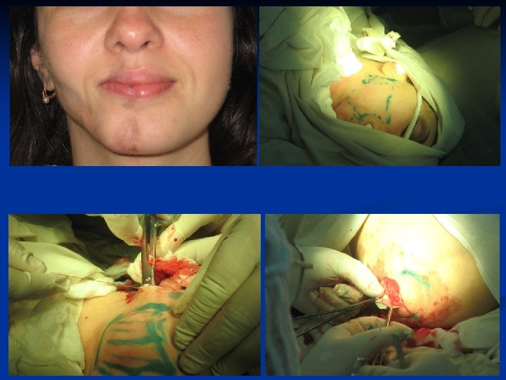
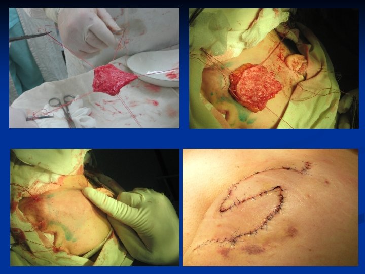
Set of chin-mandibular implants
Set of front mandibular implants
Tools are used for increasing of chin and bath with antibiotic
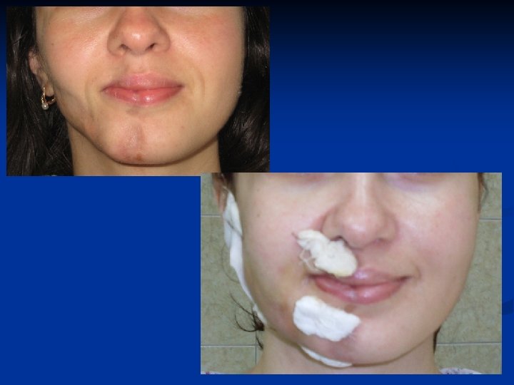
A - patient with underdeveloped chin, photos before surgery. B - photo of the patient 3 months after contouring plastic.
A - Patient with underdeveloped chin, photo before surgery. B - photo of the patient 6 months after contouring plastic.
The soft silicone material implant for corrugated of rear surface, which reduces the risk of displacement
Photography of patient S. Diagnosis: involutional ptosis of midface skin, hypoplasia of malar eminence. Before and after contouring plastic.
Questions for discussion of the lecture 1. 2. 3. 4. 5. What are the main problems in plastic surgery at present? What is minimally invasive surgery? Basic principles of operations: blepharoplasty, rhinoplasty, facelifting How many types of rhytidectomy? Possible complications of plastic surgery and their prevention.
Thank you for attention!