Ministry of Health of Ukraine Ukrainian Medical Dental


- Slides: 54
Ministry of Health of Ukraine Ukrainian Medical Dental Academy Chair of surgical stomatology and maxillo-facial surgery with plastic and reconstructive surgery of head and neck International classification of tumors of maxillofacial area by WHO. Organization of the oncostomatological aid for patients and their medical examination. Tumors and tumor-like defeats of a germinal origin – branchial , thyroglossal and dermoid cysts. Clinic, diagnostics, treatment. Candidate of sciences, reader Lokes K. P.
Lecture plan 1. International classification of tumors of maxillofacial area by WHO. 2. Organization of the oncostomatological aid for patients and their medical examination. 3. Tumors and tumor-like defeats of a germinal origin – branchial , thyroglossal and dermoid cysts. Differential diagnosis 4. Clinic, diagnostics in dentistry, treatment methods.
Oncology is the branch of medicine dealing with tumors (cancer). A medical professional who practices oncology is an oncologist. The term originates from the Greek "Ογκολογία" derived from onkos (όγκος), meaning bulk, mass, or tumor, and the suffix -logy (-λογία), meaning "study of" or "to talk about". The oncology – science which studies the origin, development, prevalence of tumours, opportunities of their diagnostics, treatment and preventive measures.
interconnected directions of oncology 1. Biological - experimental оncology. It studies reasons, patterns and mechanisms of growth of tumours. 2. Individual - clinical oncology. Studies reasons of occurrence, pathogenesis, clinical displays of tumour development, develops methods of diagnostics, treatment and preventive measures of concrete displays of tumours of various localizations. 3. Social - study of prevalence and character of tumours (epidemiology), reasons of their occurrence and development, sexual as well as age structure of patients, etc.
Risk factors Smoking is a risk factor. Leaving a cigarette on the lip is predictive of lip cancer risk irrespective of cumulative tobacco consumption. Chewing tobacco is a risk factor for cancer of the oral cavity. Alcohol consumption strongly increases the risk of developing cancers of the oral cavity, pharynx and larynx. Poor diet the presence of gastro-oesophageal reflux disease genetic susceptibility to head and neck cancer Human papillomavirus (HPV) 16 sero-positivity is associated with an increased risk of oral/ pharyngeal cancer.
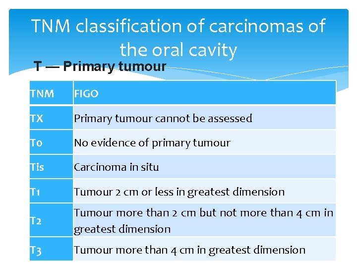
TNM classification of carcinomas of the oral cavity T — Primary tumour TNM FIGO TX Primary tumour cannot be assessed T 0 No evidence of primary tumour Tis Carcinoma in situ T 1 Tumour 2 cm or less in greatest dimension T 2 Tumour more than 2 cm but not more than 4 cm in greatest dimension T 3 Tumour more than 4 cm in greatest dimension
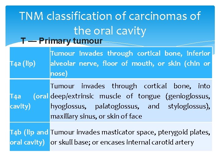
TNM classification of carcinomas of the oral cavity T — Primary tumour T 4 a (lip) Tumour invades through cortical bone, inferior alveolar nerve, floor of mouth, or skin (chin or nose) Tumour invades through cortical bone, into T 4 a (oral deep/extrinsic muscle of tongue (genioglossus, cavity) hyoglossus, palatoglossus, and styloglossus), maxillary sinus, or skin of face T 4 b (lip and Tumour invades masticator space, pterygoid plates, oral cavity) or skull base; or encases internal carotid artery
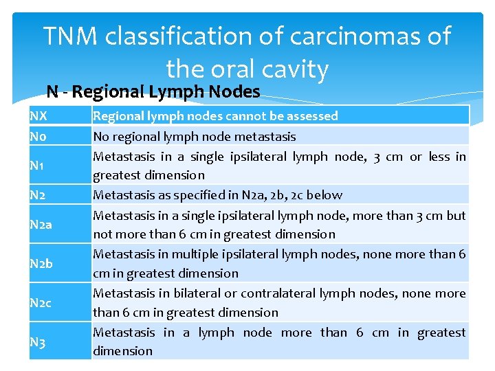
TNM classification of carcinomas of the oral cavity N - Regional Lymph Nodes NX N 0 N 1 N 2 a N 2 b N 2 c N 3 Regional lymph nodes cannot be assessed No regional lymph node metastasis Metastasis in a single ipsilateral lymph node, 3 cm or less in greatest dimension Metastasis as specified in N 2 a, 2 b, 2 c below Metastasis in a single ipsilateral lymph node, more than 3 cm but not more than 6 cm in greatest dimension Metastasis in multiple ipsilateral lymph nodes, none more than 6 cm in greatest dimension Metastasis in bilateral or contralateral lymph nodes, none more than 6 cm in greatest dimension Metastasis in a lymph node more than 6 cm in greatest dimension
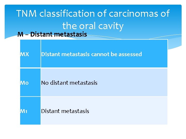
TNM classification of carcinomas of the oral cavity M – Distant metastasis MX Distant metastasis cannot be assessed M 0 No distant metastasis M 1 Distant metastasis
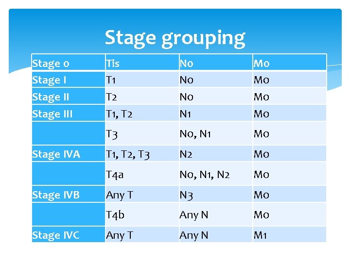
Stage grouping Stage 0 Stage III Stage IVA Stage IVB Stage IVC Tis T 1 T 2 T 1, T 2 N 0 N 0 N 1 M 0 M 0 T 3 N 0, N 1 M 0 T 1, T 2, T 3 N 2 M 0 T 4 a N 0, N 1, N 2 M 0 Any T N 3 M 0 T 4 b Any N M 0 Any T Any N M 1
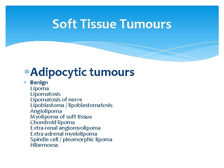
Soft Tissue Tumours Adipocytic tumours Benign Lipomatosis of nerve Lipoblastoma / lipoblastomatosis Angiolipoma Myolipoma of soft tissue Chondroid lipoma Extra-renal angiomyolipoma Extra-adrenal myelolipoma Spindle cell / pleomorphic lipoma Hibernoma
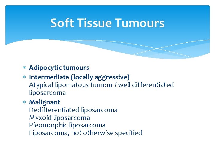
Soft Tissue Tumours Adipocytic tumours Intermediate (locally aggressive) Atypical lipomatous tumour / well differentiated liposarcoma Malignant Dedifferentiated liposarcoma Myxoid liposarcoma Pleomorphic liposarcoma Liposarcoma, not otherwise specified
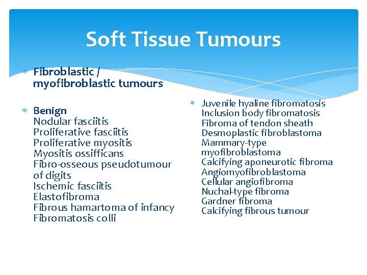
Soft Tissue Tumours Fibroblastic / myofibroblastic tumours Benign Nodular fasciitis Proliferative myositis Myositis ossifficans Fibro-osseous pseudotumour of digits Ischemic fasciitis Elastofibroma Fibrous hamartoma of infancy Fibromatosis colli Juvenile hyaline fibromatosis Inclusion body fibromatosis Fibroma of tendon sheath Desmoplastic fibroblastoma Mammary-type myofibroblastoma Calcifying aponeurotic fibroma Angiomyofibroblastoma Cellular angiofibroma Nuchal-type fibroma Gardner fibroma Calcifying fibrous tumour
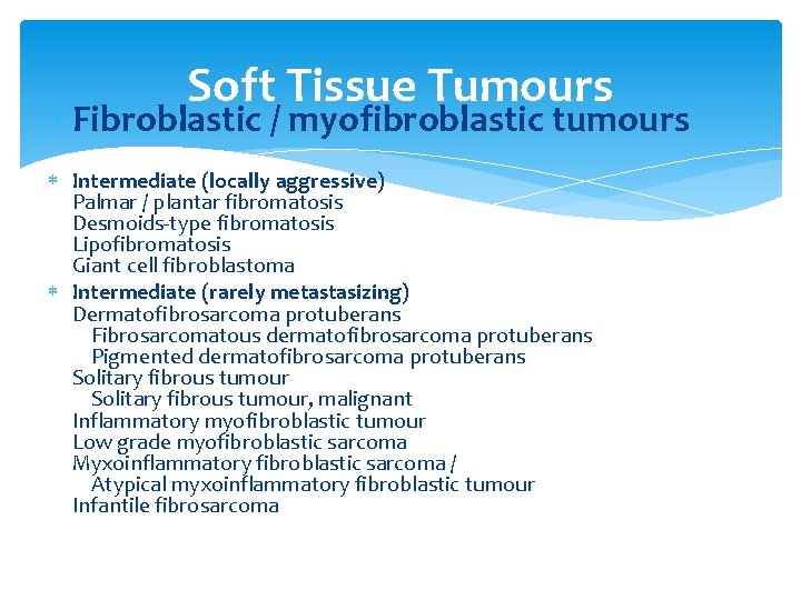
Soft Tissue Tumours Fibroblastic / myofibroblastic tumours Intermediate (locally aggressive) Palmar / plantar fibromatosis Desmoids-type fibromatosis Lipofibromatosis Giant cell fibroblastoma Intermediate (rarely metastasizing) Dermatofibrosarcoma protuberans Fibrosarcomatous dermatofibrosarcoma protuberans Pigmented dermatofibrosarcoma protuberans Solitary fibrous tumour, malignant Inflammatory myofibroblastic tumour Low grade myofibroblastic sarcoma Myxoinflammatory fibroblastic sarcoma / Atypical myxoinflammatory fibroblastic tumour Infantile fibrosarcoma
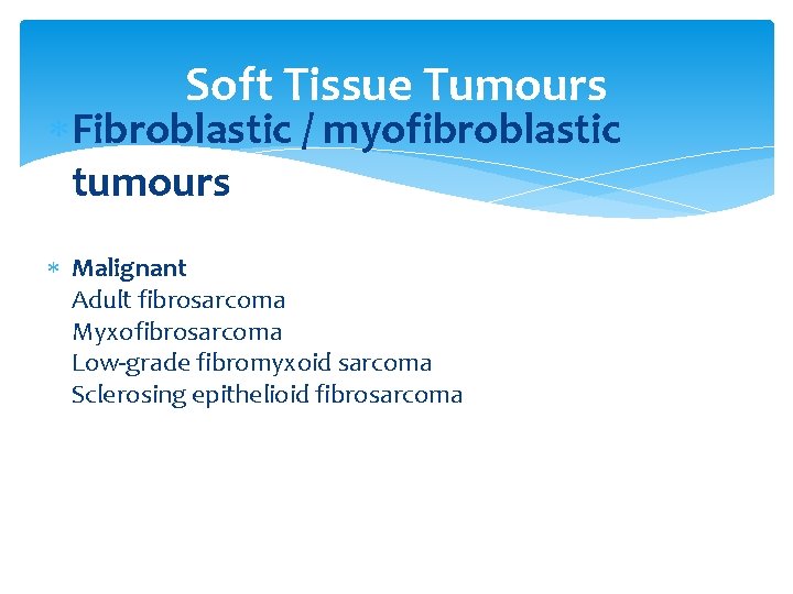
Soft Tissue Tumours Fibroblastic / myofibroblastic tumours Malignant Adult fibrosarcoma Myxofibrosarcoma Low-grade fibromyxoid sarcoma Sclerosing epithelioid fibrosarcoma
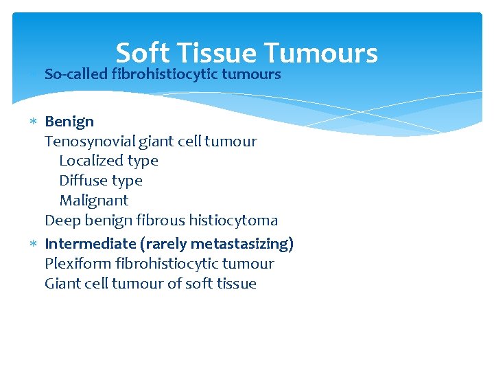
Soft Tissue Tumours So-called fibrohistiocytic tumours Benign Tenosynovial giant cell tumour Localized type Diffuse type Malignant Deep benign fibrous histiocytoma Intermediate (rarely metastasizing) Plexiform fibrohistiocytic tumour Giant cell tumour of soft tissue
Soft Tissue Tumours So-called fibrohistiocytic tumours Benign Tenosynovial giant cell tumour Localized type Diffuse type Malignant Deep benign fibrous histiocytoma Intermediate (rarely metastasizing) Plexiform fibrohistiocytic tumour Giant cell tumour of soft tissue
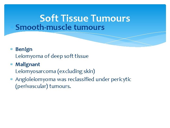
Soft Tissue Tumours Smooth-muscle tumours Benign Leiomyoma of deep soft tissue Malignant Leiomyosarcoma (excluding skin) Angioleiomyoma was reclassified under pericytic (perivascular) tumours.
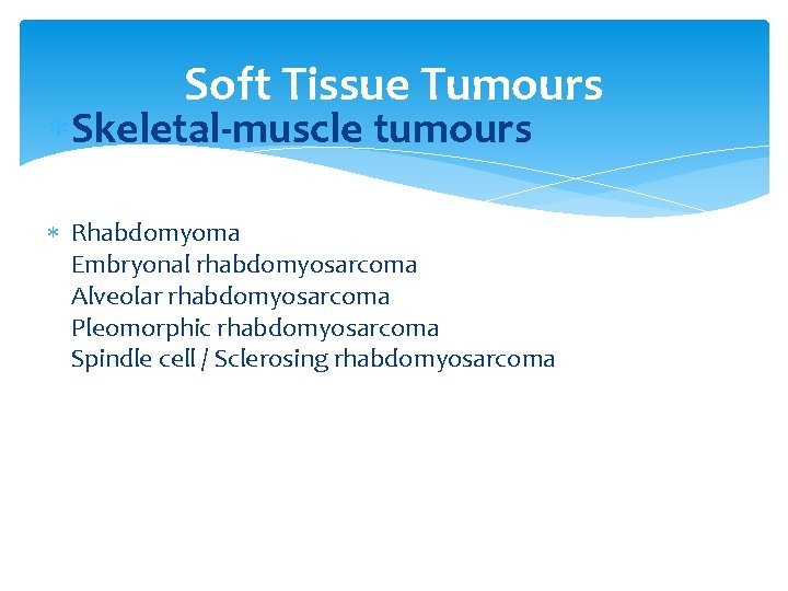
Soft Tissue Tumours Skeletal-muscle tumours Rhabdomyoma Embryonal rhabdomyosarcoma Alveolar rhabdomyosarcoma Pleomorphic rhabdomyosarcoma Spindle cell / Sclerosing rhabdomyosarcoma
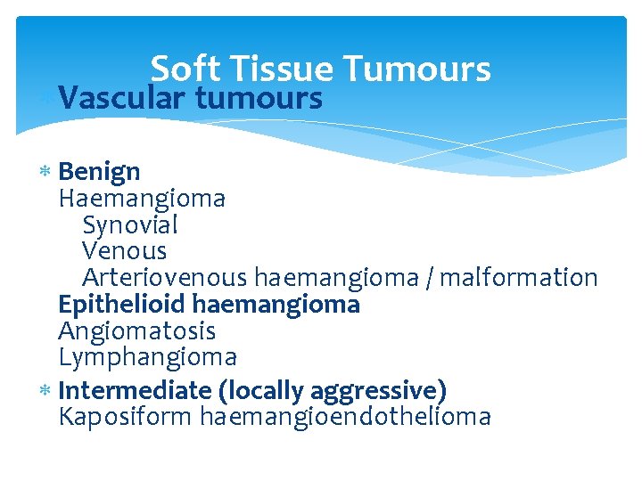
Soft Tissue Tumours Vascular tumours Benign Haemangioma Synovial Venous Arteriovenous haemangioma / malformation Epithelioid haemangioma Angiomatosis Lymphangioma Intermediate (locally aggressive) Kaposiform haemangioendothelioma
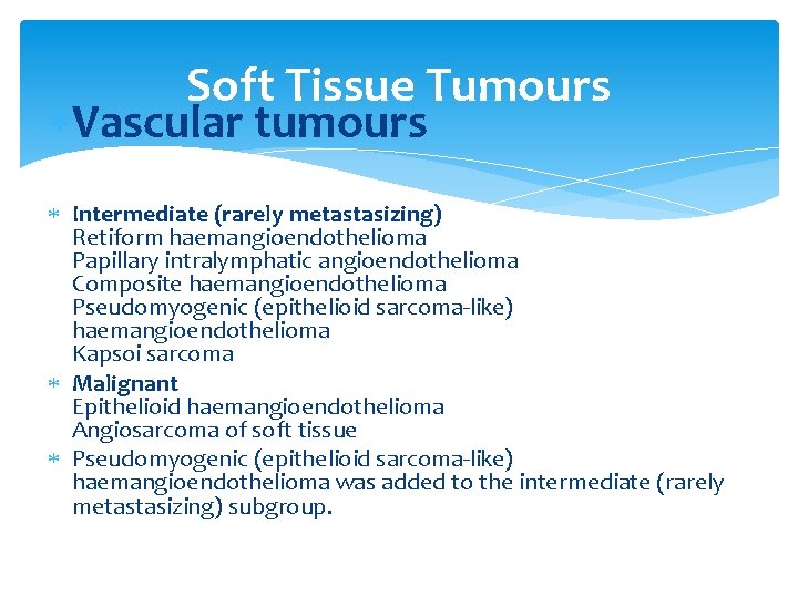
Soft Tissue Tumours Vascular tumours Intermediate (rarely metastasizing) Retiform haemangioendothelioma Papillary intralymphatic angioendothelioma Composite haemangioendothelioma Pseudomyogenic (epithelioid sarcoma-like) haemangioendothelioma Kapsoi sarcoma Malignant Epithelioid haemangioendothelioma Angiosarcoma of soft tissue Pseudomyogenic (epithelioid sarcoma-like) haemangioendothelioma was added to the intermediate (rarely metastasizing) subgroup.
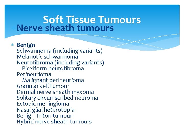
Soft Tissue Tumours Nerve sheath tumours Benign Schwannoma (including variants) Melanotic schwannoma Neurofibroma (including variants) Plexiform neurofibroma Perineurioma Malignant perineurioma Granular cell tumour Dermal nerve sheath myxoma Solitary circumscribed neuroma Ectopic meningioma Nasal glial heterotopia Benign Triton tumour Hybrid nerve sheath tumours
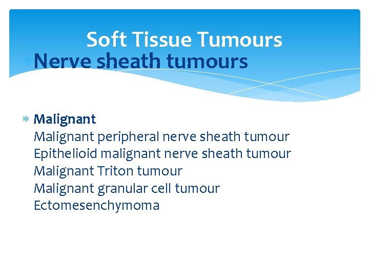
Soft Tissue Tumours Nerve sheath tumours Malignant peripheral nerve sheath tumour Epithelioid malignant nerve sheath tumour Malignant Triton tumour Malignant granular cell tumour Ectomesenchymoma
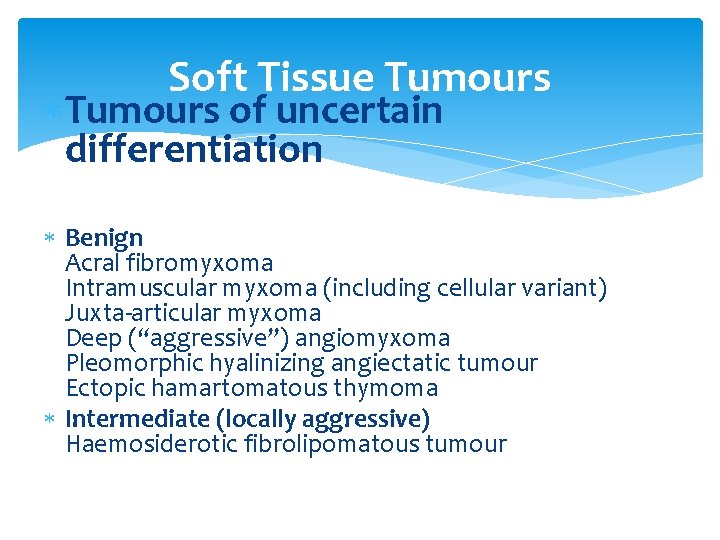
Soft Tissue Tumours of uncertain differentiation Benign Acral fibromyxoma Intramuscular myxoma (including cellular variant) Juxta-articular myxoma Deep (“aggressive”) angiomyxoma Pleomorphic hyalinizing angiectatic tumour Ectopic hamartomatous thymoma Intermediate (locally aggressive) Haemosiderotic fibrolipomatous tumour
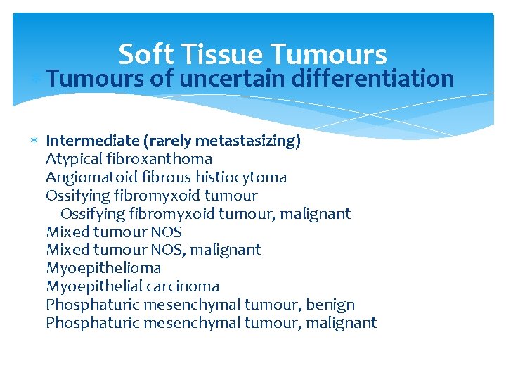
Soft Tissue Tumours of uncertain differentiation Intermediate (rarely metastasizing) Atypical fibroxanthoma Angiomatoid fibrous histiocytoma Ossifying fibromyxoid tumour, malignant Mixed tumour NOS, malignant Myoepithelioma Myoepithelial carcinoma Phosphaturic mesenchymal tumour, benign Phosphaturic mesenchymal tumour, malignant
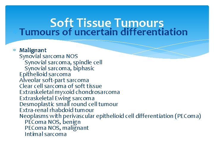
Soft Tissue Tumours of uncertain differentiation Malignant Synovial sarcoma NOS Synovial sarcoma, spindle cell Synovial sarcoma, biphasic Epithelioid sarcoma Alveolar soft-part sarcoma Clear cell sarcoma of soft tissue Extraskeletal myxoid chondrosarcoma Extraskeletal Ewing sarcoma Desmoplastic small round cell tumour Extra-renal rhabdoid tumour Neoplasms with perivascular epithelioid cell differentiation (PEComa) PEComa NOS, benign PEComa NOS, malignant Intimal sarcoma
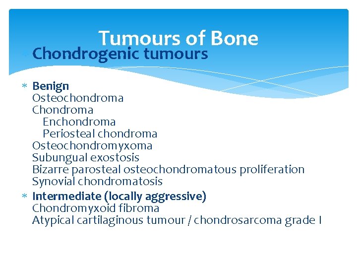
Tumours of Bone Chondrogenic tumours Benign Osteochondroma Chondroma Enchondroma Periosteal chondroma Osteochondromyxoma Subungual exostosis Bizarre parosteal osteochondromatous proliferation Synovial chondromatosis Intermediate (locally aggressive) Chondromyxoid fibroma Atypical cartilaginous tumour / chondrosarcoma grade I
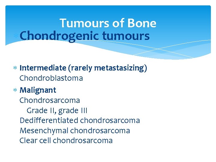
Tumours of Bone Chondrogenic tumours Intermediate (rarely metastasizing) Chondroblastoma Malignant Chondrosarcoma Grade II, grade III Dedifferentiated chondrosarcoma Mesenchymal chondrosarcoma Clear cell chondrosarcoma
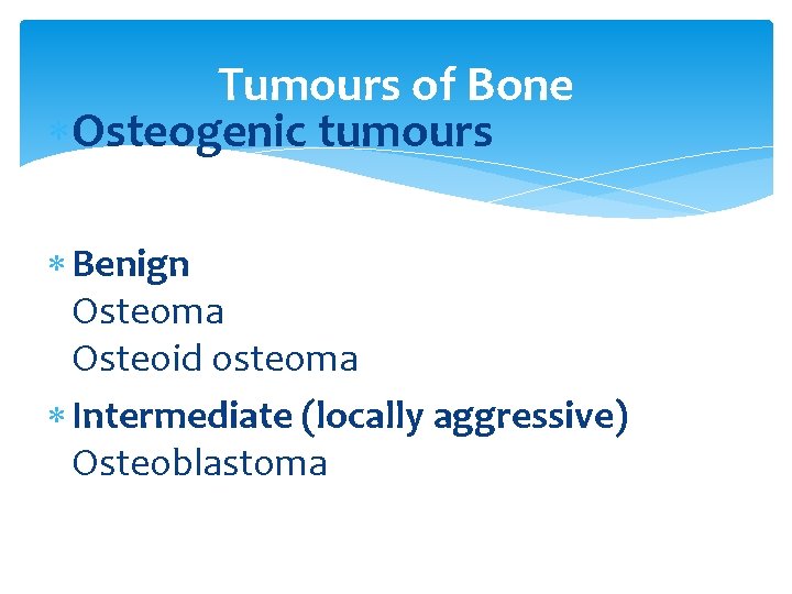
Tumours of Bone Osteogenic tumours Benign Osteoma Osteoid osteoma Intermediate (locally aggressive) Osteoblastoma
Tumours of Bone Osteogenic tumours Malignant Low-grade central osteosarcoma Conventional osteosarcoma Chondroblastic osteosarcoma Fibroblastic osteosarcoma Osteoblastic osteosarcoma Telangiectatic osteosarcoma Small cell osteosarcoma Secondary osteosarcoma Parosteal osteosarcoma Periosteal osteosarcoma High-grade surface osteosarcoma
Tumours of Bone Osteoclastic giant cell rich tumours Benign Giant cell lesion of the small bones Intermediate locally aggressive, rarely metastasizing Giant cell tumour of bone Malignant Malignancy in giant cell tumor of bone Giant cell tumor of bone is now considered a locally aggressive, very rarely metastasizing lesion.
Tumours of Bone Fibrohistiocytic tumours Benign fibrous histiocytoma / nonossifying fibroma Malignant fibrous histiocytoma of bone was removed from the current classification. MFH of bone was renamed undifferentiated high-grade pleomorphic sarcoma of bone.
Tumours of Bone Notochordal tumours Benign notochordal tumour Malignant Chordoma
Tumours of Bone Vascular tumors Benign Haemangioma Intermediate locally aggressive rarely metastasizing epithelioid hemangioma Malignant epithelioid hemangioendothelioma angiosarcoma
Tumours of Bone Tumour syndromes Beckwith-Wiedemann syndrome Cherubism Enchondromatosis: Ollier disease and Maffucci syndrome Li-Fraumeni syndrome Mc. Cune-Albright syndrome Multiple osteochondromas Neurofibromatosis type 1 Retinoblastoma syndrome Rothmund-Thomson syndrome Werner syndrome
The main categories of cancer Carcinoma – cancer that begins in the skin or in tissues that line or cover internal organs. There a number of subtypes, including adenocarcinoma, basal cell carcinoma, squamous cell carcinoma, and transitional cell carcinoma Sarcoma – cancer that begins in the connective or supportive tissues such as bone, cartilage, fat, muscle, or blood vessels Leukaemia – cancer that starts in blood forming tissue such as the bone marrow and causes large numbers of abnormal blood cells to be produced and go into the blood Lymphoma and myeloma – cancers that begin in the cells of the immune system Brain and spinal cord cancers – these are known as central nervous system cancers
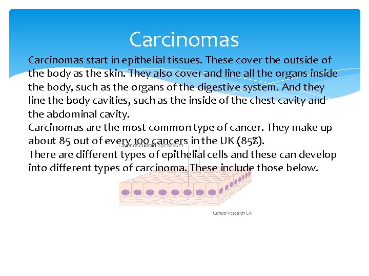
Carcinomas start in epithelial tissues. These cover the outside of the body as the skin. They also cover and line all the organs inside the body, such as the organs of the digestive system. And they line the body cavities, such as the inside of the chest cavity and the abdominal cavity. Carcinomas are the most common type of cancer. They make up about 85 out of every 100 cancers in the UK (85%). There are different types of epithelial cells and these can develop into different types of carcinoma. These include those below.
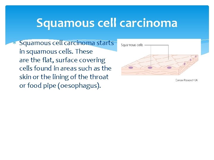
Squamous cell carcinoma starts in squamous cells. These are the flat, surface covering cells found in areas such as the skin or the lining of the throat or food pipe (oesophagus).
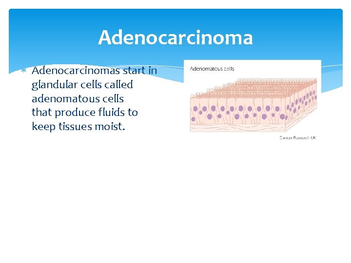
Adenocarcinoma Adenocarcinomas start in glandular cells called adenomatous cells that produce fluids to keep tissues moist.
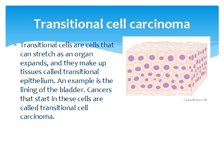
Transitional cell carcinoma Transitional cells are cells that can stretch as an organ expands, and they make up tissues called transitional epithelium. An example is the lining of the bladder. Cancers that start in these cells are called transitional cell carcinoma.
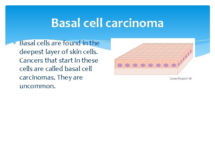
Basal cell carcinoma Basal cells are found in the deepest layer of skin cells. Cancers that start in these cells are called basal cell carcinomas. They are uncommon.
Sarcomas start in connective tissues, which are the supporting tissues of the body. Connective tissues include the bones, cartilage, tendons and fibrous tissue that support the body organs. Sarcomas are much less common than carcinomas. They are usually grouped into two main types – bone sarcomas (osteosarcoma) and soft tissue sarcomas. Altogether, these make up less than 1 in every 100 cancers diagnosed (1%).
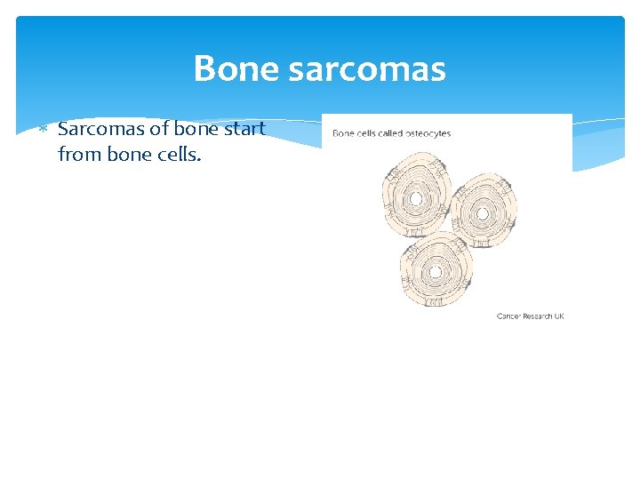
Bone sarcomas Sarcomas of bone start from bone cells.
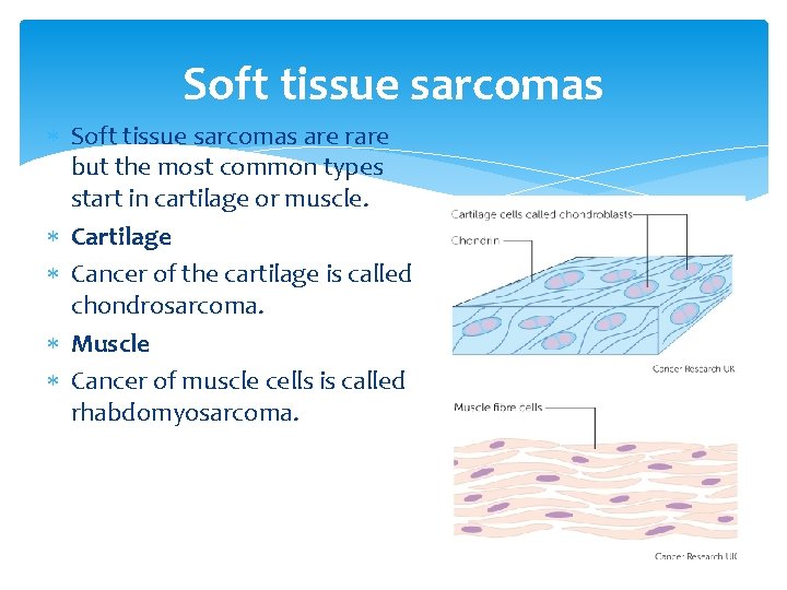
Soft tissue sarcomas are rare but the most common types start in cartilage or muscle. Cartilage Cancer of the cartilage is called chondrosarcoma. Muscle Cancer of muscle cells is called rhabdomyosarcoma.
The physical examination inspection; palpation; auscultation.
Additional examination Endoscopy. Cytology (swabs, aspirates). Histology (biopsy). X-ray investigations. Radioisotope mthods (scanning, scintigraphy). Ultrasonography. Computerised axial tomography. Laboratory tests (blood cell morphology, enzyme activity etc, as indicated).
Theories throughout history (Cancer causes) Humoral theory. Hippocrates believed that the body had 4 humors (body fluids): blood, phlegm, yellow bile, and black bile. When the humors were balanced, a person was healthy. Too much or too little of any of the humors caused disease. An excess of black bile in various body sites was thought to cause cancer. This theory of cancer was passed on by the Romans and was embraced by the influential doctor Galen’s medical teaching, which remained the unchallenged standard through the Middle Ages for over 1, 300 years. During this period, the study of the body, including autopsies, was prohibited for religious reasons, which limited progress of medical knowledge.
Theories throughout history (Cancer causes) Lymph theory. Among theories that replaced the humoral theory of cancer, was the formation of cancer by another body fluid, lymph. Life was believed to consist of continuous and appropriate movement of the fluid parts of the body through the solid parts. Of all the fluids, the most important were blood and lymph. Stahl and Hoffman theorized that cancer was composed of fermenting and degenerating lymph varying in density, acidity, and alkalinity. The lymph theory gained rapid support. The eminent Scottish surgeon John Hunter (1728− 1793) agreed that tumors grow from lymph constantly thrown out by the blood.
Theories throughout history (Cancer causes) Blastema theory. In 1838, German pathologist Johannes Muller demonstrated that cancer is made up of cells and not lymph, but he believed that cancer cells did not come from normal cells. Muller proposed that cancer cells developed from budding elements (blastema) between normal tissues. His student, Rudolph Virchow (1821− 1902), the famous German pathologist, determined that all cells, including cancer cells, are derived from other cells.
Theories throughout history (Cancer causes) Chronic irritation theory. Virchow proposed that chronic irritation was the cause of cancer, but he falsely believed that cancers “spread like a liquid. ” In the 1860 s, German surgeon, Karl Thiersch, showed that cancers metastasize through the spread of malignant cells and not through some unidentified fluid.
Theories throughout history (Cancer causes) Trauma theory. Despite advances in the understanding of cancer, from the late 1800 s until the 1920 s, trauma was thought by some to cause cancer. This belief was maintained despite the failure of injury to cause cancer in experimental animals.
Theories throughout history (Cancer causes) Infectious disease theory. Zacutus Lusitani (1575− 1642) and Nicholas Tulp (1593− 1674), 2 doctors in Holland, concluded at almost the same time that cancer was contagious. They made this conclusion based on their experiences with breast cancer in members of the same household. Lusitani and Tulp publicized the contagion theory in 1649 and 1652, respectively. They proposed that cancer patients should be isolated, preferably outside of cities and towns, in order to prevent the spread of cancer. Throughout the 17 th and 18 th centuries, some believed that cancer was contagious. In fact, the first cancer hospital in France was forced to move from the city in 1779 because people feared cancer would spread throughout the city. Although human cancer, itself, is not contagious, we now know that certain viruses, bacteria, and parasites can increase a person’s risk of developing cancer.
Questions for discussion of the lecture 1. Theories of carcinogenesis. 2. Antitumor resistance of the body. 3. Biological characteristics of tumor tissue. 4. Deontological aspects of the diagnosis of cancer. 5. Features of the use of stereolithographic models of the skull for topical diagnosis of tumors.
Thank you for attention!