Ministry of Health of Ukraine Ukrainian Medical Dental


 Odontogenic keratocyst (“primordial” cysts)")

 cyst is the most common odontogenic cyst. The usual etiology is")


 – Form in bone in relation to the root of a")


 Carcinomatous/Neoplastic Changes: Squamous Cell Carcinoma or Epidermoid Carcinoma may occasionally arise")


 is the most important of the odontogenic cysts. This cyst")


- Slides: 40
Ministry of Health of Ukraine Ukrainian Medical Dental Academy Chair of surgical stomatology and maxillo-facial surgery with plastic and reconstructive surgery of head and neck Odontogenic tumors of jaws – ameloblastoma, odontoma, cementoma, epulids, odontogenic fibroma. Odontogenic cysts of the jaws arising owing to developmental anomalies and inflammatory genesis. Candidate of sciences, reader Skikevich M. G.
Lecture plan 1. Odontogenic tumors of jaws. 2. The clinical picture of tumors: ameloblastoma, odontoma, cementoma, epulids, odontogenic fibroma. 3. Modern methods of surgical treatment 4. Odontogenic cysts of the jaws arising owing to developmental anomalies 5. Odontogenic cysts of inflammatory genesis.
A cyst is a closed sac having a distinct membrane and division on the nearby tissue. It may contain air, fluids, or semi-solid material. A collection of pus is called an abscess, not a cyst. Once formed, a cyst could go away on its own or may have to be removed through surgery.
Cysts are pathological fluid-filled cavities lined by epithelium. Cysts are the most common cause of chronic swellings of the jaws. They are more common in the jaws than in any other bone because of the many rests of odontogenic epithelium remaining in the tissues. Cysts formed from this epithelium (odontogenic cyst) account for most cysts of the jaws. By far the most common is the radicular (periodontal or “dental”) cyst.
Key features of jaw cysts Form sharply -defined radiolucencies with smooth borders Fluid may be aspirated and thin- walled cysts may be transilluminated Grow slowly, displacing rather than resorbing teeth Symptomless unless infected and are frequently chance radiographic findings Rarely large enough to cause pathological fracture From compressible and fluctuant swellings if extending into soft tissues Appear bluish when close to the mucosal surface
DEVELOPMENTAL CYSTS ODONTOGENIC “Gingival cysts” of infants (Epstein pearls) Odontogenic keratocyst (“primordial” cysts) Dentigerouse (follicular) ERUPTION CYSTS Lateral periodontal cyst Gingival cyst of adults Glandular odontogenic NON-ODONTOGENIC CYSTS Nasopalatine duct cyst Nasolabial cyst
Odontogenic Mandibular Cysts Odontogenic cysts are defined as epithelial-lined structures derived from odontogenic epithelium. Most odontogenic cysts are defined more by their location than by any histologic characteristics. Accordingly, the surgeon must provide the pathologist with appropriate history and radiographs when submitting such specimens for examination.
A periapical (radicular) cyst is the most common odontogenic cyst. The usual etiology is a tooth that becomes infected, leading to necrosis of the pulp. Toxins exit the apex of the tooth, leading to periapical inflammation. This inflammation stimulates the Malassez epithelial rests, which are found in the periodontal ligament, resulting in the formation of a periapical granuloma that may be infected or sterile. Eventually, this epithelium undergoes necrosis caused by a lack of blood supply, and the granuloma becomes a cyst. The lesions are not usually clinically detectable when small but most often are discovered as incidental findings on radiographic survey.
RADICULAR CYSTS Major factors in the pathogenesis of cyst formation: Proliferation of epithelial lining and fibrous capsule Hydrostatic pressure of cyst fluid Resorption of surrounding bone
Hydrostatic effects of cyst fluids That radicular and many other cysts expend in balloon-like fashion, wherever the local anatomy permits, indicates that internal pressure is a factor in their growth. The hydrostatic pressure within cysts is about 70 cm of water therefore higher than the capillary blood pressure.
Radicular cyst (radiography) – Form in bone in relation to the root of a non-vital tooth – Are usually asymptomatic unless infected – Diagnosis is by the combination of radiographic appearances, a non-vital tooth and appropriate histological appearances – Clinical and radiographic features are usually adequate for planning treatment – Do not recur after competent enucleation – Residual cysts can remain after the causative tooth has been extracted and diagnosis is then less obvious – Cholesterol crystals often seen in the cyst fluid but not specific to radicular cysts
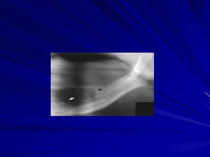
This is a typical appearance of apical radiolucency. Note the well delineated cavity with a marked radiopaque rim. The biopsy proved this to be a radicular cyst. Note the radicular rest corresponding to the roots of a first mandibular molar.
Differential Diagnosis: Radicular Cyst Periapical Granuloma Traumatic Bone Cyst Periapical Scar Periapical Cemental Dysplasia Periapical Surgical Defect Globulomaxillary Cyst Aneurysmal Bone Cyst Mandibular Infected Buccal Cyst Periapical Cemento-osseous dysplasia
Prognosis: Prognosis depends on particular tooth, the extent of bone destroyed & accessibility for treatment.
Expected Complications 1) Carcinomatous/Neoplastic Changes: Squamous Cell Carcinoma or Epidermoid Carcinoma may occasionally arise from epithelial lining of Radicular Cyst. 2) Pathologic Jaw Fracture: If Cyst have completely eroded the bone specially if it is present in posterior region which is very rare in case of Radicular Cyst it may cause pathologic jaw bone fracture. 3) Secodary Infection: Cyst may get secondarily infected and create further complications.
Advantages and limitations of enucleation of cysts Advantages. The cavity usually heals without complications
Several treatment options exist for such cysts. Many cysts resolve with endodontic therapy of the involved tooth. Those lesions should be monitored radiographically to ensure such resolution. Lesions that fail to resolve with such therapy should be surgically removed and histopathologically examined. Although these cysts arise from a mature resting epithelium and thus have a relatively low growth potential, a squamous cell carcinoma occasionally may arise de novo in a radicular cyst, thus the recommendation for histopathologic examination of all tissues removed.
The second most common odontogenic cyst is the dentigerous cyst, which develops within the normal dental follicle that surrounds an unerupted tooth. The dentigerous cyst is not thought to be neoplastic. It most frequently is found in areas where unerupted teeth are found: mandibular third molars, maxillary third molars, and maxillary canines, in decreasing order of frequency. These cysts can grow very large and can move teeth, but, more commonly, they are relatively small. Most dentigerous cysts are asymptomatic, and their discovery is usually an incidental finding on radiography.
The usual radiographic appearance is that of a well. Demarcated radiolucent lesion attached at an acute angle to the cervical area of an unerupted tooth. The border of the lesion may be radiopaque. The radiographic differentiation between a dentigerous cyst and a normal dental follicle is based merely on size.
By definition, the primordial cyst develops instead of a tooth. Presumably, the dental follicle forms and subsequently undergoes cystic degeneration without ever completing odontogenesis. This is the rarest odontogenic cyst, and lesions designated as primordial cysts may represent residual cysts. The histology of these lesions is a nondescript stratified squamous epithelium. A complete dental history is important to establish a diagnosis of primordial (versus residual) cyst, although such a diagnosis often has little clinical significance in terms of treatment planning and decision making.
Residual cyst is a term of convenience because no teeth are left by which to identify the lesion. Most commonly, these are actually retained periapical cysts from teeth that have been removed. The histology is a nondescript stratified squamous epithelium.
The name lateral periodontal cyst is a misnomer. These cysts are not inflammatory, they do not arise from periodontitis, and they are not a phenomenon associated with lateral canals within the tooth structure. These cysts are always well demarcated, relatively small, and radiolucent (sometimes with a radiopaque roof). They are most commonly associated with the mandibular premolar area and are occasionally found in the maxillary anterior. They are usually not clinically apparent but, rather, are detected through radiographic examination.
Gingival cysts of the adult are found only in soft tissue in the lower premolar areas. These cysts present as tense, fluctuant, vesicular, or bullous lesions. Histologically, they look like lateral periodontal cysts, and they probably represent the same lesion when found in soft tissue.
The ODONTOGENIC KERATOCYST (OKC) is the most important of the odontogenic cysts. This cyst may have any clinical appearance; it is a great mimic, and the diagnosis is a histologic one. These lesions are different from other cysts; they are aggressive and can be difficult to remove. OKCs can grow quite rapidly, and recurrences are frequent. This is the third most common odontogenic cyst and belongs in the differential diagnoses of any radiolucency of the jaws.
The variant of OKC that produces only orthokeratin acts somewhat differently than other OKCs. These almost always are found in a dentigerous association, usually around the mandibular third molar, and they are much less aggressive. They do not have a hyperchromatic basal layer; in fact, the basal layer is flattened. They are not associated with basal cell nevus syndrome.
AMELOBLASTOMA Ameloblastoma (from the early English word amel, meaning enamel + the Greek word blastos, meaning germ) is a rare, benign tumor of odontogenic epithelium (ameloblasts, or outside portion, of the teeth during development) much more commonly appearing in the mandible than the maxilla.
It was recognized in 1827 by Cusack. This type of odontogenic neoplasm was designated as an adamantinoma in 1885 by the French physician Louis-Charles Malassez. It was finally renamed to the modern name ameloblastoma in 1930 by Ivey and Churchill.
While these tumors are rarely malignant or metastatic (that is, they rarely spread to other parts of the body), and progress slowly, the resulting lesions can cause severe abnormalities of the face and jaw. Additionally, because abnormal cell growth easily infiltrates and destroys surrounding bony tissues, wide surgical excision is required to treat this disorder.
Subtypes There are three main clinical subtypes of ameloblastoma: unicystic, multicystic, peripheral. The peripheral subtype composes 2% of all ameloblastomas.
Clinical features Ameloblastomas are the most common neoplasm’s of the jaws. They are usually first recognized between the ages of 30 and 50, and rare in children and old people. Eighty percent form in the mandible; of these, 70% develop in the posterior molar region, and often involve the ramus. They are symptomless until the swelling becomes obtrusive.
Radiographically, ameloblastomas typically form rounded, cyst-like, radiolucent areas with moderately well-defined margins and typically appear multilocular. Lingual expansion may sometimes be seen, but is not pathognomonic of ameloblastoma. Other variants are a honeycomb pattern, a single welldefined cavity indistinguishable from a ridiculer or, rarely, a dentigerous cyst. However, differentiation from non-neoplastic cysts and other tumours or tumour-like lesions of the jaws is not possible by radiography alone.
Ameloblastomas are often associated with the presence of unerupted teeth. Symptoms include: painless swelling, facial deformity if severe enough, pain if the swelling impinges on other structures, loose teeth, ulcers, and periodontal (gum) disease. Lesions will occur in the mandible and maxilla, although 75% occur in the ascending ramus area and will result in extensive and grotesque deformitites of the mandible and maxilla. In the maxilla it can extend into the maxillary sinus and floor of the nose. The lesion has a tendency to expand the bony cortices because slow growth rate of the lesion allows time for periosteum to develop thin shell of bone ahead of the expanding lesion
This shell of bone cracks when palpated and this phenomenon is referred to as "Egg Shell Cracking" or crepitus, an important diagnostic feature. Ameloblastoma is tentatively diagnosed through radiographic examination and must be confirmed by histological examination (e. g. , biopsy). Radiographically, it appears as a lucency in the bone of varying size and features—sometimes it is a single, well-demarcated lesion whereas it often demonstrates as a multiloculated "soap bubble" appearance.
Resorption of roots of involved teeth can be seen in some cases, but is not unique to ameloblastoma. The disease is most often found in the posterior body and angle of the mandible, but can occur anywhere in either the maxilla or mandible.
Treatment Tracheal intubation is anticipated to be difficult in this child with a massive ameloblastoma. While chemotherapy, radiation therapy, curettage and liquid nitrogen have been effective in some cases of ameloblastoma, surgical resection or enucleation remains the most definitive treatment for this condition.
Recurrence is common, although the recurrence rates for block resection followed by bone graft are lower than those of enucleation and curettage.
Cementoma is benign, dysplastic cementumproducing tumor associated with the apices of teeth. Itmay be present as a mass of fibrous connective tissue, as fibrous conne ctive tissue withspicules of cementum, or as a calcified mass r esembling cementum. Radiographically, early cementomas app ear as welldefined radiolucencies at the apex of mandibular ant eriorteeth that are often mistaken for periapical lesions. Older l esions may appear to calcifywith time. An important diagnosti c indication is that these teeth test vital to vitality testing. Examples are cementoblastoma and cementifying fibroma.
Questions for discussion of the lecture 1. Surgical treatment of ameloblastoma. 2. What is a cyst? Define this concept. Odontogenic cysts. 3. Features of radiation diagnostics odont. 4. Non-epithelial cysts of the bones of the facial skeleton. 5. Features of the clinic and treatment of globulomaxillary cysts.
Thank you for attention!