LOGO Endocrine system Yao Yang Physiology department of


")

 and the adrenal medullae (epinephrine")

, the ovaries (estrogen")


 Great regulatory function Stronger biological amplification of")


Hypothalamus pituitary target glands axis (2)Feed back control Glucose Ca 2+")


 w")


 Stimulates")
-stimulate secretion of MSH v Growth hormone inhibiting hormone")


")

 v. Having 191 amino acids. v. Variant species are present")
 (also called")
 Growth effect Growth hormone stimulates cell division, especially")
 Epiphyses Cartilage")


 Metabolic effects of GH A, On Protein metabolism Enhance amino acid transport to")

 Regulation of GH secretion The plasma concentration of GH changes with age. 5")
 Role of hypothalamus and feedback mechanism - Hypothalamus - - GHIH GHRH +")
 Other factors influencing GH secretion Stimulatory factors Inhibitory factors Low blood glucose, free")
 v. The hormone prolactin is a protein. v. Having 198 amino acids.")
 On breast: stimulate the development and milk secretion")
 Effect on sexual organs In women, PRL combined with PRL receptors in granulosa")

 Regulation of PRL secretion 1) Hypothalamic hormones and feedback mechanism Hypothalamus: PIF +")
 Milk ejection reflex Sucking, tactile stimulation Afferent nerve (somatic nerve) Centers including spinal")

Roles of ADH 1) Antidiuretic effect (refer to chapter 8) 2) Pressure effect.")

 1) Effect on mammary glands. Cause the contraction of the")


 Effect on uterus OXT powerful stimulate the smooth muscle contraction, especially that towards")


- Slides: 71
LOGO Endocrine system Yao. Yang, Physiology department of SMC
Introduction v. Endocrine and nervous systems coordinate complex body functions. v. Classic distinction of these two is that endocrine system communicates to distant tissues through blood-borne chemicals while the nervous system communicates to adjacent tissues by local chemical release.
Introduction ⅠEndocrine and Endocrine system ⒈ Endocrine glands vs Exocrine glands : Endocrine glands do not have ducts. Exocrine glands have ducts that carry their secretions to specific locations.
Hormone: Hormones are defined as bioactive chemical messengers which are secreted by the endocrine glands and the endocrine cells , and exert regulating effects by the transmission of the blood stream and the extracellular fluid.
Hormones are carried to receptors in four manners ③ Autocrine ① Telecrine ④ Neuroendocrine ② Paracrine
Endocrine, or telecrine: glands or specialized cells release hormones into the circulating blood that influence the function of cells at another location in the body. Neuroendocrine: neurons secrete substances (neurohormones) that reach the circulating blood and influence the function of cells at another location of the body.
Paracrine, in which cells secret substances that diffuse into the extracellular fluid and affect neighboring cells. Autocrine: a cell secretes substances that affect the function of the same cell.
⒉ Endocrine system The endocrine system consists of endocrine glands and cells that secrete hormones in various tissues. Functions: § Maintain the homeostasis of internal environment to play a role in regulation of BP, temperature and stress, etc. § Regulate metabolism § Regulate growth and development § Regulate water and electrolyte balance § Control the reproduction process
Hypothalamus and pituitary Non-Classical Organs Thyroid and parathyroid Heart Adrenal gland (Medulla and cortex) Pancreas (islet) Classical Glands Digestive tract Ovary Kidney Testis
Ⅱ Classification of Hormones Amine hormones Protein and polypeptide hormones Lipid hormones
Amine hormones: secreted by the thyroid (thyroxine and triiodothyronine) and the adrenal medullae (epinephrine and norepinephrine) and pineal gland (melatonin) v Synthesized from a single amino acid v Melatonin from tryptophan v Thyroid hormone from tyrosine v Catecholamines (EPI, DA) from tyrosine
Protein and polypeptide hormones: including hormones secreted by the anterior and posterior pituitary gland, the pancreas (insulin and glucagon), the parathyroid gland (parathyroid hormone), and many others. n n Hydrophilic Large Can't fit through membrane Second messenger mechanism of action n Most hormones
Lipid hormones: Steroids secreted by the adrenal cortex (cortisol and aldosterone), the ovaries (estrogen and progesterone), the testes (testosterone), and the placenta (estrogen and progesterone), and VD 3 v Small v Hydrophobic/Lipophilic v Travel in blood with carrier v Cytoplasmic or nuclear receptors
Ⅲ Mechanisms of Hormonal Action v. The first step of a hormone’s action is to bind to specific receptors at the target cell. 1. Hormone receptor of target cell: Group Ⅰ: in the cell cytoplasm or nucleus. For the Steroids and thyroid hormones. Group Ⅱ: in or on the surface of cell membrane. For the protein, peptide, catecholamine hormones
Hormone + Receptor
4. termination of hormone action 1. Regulation system stops the secretion of hormone. 2. Hormone isolate from the receptor to stop the signal transducer process. 3. Hormone are degraded by endocytosis at their target cells. 4. Hormone are degraded in liver and blood circulation. 5. Hormone are degraded by oxidation-reduction or deaminization.
Ⅳ Properties of the hormone effect Signal transmission Enlargement Relatively specific binding with receptors Interaction between the hormones
Signal Transmission The role of the hormones is to transit the regulatory signals from the control (endocrine) system to the target cells (organs or glands). It could enhance or inhibit some function of the target.
Enlargement Low plasma concentration (nmol – pmol/L) Great regulatory function Stronger biological amplification of hormones in hypothalamus pituitary target glands axis regulation system 0. 1 μg CRH→ 1. 0μg ACTH release → 40μg Cortisol → 6000μg glycogen
Signal amplification during the transmembrane and intracellular transmission
Specificity The special feature of the target cells is the presence of receptors which can “attract” and interact with the hormone.
Interaction Between the Hormones Synergistic effect When two or more hormones working together produces an effect greater than the sum of their individual effects. Permissive effect A hormone is said to have a permissive effect on the action of a second hormone when it enhances the responsiveness of a target organ to the second hormone or when it increases the activity of the second hormone. Antagonist Effects In some situations the actions of one hormone antagonize the effects of another.
Synergistic effect
Permissive effect Cortisol enhances the responsiveness of vessel to E and NE cortisol E NE cortisol+E/NE
Ⅴ Control 1. Biorhythm regulation Suprachiasmatic nucleus of hypothalamus cortisol secretion rhythm
2. Humoral regulation (1)Hypothalamus pituitary target glands axis (2)Feed back control Glucose Ca 2+ (3) Interaction Between the Hormones 3. Nervous regulation
Section II Hypothalamus-Pituitary System and Their Hormones
Location and Structure of Pituitary
Blood Connection to Anterior Pituitary Secreting tropic hormones secrete into the hypothalamus-pituitary portal system Portal vessels: ▪ link 2 capillary beds ▪ In capillaries is where exchange occurs between blood and tissue Figure 6. 4
Neural Connection between hypothalamus and Posterior Pituitary w Antidiuretic Hormone (ADH or vasopressin) w Oxytocin ▪ Synthesized in the supraoptic nucleus and paraventricular nucleus
Hormone Secreted from Hypothalamus and Pituitary TRH CRH Gn. RH GHRH//GHRIH PRF//PIF MRF//MIF TSH ACTH LH/FSH GH PRL MSH ADH OXT
Hypothalamus pituitary target glands axis Neural input Hormonal - negative feedback loops Long loop negative feedback Short loop negative feedback
What the letters stand for… v TSH: thyroid-stimulating hormone v ACTH: adrenocorticotropic hormone v FSH: follicle-stimulating hormone v LH: luteinizing hormone v GH: growth hormone v PRL: prolactin v MSH: melanocyte-stimulating hormone v ADH: antidiuretic hormone v Oxytocin
Hypothalamus controls anterior pituitary hormone release hypothalamic regulatory peptide v Corticotropin-releasing hormone (CRH) Stimulates secretion of ACTH v Gonadotropin-releasing hormone (Gn. RH) Stimulates secretion of FSH and LH) v Thyrotropin-releasing hormone (TRH) stimulates secretion of TSH v Melanocyte-stimulating hormone release inhibiting factor (MIF) inhibits secretion of MSH
v Melanocyte-stimulating hormone releasing factor (MRF)-stimulate secretion of MSH v Growth hormone inhibiting hormone (GHIH) or Somatostatin (SS) – inhibits secretion of growth hormone v Growth hormone-releasing hormone (GHRH)– stimulates growth hormone secretion v Prolactin-inhibiting factor (PIF)- inhibits prolactin secretion v Prolactin-releasing factor (PRF)-stimulates prolactin section
Tropic Hormones of Hypothalamus & Anterior Pituitary
So what do the pituitary hormones do? The four tropic ones regulate the function of target gland v TSH stimulates the thyroid to produce thyroid hormone v ACTH stimulates the adrenal cortex to produce corticosteroids: aldosterone and cortisol v FSH stimulates follicle growth and ovarian estrogen production; stimulates sperm production androgen-binding protein v LH has a role in ovulation and the growth of the corpus luteum; stimulates androgen secretion by interstitial cells in testes
The three ones regulate the function of target cell v. GH (aka somatrotropic hormone) stimulates growth of skeletal epiphyseal plates and body to synthesize protein v. PRL stimulates mammary glands in breast to make milk v. MSH stimulates melanocytes; may increase mental alertness
OXT ADH
Introduction of GH (Somatotropin) v. Having 191 amino acids. v. Variant species are present Normal human, bovine and porcine growth hormones. vsecreted in pulse. vbound to a protein in plasma vmetabolized rapidly, in the liver. v. The basal plasma growth hormone level in adult humans is normally less than 3 ng/m. L.
mechanism of the growth hormone effect 1. GH receptor 2. somatomedins (SM) (also called insulin-like growth factor, IGF) in the liver growth of bone and other peripheral tissues.
Physiological functions of growth hormone 1) Growth effect Growth hormone stimulates cell division, especially in muscle and epiphyseal cartilage of long bones. The result is muscular growth as well as linear growth.
(Most importantly, GH stimulates cartilage and bone growth in length and thickness) Epiphyses Cartilage Diaphysis (shaft)
n Promotes growth of many body tissues skeletal muscle, heart, skin, connective tissue, liver, kidney, pancreas, intestines, adrenals and parathyroids. Increase in cell size and number;
Hypersecretion of GH leads to cause gigantism in children and acromegaly in adult. Hyposecretion of GH results in dwarfism during childhood.
Dwarfism no i N m c 70 Jyoti Amge 68. 2 cm
The tallest male 271 cm gigantism Gigantism
9 Y 33 Y 16 Y 52 Y Acromegaly
acromegaly after the epiphysis of the long bones have fused with shafts – the person cannot grow taller, but the soft tissue can continue to grow and the bones can grow in thickness. This condition is known as acromegaly.
Effect of hypophysectomy on growth of the immature rhesus monkey. Both monkeys were the same size and weight 2 years previously, when the on the left was hypophysectomi zed.
2) Metabolic effects of GH A, On Protein metabolism Enhance amino acid transport to the interior of the cells and increase RNA translation and nuclear transcription of DNA to form m. RNA, and so increase rate of protein synthesis. GH also reduces the breakdown of cell proteins by decreasing catabolism of protein.
B, On fat metabolism Cause release of fatty acids from adipose tissue and then increasing the concentration of fatty acids. Therefore, utilization of fat is used for providing energy in preference to both carbohydrates and proteins. C. On glucose metabolism Decreases cellular uptake of glucose and glucose utilization, leads to increase of the blood glucose concentration.
(2) Regulation of GH secretion The plasma concentration of GH changes with age. 5 – 20 years old, 6 ng/ml; 20 – 40 years old, 3 ng/ml; 40 – 70 years old, 1. 6 ng/ml. The change of GH concentration within one day.
1) Role of hypothalamus and feedback mechanism - Hypothalamus - - GHIH GHRH + Pituitary - GH Liver SM Target tissues + increase the secretion; - inhibit the secretion
2) Other factors influencing GH secretion Stimulatory factors Inhibitory factors Low blood glucose, free fatty acids High blood glucose, free fatty acids Protein deficiency Aging, Obesity etc Excitement, excise, stress, slowwave sleep etc GH & IGF GHRH GHIH
PROLACTIN (PRL) v. The hormone prolactin is a protein. v. Having 198 amino acids. v. In human beings it has been shown to process some activity of the growth hormone as well; this because its structure resembles that of GH.
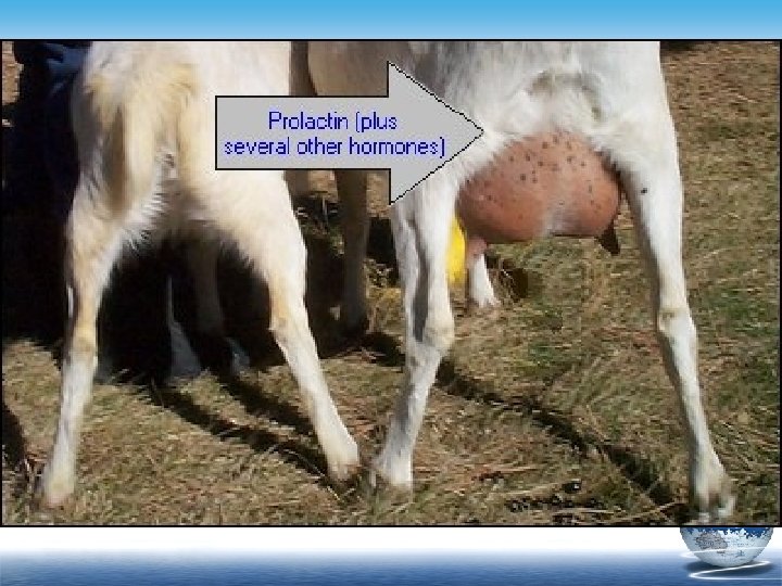
1. Physiological function of PRL 1) On breast: stimulate the development and milk secretion puberty E, P, GH, cortisol, insulin, thyroid hormones and prolactin pregnancy After baby born estrogen, Click to add Text prolactin progesterone prolactin
2) Effect on sexual organs In women, PRL combined with PRL receptors in granulosa cells stimulates production of LH receptors. Through LH receptors, LH promotes ovulation and then formation of corpus luteum. (permissive effect) But high PRL→Gn. RH↓→FSH、LH↓ In male, PRL promotes growth of prostate glands and seminal vesicle, enhancing the effect of LH on the interstitial cells producing testosterone.
Prolactin Excess v. The most common secretory neoplasm of the interior pituitary gland is prolactin secreting adenoma (prolactinoma). v. Increase plasma level of prolactine decreases plasma testosterone leading to decrease in both libido and potency. v. Symptoms: Galactorrhea Males: Decrease Libido Menstrual irregularity Sexual Impotence Amenorrhea Infertility
(2) Regulation of PRL secretion 1) Hypothalamic hormones and feedback mechanism Hypothalamus: PIF + Anterior pituitary: - PRF + Prolactin + increase the secretion; - inhibit the secretion
2) Milk ejection reflex Sucking, tactile stimulation Afferent nerve (somatic nerve) Centers including spinal cord and hypothalamus PRF secretion PRL secretion Milk production increase Oxytocin secretion Myoepithelial cells contraction of mammary glands Milk flows
PROLACTIN SECRETION
ADH (1)Roles of ADH 1) Antidiuretic effect (refer to chapter 8) 2) Pressure effect. High concentration of ADH have a potent effect of constricting the arterioles everywhere in the body, raise the resistance blood flow and blood pressure
ADH l Antidiuretic effect Antidiuretic hormone V 2 -receptor: collecting duct l Pressure effect Vasopressor hormone V 1 -receptor: vascular smooth muscle
Role of Oxytocin (OXT) 1) Effect on mammary glands. Cause the contraction of the myoepithelial cells that surround the outer walls of the alveoli of the mammary glands, press the milk from the alveoli to the duct and make it flow out --milk ejection
OXYTOCIN
OXT & PRL
2) Effect on uterus OXT powerful stimulate the smooth muscle contraction, especially that towards the end of gestation. It is believed that OXT is at least partially responsible for causing birth of the baby
Pineal Gland § Glandular tissue in brain § Secretes melatonin w Function unknown w May be involved in circadian rhythms ▪ Melatonin secretion rises at night & falls during the day ▪ Melatonin is a potent sleepinducing agent ▪ Melatonin also enhances immune function and exerts a suppressive effect on reproduction function by interfering with the activity of certain hormones.
Key point v. Hormone v. Paracrine vneuroendocrine v. Permissive effect v. Properties of the hormone effect v. Physiological functions of growth hormone v. Role of Prolactin (PRL) v. Role of Oxytocin (OXT)