Kidney Anatomy of the Urinary System Kidneys urine

 • Lower Urinary Tract Ureters")


, glucose, amino acids, vitamins,")
")


 is entirely reabsorbed back into the")
 and other ions are only partially reabsorbed from")


: • Osmoreceptors (hypothalamus) • Trigger release")


 Na+ &")

- Slides: 45
Kidney
Anatomy of the Urinary System • Kidneys (urine formation) • Lower Urinary Tract Ureters (2) Bladder (1) Urethra (1) (urine collection, storage, excretion)
The Kidney
THE URINARY SYSTEM • • • Kidneys Blood supply: Renal arteries and veins Ureter Urinary bladder Urethra
Kidney Functions • • • Water balance Electrolyte balance Plasma volume Acid-base balance Removal of waste from blood and excretion of urine. • Secretion of hormones, Erythropoietin, Renin, Vitamin D 3
Cortex Medulla Cortex Glomeruli Medulla Renal tubules Renal artery Renal vein Ureter Takes urine to bladder Blood carried to the kidney by the renal artery and taken away by the renal vein.
Each KIDNEY consists of 1 million NEPHRONS Each nephron consists of a: • GLOMERULUS (found in cortex) forms a protein-free filtrate from blood • TUBULE (found in medulla) processes the filtrate to form urine Each TUBULE consists of several segments: • Proximal tubule • Loop of Henle • Distal Tubule • Collecting Ducts.
Functional Unit of the Kidney is the NEPHRON 1. Glomerulus 2. Proximal Tubule 2. Loop of Henle 3. Distal Tubule 4. Collecting Duct
Structure of the Kidney • A frontal section of the kidney reveals 3 regions: – Renal cortex – Renal medulla – Renal pelvis
Structure of the Kidney • With in the renal medulla are located the – Renal pyramid, renal papilla and renal columns. • Within the nephrons of kidneys urine is produced. • It flows from the renal papilla, to the minor calyx, to the major calyx, to the renal pelvis, and finally exits the kidney within the ureter.
The Nephron • Each kidney in human contains about 1 million nephrons, each capable of forming urine. • The kidney cannot regenerate new nephrons. • Therefore, with renal injury, disease or normal aging, there is a gradual decrease in nephron number.
The Nephron Bowman’s Descending limb Ascending limb
The Nephron • Each nephron contains – 1. a tuft of glomerular capillaries called glomerulus, through which large amount of fluid are filtered from the blood, and – 2. a long tubule in which the filtered fluid is converted into urine on its way to the pelvis of the kidney. • The glomerulus contain a network of branching and anastomosing glomerular capillaries.
The Nephron • The glomerular capillaries are covered by epithelial cells, and • The total glomerulus is encased in Bowman’s Capsule. • Fluid filtered from glomerular capillaries flows into Bowman’s capsule and • Then in to the Proximal Tubule, which lies in the cortex of the kidney.
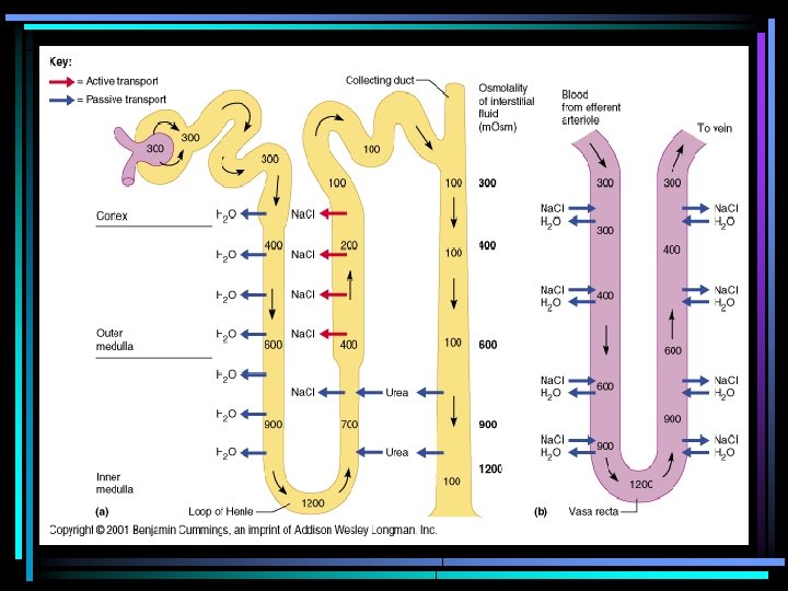
The Nephron • From proximal tubule, fluid flows into the Loop of Henly, which dips into renal medulla. • Each loop consists of a Descending and an Ascending Limb. • At the end of the ascending limb is a short segment, known as Macula Densa. • Beyond the macula densa, fluid enters the Distal Tubule, which also lies in the renal cortex. This is followed by Connecting Tubule. • And it runs downward into the medulla and becomes Collecting Duct.
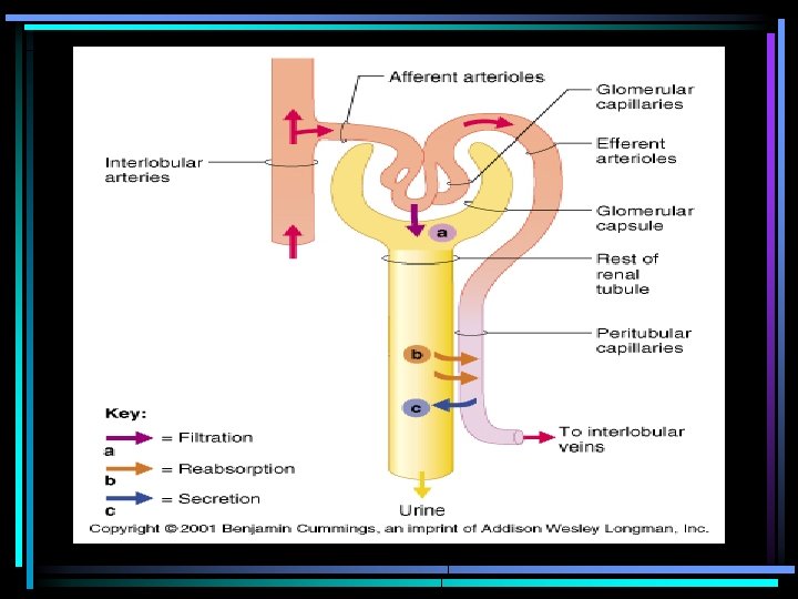
Urine Formation • Urine formation and the simultaneous adjustment of blood composition involves 3 major processes: 1) 2) 3) 4) Glomerular Filtration Tubular Reabsorption Secretion Excretion
Glomerular Filtration • Urine formation begins with the process of filtration, which goes on continually in the renal corpuscles. • As blood courses through the glomeruli, much of its fluid, containing both useful chemicals and dissolved waste materials, soaks out of the blood through the membranes (by osmosis and diffusion) where it is filtered and then flows into the Bowman's capsule. • This process is called glomerular filtration.
Glomerular Filtration • The water, electrolytes (primarily Na+ and K+), glucose, amino acids, vitamins, small proteins, creatinine, and urea that have been filtered out of the blood are known collectively as glomerular filtrate. • Urea is formed in the body to eliminate the very toxic ammonia products that are formed in the liver from amino acids.
Glomerular Filtration • The total rate of glomerular filtration (glomerular filtration rate or GFR) for the whole body (i. e. , for all of the nephrons in both kidneys) is normally about 125 ml per minute. That is, about 125 ml of water and dissolved substances are filtered out of the blood per minute. • The following calculations may help you visualize how enormous this volume is. • The GFR per hour is: 125 ml/min X 60 min/hr= 7500 ml/hr. • The GFR per day is: 7500 ml/hr X 24 hr/day = 180, 000 ml/day or 180 liters/day.
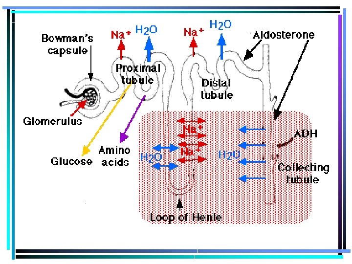
Tubular Reabsorption • Reabsorption, by definition, is the movement of substances out of the renal tubules back into the blood capillaries located around the tubules. • The proximal convoluted tubules are the most active in tubular reabsorption. All glucose, lactate, and amino acids are reabsorbed in this area. • Reabsorption begins in the proximal convoluted tubules and continues in the loop of Henle, distal convoluted tubules and collecting tubules.
Tubular Reabsorption • Large amounts of water - more than 178 liters per day - are reabsorbed back into the bloodstream from the proximal tubules because the physical forces acting on the water in these tubules actually push most of the water back into the blood capillaries. • In other words, about 99% of the 180 liters of water that leave the blood each day by glomerular filtration returns to the blood from the proximal tubule through the process of passive reabsorption.
Tubular Reabsorption • The nutrient glucose (blood sugar) is entirely reabsorbed back into the blood from the proximal tubules. • In fact, it is actively transported out of the tubules and into the peritubular capillary blood. • All the glucose that seeps out through the glomeruli into the tubules is reabsorbed into the blood. • But if too much is present, the tubules reach the limit of their ability to pass the sugar back into the bloodstream, and the tubules retain some of it.
Tubular Reabsorption • Sodium ions (Na+) and other ions are only partially reabsorbed from the renal tubules back into the blood. • For the most part, however, sodium ions are actively transported back into blood from the tubular fluid. The amount of sodium reabsorbed varies from time to time; it depends largely on how much salt we take in from the foods that we eat.
Tubular Reabsorption • About 65 % of sodium, 70 % of water, are reabsorbed. • 90% of bicarbonate ions, 50% of chloride ions and 55% potassium are reabsorbed in the proximal convoluted tubules. • Tubular reabsorption from the loop of henly results in 10% of water being reabsorbed from the descending limb, 30% of potassium ions, 20% of sodium, and 35% of chloride.
Tubular Reabsorption • Fluids enters the distal convoluted tubule at a rate of about 25 ml/min. • Because about 80% of the water in the filtrate has been reabsorbed. • As fluid flows through the distal convoluted tubule, sodium and chloride are reabsorbed. • By the time fluid reaches the end of the distal convoluted tubule, about 90% of the filtered solutes and water has been returned to the blood. • Hydrogen and potassium ions, creatinine, ammonium ion, and certain organic acids moves from the blood into the distal convoluted tubule of the nephron.
Secretion • Secretion is the process by which substances move into the distal and collecting tubules from blood in the capillaries around these tubules. • Secretion moves substances out of the blood and into the tubules where they mix with the water and other wastes and are converted into urine. • These substances are secreted through either an active transport mechanism or as a result of diffusion across the membrane. • Substances secreted are hydrogen ions (H+), potassium ions (K+), ammonia (NH 3), and certain drugs. • Kidney tubule secretion plays a crucial role in maintaining the body's acid-base balance.
Regulation of Fluid Volume Kidneys influence fluid volume by: • Altering water content of urine: » Removal of H 2 O in urine = diuresis Substances that cause diuresis = diuretics
Retention of Water is controlled by ADH: – Anti Diuretic Hormone – ADH Release Is Controlled By: – Decrease in Blood Volume – Decrease in Blood Pressure – Increase in ECF Osmolarity
The Role of ADH • Whether the water actually leaves the collecting duct (by osmosis) is determined by the hormone ADH (anti-diuretic hormone) • Osmoreceptors in the hypothalamus detect the low levels of water, so the hypothalamus sends an impulse to the pituitary gland which releases ADH into the bloodstream. • ADH makes the wall of the collecting duct more permeable to water. • Therefore, when ADH is present more water is reabsorbed and less is excreted.
• When Body Fluid Osmolarity Increases (280+): • Osmoreceptors (hypothalamus) • Trigger release of ADH • Increased permeability of CD to water • Water reabsorbion from tubule • Concentrated urine produced
ANTIDURETIC HORMONE
Regulation of Sodium: – On average, an adult takes in 9 g/Na. Cl/day • Addition of salt can raise body fluid osmolarity
Sodium Balance Is Controlled By Aldosterone: • Steroid hormone • Synthesized in Adrenal Cortex • Causes reabsorbtion of Na+ in DCT & CD » Also, K+ secretion
ALDOSTERONE
Sodium Balance is Controlled By Aldosterone – Aldosterone Release: • Triggered by increased ECF K+ Na+ reabsorbtion – Inhibition of Aldosterone: • Triggered by increased extracellular osmolarity No Na+ reabsorbtion
ALDOSTERONE
Na. HCO 3 Reabsorption in Proximal Tubule Na+-H+ antiporter Na+-HCO 3 - symporter Na+ H+ H+ Na+ HCO 3 - H 2 CO 3 CA CO 2 + H 2 O Lumen CO 2 + H 2 O Blood
Na. Cl Reabsorption in Thick Ascending Limb Na+-K+-2 Cl- symport Na+ K+ Cl- Na+ pump (Na+-K+ ATPase) Na+ K+ Cl- K+ ATPase K+ Na+ Cl- Lumen K+ Channel Cl- Channel Blood
Na. Cl Reabsorption in Disatal Convoluted Tubule Na+-Cl- symporter K+ Channel Na+ Cl. K+ Cl- K+ K+ ATPase Na+ K+ K+ Channel Lumen Na+ pump (Na+-K+ ATPase) Cl- Channel Blood
Na+ Reabsorption and Effects of Aldosterone on Late Distal Tubule and Collecting Duct MR AL MR-AL m. RNA Nucleus Na+-K+ ATPase AIP Na+ Channel Na+ K+ Lumen Na+ K+ ATPase K+ Blood
Renin Angiotensin System ANGIOTENSINOGEN RENIN ANGIOTENSIN I ACE ANGIOTENSIN II (ANGII Receptor) Na+ & • H 2 O retention by the kidney ALDOSTERONE Blood vessel constriction
Rennin-Angiotensin-Aldosterone System Fall in Na. Cl, extracellular fluid volume, arterial blood pressur Adrenal Cortex Juxtaglomerular Apparatus Liver Lungs Renin + Angiotensin Helps Correct Angiotensin Converting Enzyme Angiotensin Increased Sodium Reabsorption Aldosterone