Urinary System Two kidneys Two ureters Urinary Bladder

Urinary System Two kidneys Two ureters Urinary Bladder Urethra

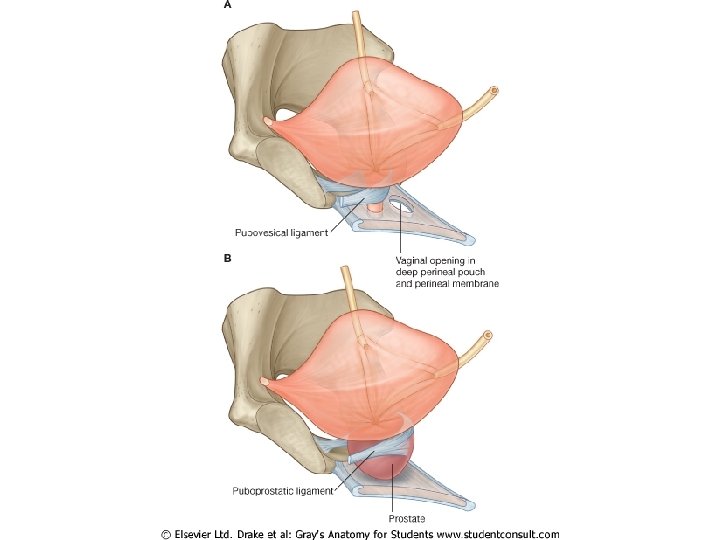
Prostate Male

Functions of Urinary System • Kidneys filter blood to keep it pure – – Toxins Metabolic wastes Excess water Excess ions • Dispose of nitrogenous wastes from blood – Urea – Uric acid – Creatinine • Regulate the balance of water and electrolytes, acids and bases 3
 Regulate p.")
Functions of Urinary System • • • Regulate electrolytes (K, Na, etc) Regulate p. H in blood Regulate blood pressure(aldestron, ADH) Regulate blood volume (removes excess fluid) Removing metabolic wastes – – 4 Urea, uric acid, and creatinine This is the least important of the kidney’s functions. You can survive for a few weeks without excreting waste products in the urine, but hour by hour, the other functions are more important.

Location of Kidneys • Retroperitoneal • Posterior wall of abdominal cavity • T 12 – L 3 • Right kidney is lower than left kidney due to the shape of the liver. • 12 cm x 6 cm x 3 cm • 150 gr

Location of Kidneys

Location of Kidneys

Sourronds of Kidneys

Sourronds of Kidneys

Sourronds of Kidneys • Superior surface – Adrenal gland • Lateral surface – Convex • Medial surface – Concave – Renal hilus

Fascia of Kidneys

Fascia of Kidneys

Fascia of Kidneys 13

Fascia of Kidneys 14

Renal capsule • renal capsule

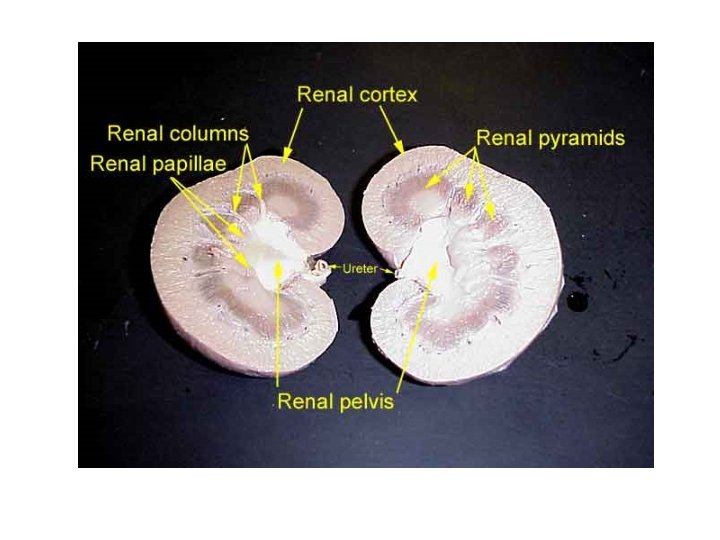
Dissected Kidney • • • Dissected Kidney 1. Renal Capsule 2. Renal Cortex 3. Renal Medulla renal capsule 4. Renal Pyramid 5. Renal Pelvis 6. Renal Column 7. Renal Calyx 8. Ureter

Dissected Kidney • Renal Sinus • Renal Pelvis • Major Calyx • minor calyx

Urine collection minor calyx major calyx renal pelvis ureter

Dissected Kidney • Cortex – Superficial layer – Light, granular • Medulla – Deep layer – Darker – Medullary pyramids • Cone shape • Base against cortex • Apex = papilla

Microscopic anatomyof kidneys Kidney anatomy

Microscopic anatomyof kidneys Lobe of Kidney

Microscopic anatomyof kidneys Nephron – – Functional Unit 1 million per kidney Renal corpuscle 1. 2. 3. 4. • Glomerulus • Bowman’s capsule Proximal convoluted tubule Loop of Henle Distal convoluted tubule Collecting tubule

Microscopic anatomyof kidneys Renal Corpuscle Glomerular filtrate collects in capsular space, flows into renal tubule

Microscopic anatomyof kidneys

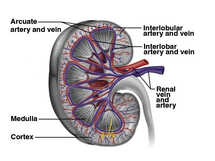
BLOOD SUPPLY OF KIDNEYS

BLOOD SUPPLY OF KIDNEYS Hilum – Renal artery – Renal nerves – Renal vein – Ureter

BLOOD SUPPLY OF KIDNEYS

BLOOD SUPPLY OF KIDNEYS

29

BLOOD SUPPLY OF KIDNEYS

ureters

Ureters • Continuation of renal pelvis • Slender tubes that transport urine from kidneys to bladder • Retroperitoneal • 25 cm long • Enters on the floor of bladder

Wall of Ureter • Adventitia • Muscularis – Smooth Muscle • Inner Longitudinal • Outer Circular • External longitudinal (on distal third) • Mucosa – Transitional epithelium

Bladder • detrusor muscle – Inner and outer longitudinal layers – Middle circular layer • Average bladder volume is 500 ml • Max capacity is 700 -800 ml • Location – Pelvic floor – Posterior to public symphysis – Anterior to • Rectum in males • Vagina & uterus in females

Bladder

Female

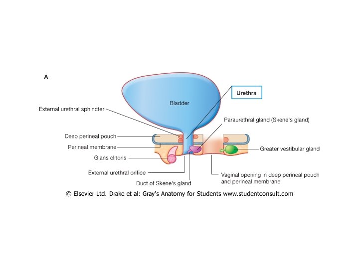
• 3 to 4 cm long • External urethral orifice – between vaginal orifice and clitoris • Internal urethral sphincter – detrusor muscle thickened, smooth muscle, involuntary control • External urethral sphincter – skeletal muscle, voluntary control

Male • 18 cm long • Internal urethral sphincter • External urethral sphincter • 3 regions – prostatic urethra • during orgasm receives semen – membranous urethra • passes through pelvic cavity – penile urethra

Urethra

Micturition Reflex Bladder with >= 200 ml of urine Sensory input to parasympathetic system Contraction of detrusor muscle and relaxation of internal urethral sphincter Relaxation of external urethral sphincter • Other brain receptors can inhibit urination by keep external urinary sphincter closed • Voluntary contraction of abdominal wall muscles increases abdominal pressure • Voluntary relaxation of external urethral sphincter

Urine Volume • Normal volume - 1 to 2 L/day • Polyuria > 2 L/day • Oliguria < 500 m. L/day • Anuria - 0 to 100 m. L

Composition of normal urine – p. H=6. 0 – water-93 -97% – volume=1200 m. L/day – color-clear yellow – odor-varies – sterile

Nephrolithiasis

 long leaving each renal")
The Ureters • Slender tubes about 25 cm (10 “) long leaving each renal pelvis • One for each kidney carrying urine to the bladder • Descend retroperitonealy and cross pelvic brim • Enter posterolateral corners of bladder • Run medially within posterior bladder wall before opening into interior • This oblique entry helps prevent backflow of urine 47

Three basic layers Ureters play an active • Transitional epithelium role in transporting urine (it’s not just by gravity) of mucosa stretches when ureters fill • Muscularis – Inner longitudinal, outer circular layers – Inferior 3 rd with extra longitudinal layer) – Stimulated to contract when urine in ureter: peristaltic waves to propel urine to bladder • Adventitia (external) 48

Urinary Tract – Urethra

Urinary Bladder See also brief atlas 50 Collapsible • muscular sac Stores and expels • urine Lies on pelvic floor • posterior to pubic symphysis Males: anterior to – rectum Females: just – anterior to the vagina and uterus

51

52

If full: bladder is spherical and extends into abdominal cavity (holds about 500 ml or 1 pt) If empty: bladder lies entirely within pelvis with shape like upside-down pyramid Urine exits via the urethra Trigone is inside area between ureters and urethra: prone to infection (see slide 38) 53 • •
 Mucosa with distensible transitional epithelium and")
Bladder wall has three layers (same as ureters) Mucosa with distensible transitional epithelium and – lamnia propria (can stretch) Thick muscularis called the detrusor muscle – 3 layers of highly intermingled smooth muscle • Squeezes urine out • Fibrous adventitia – 54

The Urethra Smooth muscle with inner mucosa • Changes from transitional through stages to stratified squamous near end – Drains urine out of the bladder and body – Male: about 20 cm (8”) long • Female: 3 -4 cm (1. 5”) long • Short length is why females have more urinary tract infections than males - – ascending bacteria from stool contamination urethra Urethra____ 55

Urethral sphincters • Internal: involuntary sphincter of smooth muscle – External: skeletal muscle inhibits urination voluntarily – until proper time (levator anni muscle also helps voluntary constriction) Males: urethra has three regions (see right) _____trigone 1. Prostatic urethra_____ 2. Membranous urethra____ 3. Spongy or penile urethra_____ 56 female

With all the labels 57

Micturition • AKA: Voiding – Urinating – Emptying the bladder – (See book for diagram explanation p 701) KNOW: Micturition center of brain: pons (but heavily influenced by higher centers) Parasympathetic: to void Sympathetic: inhibits micturition 58
 • Superior lumbar region of posterior abdominal")
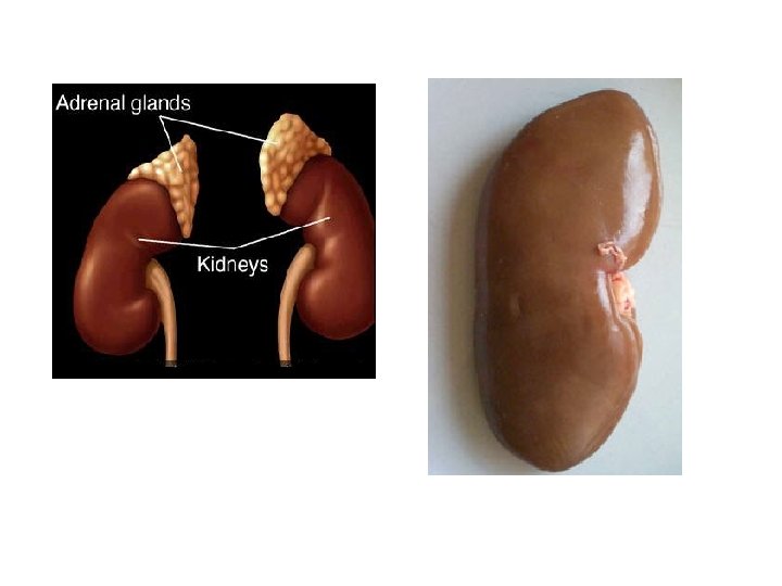
Kidneys are retroperitoneal organs (see next slide) • Superior lumbar region of posterior abdominal wall – Lateral surface is convex • Medial surface is concave • Hilus* is cleft: vessels, ureters and nerves enter and leave – Adrenal glands* lie superior to each kidney (the yellow blob in pic) * * 59 •

60

Kidney Dissection Obtain a preserved sheep kidney. Observe the kidney. You should notice adipose tissue (remnants of the adipose capsule) clinging to the renal capsule. •

Generally, the tube • with the most adipose around it is the ureter. Notice the histological differences (and similarities) between the renal arteries, renal veins, and ureter.

1. renal pyramid 4. ureter 7. renal pelvis 2. renal papilla 5. renal vein 8. renal column 3. calyx 9. cortex

Dissected Kidney
")
Nephron (2)

Microscopic anatomyof kidneys Nephron – – Functional Unit 1 million per kidney Renal corpuscle • Glomerulus • Bowman’s capsule 1. Proximal convoluted tubule 2. Loop of Henle 3. Distal convoluted tubule 4. Collecting tubule
- Slides: 69