Introduction to pathology lecture 3 Cell injury Dr


, b. Proteases")


: - Free radicals are chemical")


- Slides: 30
Introduction to pathology lecture 3/ Cell injury Dr H Awad 2017/18
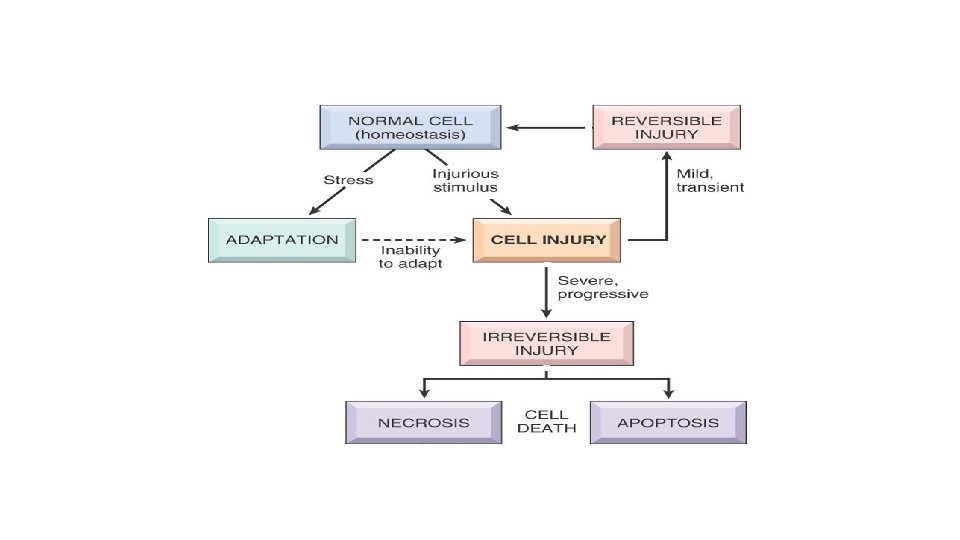
• If cells can not adapt to a change in their microenvironment. . Cell injury occurs • In this lecture we will discuss mechanisms of cell injury.
Mechanisms of cell injury. . First let’s discuss the general principles of cell injury 1. Cellular response to injury depends on the type , duration and severity of the injury. • Example: Low doses of a toxin can lead to reversible injury whereas the same toxin with a higher dose can lead to irreversible injury. • Example: ischemia lasting for a short period will be reversible whereas the same degree of ischemia lasting for a longer period can cause irreversible injury.
2. consequences of cell injury also depend on the type, status, adaptability, and genetic makeup of the injured cell • Skeletal muscle can tolerate 2 -3 hours of ischemia but cardiac muscle die after 20 -30 minutes of ischemia • Glycogen depleted hepatocyte cannot tolerate ischemia as a hepatocyte with loads of glycogen • Same dose of toxin in 2 individuals have different effects. . Due to variant of cytochrome p 450 catabolize toxins at different rates. .
3. Cell injury results from functional and biochemical abnormalities in one or more of essential cellular components which are : 1. Mitochondria and their ability to generate ATP 2. Disturbance in calcium homeostasis 3. Damage to cellular (plasma and lysosomal) membranes 4. Damage to DNA and misfolding of proteins
Biochemical mechanisms of cell injury: 1. ATP depletion -ATP is the main energy store of cells. It is produced by oxidative phosphorylation The major causes of ATP depletion in cell injury are: a. Reduced supply of oxygen and nutrients, b. Mitochondrial damage, c. The actions of some toxins (e. g. , cyanide)
Effects of ATP depletion 1. The activity of plasma membrane ATP-dependent sodium pump is reduced, resulting in Intracellular accumulation of Na and efflux of Ka accompanied by osmotic gain of water, causing cell swelling 2. Compensatory increase in anaerobic glycolysis resulting in rapid depletion of intracellular glycogen stores , and lactic acid accumulates, leading to decreased intracellular p. H and decreased activity of many cellular enzymes 3. Failure of ATP-dependent Ca 2+ pumps leads to influx of Ca 2+ with damaging effects on many cell components 4. Structural disruption of the protein synthetic apparatus, manifested as detachment of ribosomes from the rough ER with a consequent reduction in protein synthesis 5. Ultimately, there is irreversible damage to mitochondrial and lysosomal membranes, and the cell undergoes necrosis.
Biochemical mechanisms of cell injury 2. Mitochondrial Damage and Dysfunction - Mitochondria are sensitive to many types of injurious stimuli, including hypoxia, chemical toxins, and radiation. - Mitochondrial injury may result in several abnormalities: A. Failure of oxidative phosphorylation leads to progressive depletion of ATP, culminating in cell necrosis B. Abnormal oxidative phosphorylation leads to formation of reactive oxygen species with deleterious effects
Mechanism of mitochondrial dysfunction 1. Damage to mitochondria is often associated with formation of a high-conductance channel in the mitochondrial membrane, called the mitochondrial permeability transition pore 2. The opening of this channel leads to the loss of mitochondrial membrane potential and p. H changes, further compromising oxidative phosphorylation. 3. The mitochondria also contain several proteins that, when released into the cytoplasm, activating apoptosis
Mitochondrial dysfunction
Biochemical mechanisms of cell injury 3. Influx of Calcium - Cytosolic free calcium is normally maintained at concentrations as much as 10, 000 times lower than the concentration of extracellular calcium or of sequestered intracellular mitochondrial and ER calcium - Ischemia and certain toxins cause an increase in cytosolic calcium , initially because of release of Ca 2+ from the intracellular stores, and later resulting from increased influx across the plasma membrane. - Increased cytosolic Ca 2+ activates a number of enzymes, with potentially deleterious cellular effects
Enzymes activated by increased cytosolic calcium a. Phospholipases (which cause membrane damage), b. Proteases (which break down both membrane and cytoskeletal proteins) c. Endonuclease (cause DNA and chromatin fragmentation), d. Adenosine triphosphatases (ATPases) (thereby hastening ATP depletion).
Calcium influx
Biochemical mechanisms of cell injury. 4. Disturbed membrane integrity - Increased membrane permeability leading ultimately to overt membrane damage is a consistent feature of most forms of cell injury that culminate in necrosis. - The plasma membrane can be damaged by ischemia, microbial toxins, physical and chemical agents.
Several biochemical mechanisms may contribute to membrane damage 1. Decreased phospholipid synthesis: due to fall in ATP levels. The reduced phospholipid synthesis may affect all cellular membranes, including the membranes of mitochondria, thus exacerbating the loss of ATP. 2. Increased phospholipid breakdown- by activation of endogenous phospholipases by increased levels of cytosolic Ca 2+. 3. Cytoskeletal abnormalities- Cytoskeletal filaments act as anchors connecting the plasma membrane to the cell interior, and maintain normal cellular architecture , motility, and signaling. Activation of proteases by increased Ca 2+ cause damage to cytoskeletal elements leading to membrane damage 4. ROS. : Cause cell injury to by lipid peroxidation, 5. Lipid breakdown products: . These may insert into the lipid bilayer of the membrane or exchange with membrane phospholipids causing changes in permeability.
Membrane damage
DNA and protein damage • Abnormalities in DNA and misfolded proteins cause apoptosis. • This will discussed in detail later.
Oxidative stress Accumulation of Oxygen-Derived Free Radicals (Oxidative Stress): - Free radicals are chemical species with a single unpaired electron in an outer orbital. , are extremely unstable, and: A. They readily react with inorganic and organic chemicals; B. They attack nucleic acids , cellular proteins and lipids C. They initiate reactions in which molecules that react with free radicals are themselves converted into other types of free radicals, thereby propagating the chain of damage.
Damage by free radicals mainly occurs in a. Ischemia-reperfusion injury b. Chemical and radiation injury c. Toxicity from oxygen and other gases d. Cellular aging, e. Microbial killing by phagocytic cells, f. Tissue injury caused by inflammatory cells
From where oxygen radicals originate? I. ROS are produced normally in small amounts in cells During the reduction-oxidation reactions that occur during mitochondrial respiration and energy generation. - when O 2 is only partially reduced oxygen radicals are produced and they include : 1. Superoxide radicals O 2 • 2. Which is converted to hydrogen peroxide (H 2 O 2) spontaneously and by the action of the enzyme superoxide dismutase II. ROS are produced in neutrophils and macrophages, to destroy ingested microbes during inflammation
- When the production of ROS increases or the scavenging systems( that neutralize these ROS) are ineffective, the result is an excess of these free radicals, leading to a condition called oxidative stress
- The generation of free radicals is increased under several circumstances: a. The absorption of radiant energy (e. g. , ultraviolet light, x-rays). Ionizing radiation can hydrolyze water into hydroxyl ( • OH) and hydrogen (H • ) free radicals. b. The enzymatic metabolism of chemicals (e. g. , CCl 4) c. Inflammation(the free radicals are produced by leukocytes
- Reactive oxygen species cause cell injury by : a. Lipid peroxidation of membranes. Double bonds in membrane polyunsaturated lipids are vulnerable to attack by oxygen-derived free radicals b. Free radicals promote sulfhydryl-mediated protein cross-linking resulting in enhanced degradation or loss of enzymatic activity -Free radicals may directly cause polypeptide fragmentation. c. DNA damage. Free radical reactions with thymine in nuclear and mitochondrial DNA produce single-strand breaks. Such DNA damage has been implicated in cell death, aging, and malignant transformation of cells
Oxidative stress
summary
Reperfusion injury • If cells are reversibly injured, restoration of blood flow can cause recovery. • However, restoration of blood flow in ischemic tissue can cause more damage and even death to the cells. • This is called reperfusion injury • HOW does it happen ? ? Se next slide
Mechanisms of reperfusion injury • Re-oxygenation that happens during reperfusion can increase ROS production if the mitochondria is damaged. These ROS will cause more damage • Inflammation which occurs due to ischemia can increase during reperfusion because the restoration of blood supply recruits more inflammatory cells to the site of injury. • Complement component present in the blood also can contribute to reperfusion injury… complement proteins are important mediators of inflammation that react with the damaged tissue.
Reperfusion injury