Introduction to pathology lecture 5 Cell injury apoptosis


- Slides: 31

Introduction to pathology lecture 5/ Cell injury apoptosis Dr H Awad 2017/18

Apoptosis = programmed cell death = cell suicide= individual cell death

Apoptosis • cell death induced by a tightly regulated suicide program in which cells activate enzymes capable of degrading the cells' own nuclear DNA and nuclear and cytoplasmic proteins.

• Fragments of the apoptotic cells break off, giving the appearance that is responsible for the name (apoptosis, "falling off").

apoptosis • The plasma membrane remains intact. • Apoptotic bodies (contain portions of the cytoplasm and nucleus) become targets for phagocytosis before their contents leak out and so there would be no inflammatory reaction. • So in apoptosis there is no damade to surrounding cells.

Causes of Apoptosis • Physiologic situations: To eliminate cells that are no longer needed OR to maintain a steady number of various cell populations in tissues.

Physiologic apoptosis • Embryogenesis. • involution of hormone-dependent tissues upon hormone withdrawal. (endometrium and breast after pregnancy) • Cell loss in proliferating cell populations. (gastrointestinal tract, skin…) • Death of host cells after serving their useful function. (neutrophils and lymphocytes in inflammation) • Elimination of potentially harmful self-reactive lymphocytes. • Cell death induced by cytotoxic T lymphocytes (tumor cells and virally infected cells)

Pathologic situations • DNA damaged cells, if DNA damage is severe and cannot be repaired the cell dies by apoptosis. • Cells with accumulation of misfolded proteins, • Certain infections (viral ones): may be induced by the virus (as in human immunodeficiency virus infections) or by the host immune response (as in viral hepatitis). • Pathologic atrophy in parenchymal organs after duct obstruction (pancreas, parotid and kidney)

Morphology • Cell shrinkage: dense cytoplasm, tightly packed organelles. • Chromatin condensation: peripherally under the nuclear membrane. • Formation of cytoplasmic blebs • apoptotic bodies: blebbing then fragmentation into membrane bound apoptotic bodies composed of cytoplasm and tightly packed organelles with or without nuclear fragments.

Morphology • Phagocytosis of apoptotic cells or cell bodies by macrophages (quickly hence no inflammation).


Mechanisms of Apoptosis • Activation of enzymes called caspases. • Two main pathways: • 1 - Mitochondrial pathway (intrinsic) • 2 - Death receptor pathway (extrinsic)

• 1 - mitochondrial pathway (intrinsic) • Leak of cytochrome c out of mitochondria and activation of caspase 9… • 2 - death receptor pathway (extrinsic) • Involved in elimination of self-reactive lymphocytes and in killing of target cells by some cytotoxic T lymphocytes. • Activation of caspase 8.

Intrinsic pathway = mitochondrial pathway • Mitochondria contains several proteins that can induce apoptosis • The most important of these is cytochrome C • Stimulation of apoptosis depends on mitochondrial permeability • Mitochondrial permeability is controlled by a family of more than 20 proteins ( Bcl 2 family)

• When cells are deprived of growth signals, or exposed to severe DNA damage or have misfolded proteins. . In all these situations certain sensors are activated • These sensors are called BH 3 proteins ( they are part of the bcl 2 family) • BH 3 now activate proapoptotic members of the family= Bax and Bak

• When bax and bak are stimulated they dimerize and insert into the mitochondrial membrane • They form channels through which cytochrome c escapes into cytosol • BH 3 also inhibit anti inhibitory members of the family (BCL-2 and BCL-xl) • So BH 3 stimulate proapptotic and inhibit antiapoptotic signals. . Net result it leakage of cytochrome c from the mitochondria to cytosol

• Once cytochrome c is in the cytosol it stimulates caspase 9 • Caspase cascade is stimulated leading to nuclear fragmentation by executioner caspases.

Summary of intrinsic pathway • BH 3 stimulates pro-apoptotic (bax, bak), and inhibit anti apoptotic proteins (bcl-2, bcl-xl) • Cytochrome c leaks out • Stimulates caspase 9 • Stimulates executioner caspases that degrade cell components


Extrinsic pathway= death receptor pathway • This pathway is triggered by death receptors, which are members of the TNF ( tumor necrosis factor) receptor family • The most important types of death receptors are: TNF type 1 receptor and Fas receptor (CD 95) • Fas. L = fas ligand is a membrane protein expressed mainly on T lymphocytes • When T cells recognize fas expressing target , fas molecules are cross linked by fasl to activate caspase 8 • Caspase 8 activates executioner caspases that degrade cell components


FLIP • FLIP is a protein that is a Caspase antagonist which block activation of caspases. . So it inhibit apoptosis. • Some viruses produce FLIP like molecule to keep infected cells alive. ( so the virus can survive within that cell)

Clearance of apoptotic cells • When apoptotic cells fragment they are phagocytosed without eliciting inflammation • In normal cells phosphatidyl serine is present in the inner surface of cell membrane. in apoptotic cells it flips to outside the membrane and acts as a signal recognized by macrophages to phagocytose the apoptotic cell fragment • So the apoptotic body is phagocytosed without inflammation


note • In some situations both apoptosis and necrosis occur • necroptosis

P 53 and apoptosis • DNA damage causes accumulation of p 53 in cells • It arrests cells in G 1 phase of cell cycle to give the cell a chance to repair itself • If no repair, p 53 triggers apoptosis by stimulating bax and bak • P 53 can be mutated in cancer cells. . If mutated it cannot initiate apoptosis, so the cell survives even if its DNA is damaged. . Longer survival of a cell with damaged DNA increases the chances of accumulating more mutations. . So this cell can become malignant

Accumulation of abnormal proteins ER ( endoplasmic reticulum) stress • Chaperons in ER control proper folding of proteins • Misfolded proteins are degraded. • If there is too much of unfolded protein. . Then the cell starts an unfolded protein response which is an adaptive response aiming at increasing chaperons and decreasing protein translation • If more unfolded proteins accumulate : the situation is called ER stress. • ER stress causes caspase activation and apoptosis.

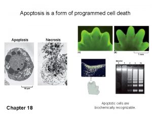
Feature Necrosis Apoptosis Cell size Enlarged (swelling) Reduced (shrinkage) Nucleus Pyknosis → karyorrhexis → karyolysis Fragmentation into nucleosome-size fragments Plasma membrane Disrupted Intact; altered structure, especially orientation of lipids Cellular content Enzymatic digestion; may leak out of cell Adjacent inflammation Frequent Intact; altered structure, especially orientation of lipids No Physiologic or pathologic role Invariably pathologic (culmination of irreversible cell injury) Often physiologic, means of eliminating unwanted cells; may be pathologic after some forms of cell injury, especially DNA damage

 Intentional injury and unintentional injury
 Definition of apoptosis
 Harrison sulcus
 Via intrinseca de la apoptosis
 Apoptosis meaning
 Emphysema
 Apoptosis definition biology
 Apoptosis greek meaning
 Apoptosis defination
 Apoptosis
 Apoptosis and necrosis
 Apoptosis assay
 Apoptosis
 Caracteristicas de la apoptosis
 Apoptosis adalah
 Bcl2 family
 Apoptosis flow chart
 Cual es la importancia de la mitosis y meiosis en la vida
 01:640:244 lecture notes - lecture 15: plat, idah, farad
 Russell bodies
 Types of necrosis
 Cellular adaptation
 Cell injury and inflammation
 Cell injury and inflammation
 Myelin figures in reversible cell injury
 Example of physiological hyperplasia
 Reversible cell injury
 Introduction and importance of seed pathology
 Enteroendocrine cell
 Diapedesis
 Introduction to biochemistry lecture notes
 Introduction to psychology lecture

