Introduction to Musculoskeletal imaging Harsha Kumar Radiodiagnosis resident
")
")


 Effusion")


")


")


 Overlying soft tissue")


- Slides: 55
Introduction to Musculoskeletal imaging Harsha Kumar Radiodiagnosis ( resident)
Imaging Techniques �Plain x-rays �CT �MRI �Ultrasound �Nuclear Medicine (bone scan)
Plain x-rays For joints like the ankle, elbow or wrist we always take 3 views: AP, lateral and oblique
3 views: AP, oblique and lateral
Advantages of plain x-rays �Quick �Not expensive �Relatively low radiation
Disadvantages of plain x-rays �Not 3 dimensional �Can miss pathology �May still require other imaging studies
CT scanner
This CT shows a fracture through the cunieform
Advantages of CT scanning of the musculoskeletal system �Excellent anatomic detail �Will detect almost all pathology related to cortical bone injury �Great for showing displacement or joint involvement �Now multiplanar
Disadvantages of CT �Expensive �More radiation �Often not necessary
MRI scanner
This is an MRI of the knee There is no radiation used
Advantages of MRI �No radiation �We can slice through the body using any imaging plane �Looks “inside” bone. Marrow evaluation. �MRI is very good for looking at the soft tissues (muscles, ligaments, tendons and cartilage) �MRI is very sensitive in detecting water
MRI shows water (fluid) Effusion
Disadvantages of MRI �Very expensive �Not as good as CT for cortical bone
APPEAR DARK ON MRI � � � Air Cortical bone Tendon Ligament Flowing blood
Normal anterior cruciate ligment
Normal meniscus anterior and posterior horns “bow tie”
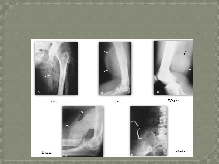
Conventional imaging Basic densities
Two views are required
Skeletal anatomy and physiology �Arises from mesoderm Intramembranous ossification ( eg parietal bone ) Enchondral ossification ( eg: femur )
Membranous ossification
Enchondral ossification
Anatomy
Parts of a long bone
Epiphysis �End of growing bone �Undergoes enchondral ossification �Eventually fuses with shaft �Supports the articular cartilage
Epiphyseal plate and physis
Physis �Cartilage growth plate between epiphysis and metaphysis. �Layers of progressively maturing cartilage and developing bone
Metaphysis �Between epiphysis and diaphysis �Most active region of bone �Most common site for tumors and infections
Parts of a long bone
Diaphysis �Between metaphysis and shaft of the bone �Has thickened cortex and decreased medullary space �Contains bone marrow and provides mechanical strength
Diaphysis
Finding a Fracture on X-Ray �Start with soft tissue, look for swelling or fat pad displacement �Examine the cortex along the entire length of the bone �Look for cortical irregularities, buckling, or evidence of impaction
Fracture Terminology � Direction • • of fracture line: Transverse Oblique Spiral Longitudinal � Alignment of fracture: Displacement � Angulation � Comminution � Articular Involvement
Fracture Terminolgy �Open vs Closed: fracture is open when exposed to air (laceration or gross exposure) �Pathologic fracture: implies fracture through weakened bone �Stress fracture: implies misuse or overuse
Path of the Fracture
Normal
Transverse Fracture
Spiral Fracture
Simple vs Comminuted Simple-2 bone fragments � Comminuted-greater then 2 fragments �
Approach to bone disease ( Location )
Osteomyelitis FEATURES � Marked bone lysis � Cortical lysis and sclerosis � Soft tissue swelling � Periosteal reaction
Osteosarcoma � Features � Codman triangle � irregular ill defined margin , wide zone of transition � Periosteal right angle speculation (sunburst) � Adjacent soft tissue mass � Osteoid matrix � Bone destruction
FEATURES � � � Ill defined destructive margins (moth eaten ) Overlying soft tissue mass Expanded cortex with displacement of periosteum ( Codman’s triangle ) Onion peel appearance ( perisoteal reaction)
Giant cell tumor of bone � Eccentric lytic lesions with sharp margins � Multiloculated soap bubble appearance � Bone destruction and cortical disruption
Points to take home � There are distinct advantages and disadvantages to plain x-rays, CT and MRI. � Become familiar with terminology: epiphysis, metaphysis, diaphysis, cortex, medullary cavity � Fracture description requires specific vocabulary � To identify common bone lesion on a plain radiograph.
�Thank you