Immunoassays ELISA What is an immunoassay The term


 are")

 for measuring insulin")
 – In immunoasay, we use antibody as a binding reagent")


, The ELISA is a rapid test used for detecting and")


 ELISA • In this format, the antigen in the sample is sandwiched")
 1) Standard solutions or assay samples are")
 After this binding reaction, the reaction mixture is discarded, and wells are washed")
 The enzyme-labeled second antibody will bind to the antigen which is bound to")
 Enzyme activity is measured by adding a chromogenic substrate of this enzyme. In")
 The standard curve is prepared from the concentration of standard solutions and their")


 is a polypeptide chain, composed of 191")


 ELISA is a solid phase sandwich")

 levels in a healthy person range as")

- Slides: 53
Immunoassays ELISA
What is an immunoassay? • The term “immunoassay” is a combined term of “immuno” (= antigen-antibody-reaction) and “assay” (= determination of the amount of any particular constituent of a mixture ). • So, immunoassay means a method to measure any particular substance in a mixture using its specific-binding antibody, for example, one constituent of blood without any purification process.
• Immunoassays are used for diagnosis, quantification (example: TSH in screening of the newborn) and monitoring (example: IGF-1 measurement before and after growth hormone therapy) of endocrine diseases
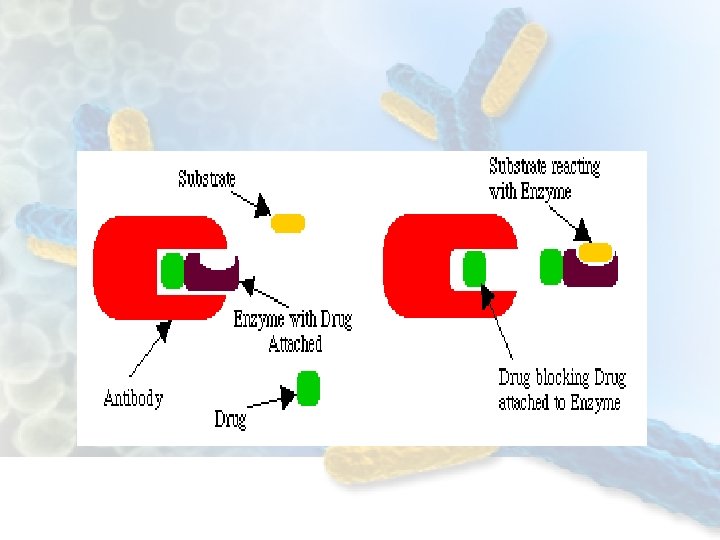
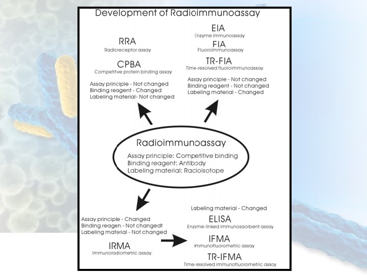
• Most immunoassay configurations can be divided into two large groups: – Competitive immunoassays: "limited reagent" methods – Noncompetitive immunoassays: "reagent excess“ methods ü Both can be divided into heterogenous and homogenous Immunoassays competitive Heterogenous RIA Compitive ELISA Homogenous EMIT Noncompetitive Heterogenous ELISA(sandwich and indirect) Homogenous
• In competitive immunoassays , the analyte and the labeled analyte (tracer) are mixed with a limited amount of anti-analyte antibody. After incubation for a certain period, the bound or the free fraction of the tracer is measured and related to the concentration of the analyte in the sample. • In noncompetitive immunoassays, an excess of immunoreactant (antibody or antigen) is added, so that all the analyte is practically in the form of an immunocomplex. Then, the immunocomplex is quantified and related to the analyte concentration in the sample.
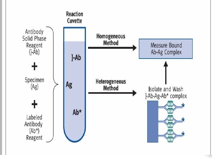
• Heterogeneous immunoassays require the separation of the immunocomplex from the free immunoreactants. – Solid phase is the most common way for separation. • But in homogeneous immunoassays, a modulation of the signal occurs as a result of the immunoreaction. Therefore the immunocomplex formation can be monitored directly without prior separation of the bound and free tracer.
Brief history…. • Berson and Yalow’s discovery of the radioimmunoassay (RIA) for measuring insulin in 1959. • The enzyme immunoassay (EIA) was introduced in the 1960 s, bringing with it significant advantages, including the replacement of radioisotopes with enzymes, faster reaction times, higher specificities, and longer shelf-life than RIA. • Other immunoassays were developed, they all have the same principle, but differ in details.
• Antibody (antiserum) – In immunoasay, we use antibody as a binding reagent of high specificity and high binding ability, that is, high affinity to the substance to be measured. – We can get antibody by immunizing an animal with such “immunogen” Antibody appears after the first inoculation is Ig. M-type which is a pentamer of the basic component of antibody. This type of antibody changes to Ig. G-type after the repeated inoculations. This is called “class switch”, and mostly Ig. G type of antibodies are used in immunoassay.
• Antigen and hapten – substances that can produce antibody and can bind to it is called “antigens”, they have large molecular size > 1000 Dalton. – Substances with smaller molecular sizes cannot produce antibody by itself, but they can bind antibody if produced. Those substances are called “haptens”. – In order to get antibody for hapten, it has to be coupled with some carrier proteins of large molecular size.
• Standard preparation is necessary for immunoassay. Using a standard preparation, we draw a standard curve from graded reaction results of various standard concentrations, and by comparison of a sample reaction result with the standard curve, we get assay value of the sample • Labeling materials • In immunoassay, it is necessary to use any marker to know the antigen-antibody binding. For such purpose, we label either antigen or antibody with some materials that do not interefere with the binding. We use radioisotopes, enzyme, fluorescent substance.
ELISA (Enzyme-linked immunosorbent assay), The ELISA is a rapid test used for detecting and quantifying antibodies or Antigens This assay method utilizes enzyme as a labeling material. v Has many configurations: – Sandwich – Indirect – competitive
• In ELISA technology, a solid phase consists of a 96 -well polystyrene plate, although other materials can be used. The function of the solid phase is to immobilize either antigens or antibodies to which the analyte in the sample will bind, After incubation, the plates are washed to remove any unbound material, finally the reaction is detected using a conjugate. • The conjugate consists of either an antigen or antibody that has been labeled with an enzyme. • The most common enzymes used as labels for ELISA are: – horseradish peroxidase – calf intestine alkaline phosphatase – E. coli ß-D-galactosidase. • These enzymes are typically used because they each meet most, if not all, of the criteria a necessary to produce a sensitive, inexpensive, and easily performed assay.
Criteria of a good enzyme for ELISA • These criteria include: – stability at typical assay temperatures: 4°C, 25°C, and 37°C, – greater than six months shelf life when stored at 4°C, – commercially available, – capable of being conjugated to an antigen or antibody, – inexpensive, – easily measurable activity, – high substrate turnover number(he maximum number of chemical conversions of substrate molecules per second) – unaffected by biological components of the assay.
Some Enzymes Used as Immunoassay Labels: Enzyme Source Acetylcholinesterase Electrophorus electricus Alkaline phosphatase Calf intestine Catalase Liver /3 -o-Galactosidase Escherichia coli Glucose oxidase Aspergi. Uus niger Glucose-6 -phosphate dehydrogenase Leuconostoc Mesenteroides /3 -Lactamase Bacillus species Peroxidase Horseradish Pyrophosphatase Escherichia coli Urease Jack bean
ELISA KIT components
ELISA Kit Components • Coated Plates The 96 -well plates are made of polystyrene and coated with either inactivated antigen or antibody. This coating is the binding site for the antibodies or antigens in the sample. • Sample Diluent Most assays require a specific dilution of the sample. Samples are added to the sample diluent and mixed prior to putting them onto the coated plates.
• Controls o The positive control is a solution that contains antibody or antigen. The negative control is a solution without antibody or antigen. o The controls help to normalize or standardize each plate. controls are also used to validate the assay and to calculate sample results. In most tests, the controls are prediluted and ready to use. • Conjugate o ELISA conjugates are enzyme-labeled antibodies or antigens that react specifically to plate-bound sample analytes. unbound conjugate is washed away after incubation and before the addition of substrate.
• Standareds • For calibration curve construction, thought quantitative measurement • Substrate • For peroxidase conjugates, the substrate is a mixture of hydrogen peroxide and a chromogen that reacts with the enzyme portion of the conjugate to produce color. • Wash Concentrate • The wash concentrate is a buffered solution containing detergent used to wash away unbound materials from the plates. • Stop Solution • The stop solution is a strong acid or base that stops the enzyme -substrate reaction and, thereby, the color development.
• Each enzyme has its coressponding substrate and stop solution • The two most common ELISA enzymes:
Antigen-Capture (Direct) ELISA • In this format, the antigen in the sample is sandwiched between antibodies coated on the plate and an enzyme-labeled conjugate. • The antibody conjugate can be either monoclonal or polyclonal. The addition of an enzyme substrate-chromogen reagent causes color to develop, This color is directly proportional to the amount of the target antigen present in the sample.
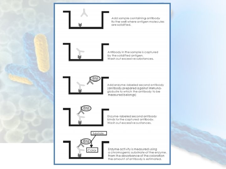
A basic procedure of direct ELISA (sandwich) 1) Standard solutions or assay samples are added to the antibody-coated wells, and incubated for several hours so as to the antigen molecules are captured by “capture antibody”.
2) After this binding reaction, the reaction mixture is discarded, and wells are washed to remove excessive materials. 3) The second antibody which recognizes another epitope in antigen is added. This second antibody has been labeled with an enzyme such as horseradish peroxidase (HRP)
4) The enzyme-labeled second antibody will bind to the antigen which is bound to the capture antibody on the bottom area of wells. This means that the enzyme (HRP) is also fixed on the bottom of wells. The amount of the antigen captured is proportional to fixed enzyme.
5) Enzyme activity is measured by adding a chromogenic substrate of this enzyme. In the case of HRP, tetramethylbenzidine (TMB) is often used. After incubation for some period, the chromogenic substrate is changed to a colored product. The reaction is stopped by adding a reaction stopper “stop solution”, e. g. diluted sulfuric acid, and absorbance is measured using a plate reader.
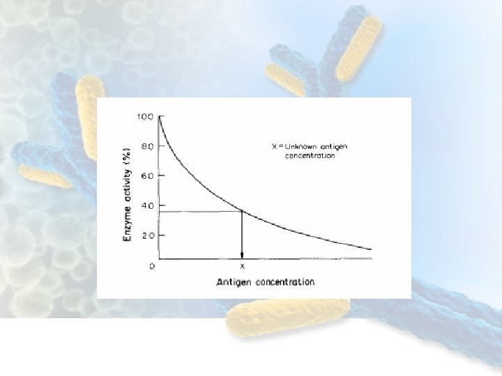
6) The standard curve is prepared from the concentration of standard solutions and their absorbance. And the sample assay values are obtained from the absorbance using the standard curve (calibration curve). – It is done by plotting standard concentration on X-axis and absorbance on Y-axis, both in normal scale, looks like a linear line signal concentration
When both X- and Y-axes are transformed to logarithmic scales, the standard curve looks nearly linear, though still curve linear strictly. By such plotting all the standard points are located with enough intervals with regard to both axes, and also third order regression fits very well due to the slight curve. Manual reading is also easy.
ELISA for measurement of antibody • ELISA system can be also set up for antibody measurement, called “ indirect ELISA”. • In this system, antigen molecule, specific to the antibody to be measured, is adsorbed on the bottom of wells, and samples containing antibody are added to the wells. For estimation of the captured antibody, the second antibody* labeled with enzyme is added and washed out after the binding reaction. Then by incubation with chromogenic substrate, the coloration is measured from the absorbance.
Competitive ELISA • Sandwich binding principle cannot be applied to small molecular substances, haptens such as steroids some hormons, olgo-peptides, neurotransmitters, etc. • Small molecular substances cannot have plural antigenic determinants to form sandwich. For these substances, competitive assay with an enzyme is applied. Sometime it is called competitive ELISA which has competitive binding principle like RIA (radioimmunoassay).
Coating Incubation L L L S Enzym. reaction P Product measurement
Competitive ELISA In this assay system an enzyme-labelled antigen is used which competes with the unlabelled antigen in the sample for binding to the immobilised antibody. The greater the concentration of the antigen in the sample the lower the amount of labelled antigen binding to the antibody.
Competitive ELISA steps 1. Antibody to the antigen of interest is immobilised on to the solid phase. This may be a plastic tube, microtitre plate, plastic strip, etc. 2. Washing is carried out to remove unbound antibody. 3. A mixture of labelled antigen and sample solution, containing the antigen of interest, is added. Both labelled and unlabelled antigen compete for binding to the antibody. Controls containing labelled antigen only and a set of standards containing known amounts of unlabelled antigen are also used. Suitable blanks are also included.
1. The plate is washed to remove all unbound antigen. 2. Enzyme substrate is added and after a suitable incubation period the levels of enzyme activity are measured. 3. A graph of enzyme activity versus antigen concentration is drawn and the levels of antigen in the sample determined. The amount of enzyme activity is inversely proportional to the concentration of unlabelled antigen in the sample
Growth hormone
• Human Growth Hormone (h. GH) is a polypeptide chain, composed of 191 amino acids and with a molecular mass of 21, 500 Da. It is released by the anterior pituitary of both men and women. • The secretion is stimulated 3 – 4 hours after a meal, about 1 hour after the beginning of sleep, and after physical exercise. • Hyposecretion of h. GH becomes apparent in infants a few months after birth and may result in dwarfism. In the opposite case, hypersecretion of h. GH results in gigantism and may be due to hypophysic tumors. • In adults, when epiphyses are closed, hypersecretion of h. GH provokes an increase in volume of soft tissues (hands, feet, lips), a proliferation of bones (acromegaly syndrome), and a limited tolerance of glucose Plays an important role in growth control.
• Its major role in stimulating body growth is to stimulate the liver and other tissues to secrete IGF-1(somatomedin C). • It stimulates both the differentiation and proliferation of myoblasts. • It also stimulates amino acid uptake and protein synthesis in muscle and other tissues. • Growth hormone secretion is regulated by three feedback loops, including both long, short and ultrashort loops. • GHRH(stomstostatin) inhibits own secretion from the hypothalamus via an ultrashort-loop feedback. • Somatomedins, which are by-products of the growth hormone action on target tissues, inhibit secretion of growth hormone by the anterior pituitary. • Both growth hormone and somatomedins inhibit the secretion of somatostatin by the hypothalamus.
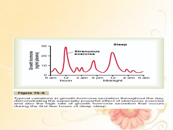
• Growth hormone is secreted in a pulsatile pattern, with bursts of secretion occurring approximately every 2 hours. • The largest secretory burst occurs within 1 hour of falling asleep (during sleep stages III and IV). The bursting pattern, in terms of both frequency and magnitude, is affected by several agents that alter the overall level of growth hormone secretion. • The normal values often increase to as high as 50 ng/ml after depletion of the body stores of proteins or carbohydrates during prolonged starvation.
• In brief, it has been found that growth hormone causes the liver (and, to a much less extent, other tissues) to form several small proteins called somatomedins that have the potent effect of increasing all aspects of bone growth. • Many of the somatomedin effects on growth are similar to the effects of insulin on growth. Therefore, the somatomedins are also called insulin-like growth factors (IGFs). At least four somatomedins have been isolated, but by far the most important of these is somatomedin C (also called IGF-1).
• Growth hormone attaches only weakly to the plasma proteins in the blood. Therefore, it is released from the blood into the tissues rapidly, having a half-time in the blood of less than 20 minutes. • By contrast, somatomedin C attaches strongly to a carrier protein in the blood that, like somatomedin C, is produced in response to growth hormone. As a result, somatomedin C is released only slowly from the blood to the tissues, with a halftime of about 20 hours. This greatly prolongs the growthpromoting effects of the bursts of growth hormone secretion shown in Figure
• The diagnosis of growth hormone defiance can’t be made in a single random growth hormone level because growth hormone is secreted in pulses. • Some pediatric endocrinologists diagnosis growth hormone defeciency based on an extremely low level of insuline like growth hormone which varies much less in the course of the day than growth hormone. • A more accurate but still imperfect way to diagnosis growth hormone deficiency is a growth hormone stimulation test. In this test, your child has a blood drawn for about 2 to 3 hours after being given medications to increase growth hormone release.
• If the child does not produce enough growth hormone after this stimulation, then the child is diagnosed with growth hormone deficiency. However, growth hormone stimulation tests can over diagnose growth hormone deficiency. Growth hormone stimulation tests vary and are complicated, so they are usually performed under the guidance of a pediatric endocrinologist. • Usually, other tests to check the pituitary or to evaluate the brain MRI are performed when treatment is considered.
Clinical significance • In children, ascertaining linear bone growth along the epiphyseal plate. Abnormally elevated levels lead to gigantism while complete absence slows the rate of growth one third to one half of normal. • In adults, the epiphyseal growth plates had fuse so h. GH excess gradually produces acromegaly, a coarse thickening of the bones of the skull, hands and feet.
Growth hormone test • The specimens shall be blood, serum in type and the usual precautions in the collection of venipuncture samples should be observed. • For accurate comparison to established normal values a fasting morning serum sample should be obtained. • The blood should be collected in a plain redtop venipuncture tube without anticoagulant or additive. Allow the blood to clot. Centrifuge the specimen to separate the serum from the cells.
Sample storage • Samples may be refrigerated at 2 -8 C for maximum period of five days. • If the specimen can not assayed within this time, the samples may be stored at temperatures of -20 C for up to 30 days. • Avoid use of contamination devices. Avoid repetitive freezing and thawing. When assayed duplicate, 0. 100 ml of the specimen is required.
Principle • The Human Growth Hormone (h. GH) ELISA is a solid phase sandwich ELISA method. The samples and antih. GH-HRP conjugate are added to the wells coated with h. GH MAb. h. GH in the serum binds to anti-h. GH MAb on the well and the anti -HGH second antibody then binds to h. GH. Unbound protein and HRP conjugate are washed off by wash buffer. Upon the addition of the substrate, the intensity of color is proportional to the concentration of h. GH in the samples. A standard curve is prepared relating color intensity to the concentration of the h. GH.
• Hyperglycemia inhibits growth hormone secretion. • Age, genes, sex, diet, exercise, stress, and other hormones is an important factors affect growth hormone concentrations. • At birth, GH is high and generally declines with the exception of a burst during the growth phase of adolestronce. • Women typically have a 50% higher level than their age-mached males.
Reference Ranges • Random growth hormone (GH) levels in a healthy person range as follows: – – Men: < 5 ng/m. L or < 226 pmol/L Women: < 10 ng/m. L or < 452 pmol/L Children: 0 -20 ng/m. L or 0 -904 pmol/L Newborns: 5 -40 ng/m. L or 226 -1808 pmol/L • GH suppression test value (using 100 g glucose) in a healthy person is as follows: – < 0 -2 ng/m. L or 0 -90 pmol/L or undetectable • GH stimulation test values (using arginine, glucagon, or insulin) are as follows: – Normal peak value: >10 ng/m. L or >452 pmol/L – Intermediate peak value: >5 ng/m. L or >226 pmol/L – Subnormal peak value: < 5 ng/m. L
e h T d n E