Histology of Spleen Largest lymphoid organ Present in


 Parenchyma Ø")


 are surrounded by an immunologically active zone")


- Slides: 39
Histology of Spleen
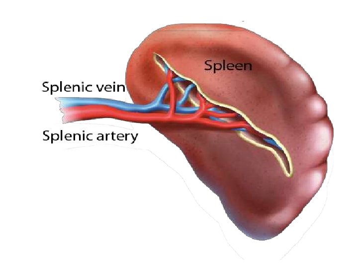
• Largest lymphoid organ • Present in the upper part of abdominal cavity behind the stomach ( Upper left quadrant ) • Covered by peritoneum
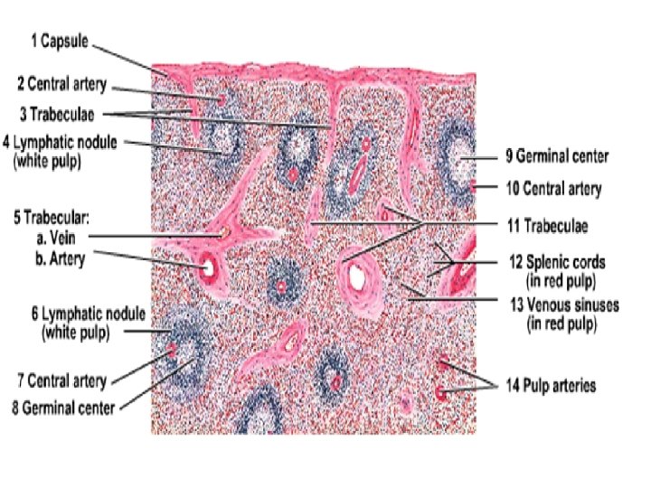
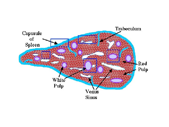
COMPONENTS Connective Tissue Framework Ø Capsule Ø Trabeculae Ø Reticular Stroma (Fibres) Parenchyma Ø White pulp Ø Red pulp
Connective Tissue Framework
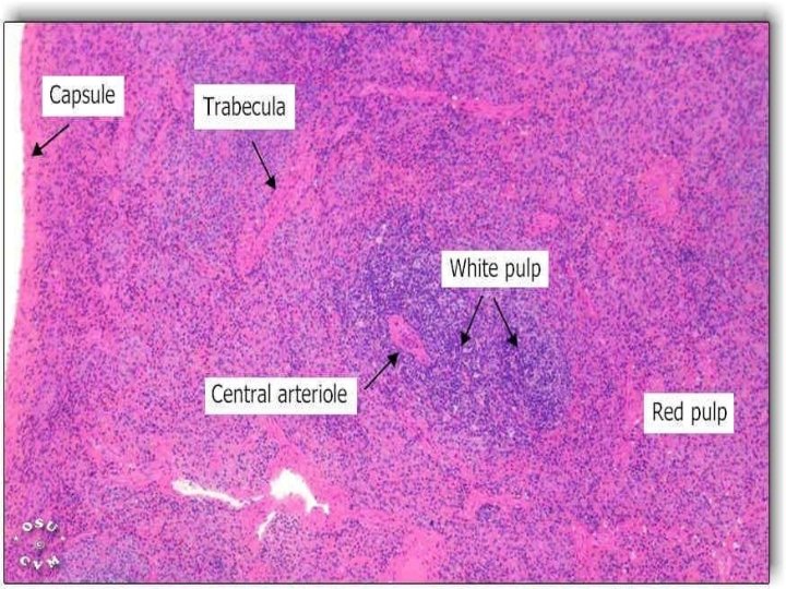
Capsule • Covers the spleen • Formed by dense irregular connective tissue and few smooth muscle fibre. • Trabeculae are given off from the capsule into the spleen and divide its parenchyma.
Reticular Stroma • Made up of reticular fibres • Forms a mesh work • Supports the cells of parenchyma
Parenchyma
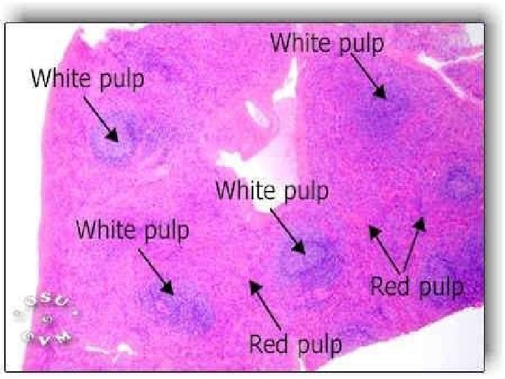
• The interior of spleen shows Rounded white/grey areas surrounded by red matrix White Pulp Dark red matrix Red Pulp
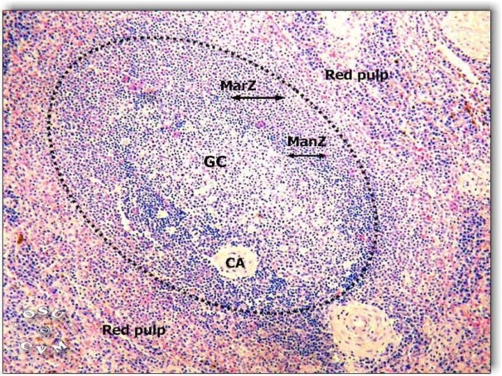
White Pulp • Made up of aggregation of lymphoid tissue around a small artery (central artery) • This artery is a branch of trabecular artery that leaves the trabeculum and enters the pulp • On entering the pulp it is surrounded by lymphoid tissue, the periarterial lymphatic sheath (PALS) • Populated by T lymphocytes, to become central artery or white pulp artery
• Around the course of PALS, there are large collection of B lymphocytes forming lymphatic nodules with germinal centres (white pulp) • In these nodules the central artery occupies an eccentric position
• The lymphatic nodules (white pulp) are surrounded by an immunologically active zone containing few T lymphocytes • This functional zone between the white and red pulp is called marginal zone
Spleen
Spleen
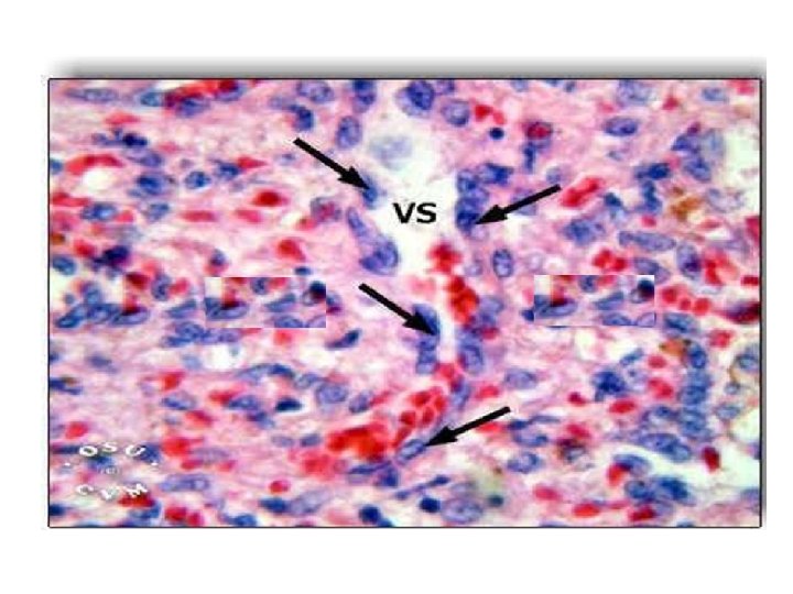
Red Pulp • Modified lymphoid tissue • Infiltrated with all the cells of circulating blood dark red colour • It is composed of splenic cords of Billroth and broad splenic venous sinuses in between the cords
• The splenic cords consists of spongy network of reticular fibres, lymphocytes, macrophages, plasma cells and all elements of the circulating blood
Spleen
Splenic Venous Sinuses • The splenic venous sinuses are lined by endothelial cells • Externally the sinuses are encircled by reticular fibres
Palatine Tonsils
• Each palatine tonsil consists of diffused lymphoid tissue in which lymphatic nodules are present • The lymphoid tissue is covered by stratified squamous nonkeratinized epithelium • This epithelium form deep invaginations into the substance of the tonsil in the form of several tonsillar crypts.
• The parenchyma consists of a dense accumulation of lymphoid follicles which show Germinal center. • The deep aspect is isolated from subjacent structures by a band of dense connective tissue called Capsule. • The lumen of the crypt usually contains some lymphocytes, food debris and bacteria.
Palatine Tonsil
dense lymphatic tissue that is accumulated in the connective tissue underlying the epithelium.
lymphoid nodules and germinal centers
Pharyngeal Tonsil
• This is a mass of lymphoid tissue present on the posterior wall of the nasopharynx in the midline. • Covered by pseudostratified columnar ciliated epithelium. • No crypts are present • Diffuse mass of lymphoid tissue is present under epithelium • The deeper surface also covered by thin capsule of fibrous connective tissue.
Lingual Tonsil • Mass of lymphoid tissue located in the root of the tongue. • Is covered by stratified squamous epithelium. • Lingual tonsil has many crypts. • Beneath the epithelium there is a layer of lymphatic nodules. • A thin capsule separates from the underlying structures.
MCQS What is the term for the entire lymphatic region of the spleen? a. Malpighian corpuscle b. Trabeculae c. White pulp d. Red pulp e. Cords of Billroth
What are the splenic cords? a. Cords of Billroth b. Cords of Paneth c. Cords of Bellini d. Cords of Rothchild e. Cords of Hassall
Where are the splenic sinuses? a. Malpighian corpuscle b. Trabeculae c. White pulp d. Red pulp e. Cords of Billroth
What does the PALS stand for? a. Papillary layer sinus b. Peyer's lymphatic sheath c. Periarterial lymphatic sheath d. Peripheral lymphatic sinus e. Parenchymal lymphatic sheath
Identify 1 3 5 2 4
Identify 1 2 3 4 5