SPLEEN The spleen is an wedgeshaped organ that


- Slides: 35

SPLEEN

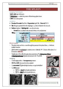
� The spleen is an wedge-shaped organ that lies in relation to the 9 th , 10 th and 11 th ribs, located in the left hypochondrium and partly in the epigastrium; thus, it is situated between the fundus of the stomach and the diaphragm. The spleen is highly vascular and reddish purple; its size and weight are variable( 75 -250 g). A normal spleen is not palpable. { �


� Although protected under the bony rib cage, the spleen remains the most commonly affected organ in blunt injury to the left lower chest and abdomen in all age groups. {7}.

Anatomy and embryology : � It consist of an encapsulated mass of vascular � and lymphoid tissue , it’s the largest reticuloendothelum organ in the body arising from the primitive mesoderm as an outgrowth of the left side of the dorsal mesogastrium. The most common anomaly of splenic embryology is the accessory of spleen, present in up to 20%of the population , one or more accessory spleen may also occur in up to 30%in patient with hematological diseases over 80%of accessory spleen found in region of the splenic hilum and vascular pedicle

Splenic artery: Its tortuous , arise from the celiac trunk and runs along the upper border of the body and tail of pancreas, a- to which it give small branches b- short gastric arteries to the stomach (greater curvature) c-left gastroepiploic branches pass between the layer of the gastro splenic ligament Main splenic artery divided to

Splenic vein Is formed from several tributaries draining the helium, the vein run behind the pancreas. and it joined by the sup. mesentric vein to form portal vein.

Functions of spleen � 1 -RBC and WBC production during fetal life 2 - removal of old or diseased RBC � 3 -maturation of reticulocyte � 4 -production of Ig. M and several opsonins 5 -removal of the fallowing from RBCs: � a-Howell –jolly bodies (nuclear remnants) b-pappenheimer bodies(iron granules) � �

Strictures related to spleen are greater curvature of stomach, the tail of pancreas, left kidney, splenic flexure of colon, the parietal peritoneum adheres firmly to the splenic capsule except at splenic hilum. peritoneum extend superior and lateral and inferior creating folds which form suspensory ligaments of spleen , splenophrenic and splenocolic ligaments and gastro splenic ligament through which the short gastric arteries and veins course. ]the splenic artery which is the largest branch from celiac trunk. enter the hilum of the spleen, branches in to trabicular arteries and then branches into the central arteries �

Functions of spleen 1 -RBC and WBC production during fetal life 2 - removal of old or diseased RBC 3 -maturation of reticulocyte 4 -production of Ig. M and several opsonins 5 -removal of the fallowing from RBCs: a-Howell –jolly bodies (nuclear remnants) b-pappenheimer bodies(iron granules)

c-Heinz bodies (denaturated Hgb) � 6 -removal of bacteria and other foreign �

Investigation of spleen; 1 - history and clinical examination 2 -laborotory test 3 -radiological imaging a- plain abdomen? ? Rare usfull some time see calcification or in truma see fracture of lower ribs B-u/s can see siz of spleen and any space accuping lesion cc. t scan with contrast enhancement. D- MRI

CONGINATAL ABNORMALITY OF SPLEEN Splenic agenesis rare about 10% of children with congenital heart disease, 2 - poly splenia rare due to failure of splenic fusion 3 - splenuculi are single or multiple accessory spleen that are found in 10 -30 % of population. (what are the significant of accessory spleen? ? )

� SPLENIC RAPTURE Spleen represent is the most common intra abdominal organ susiptable to rapture by penetrating and blunt trauma � �

susceptible to rapture by penetrating and � blunt trauma. or Iatrogenic injury to the spleen remains a � frequent complication of any surgical procedure , particularly those in the left upper quadrant when adhesion are present.

Ruptured spleen may present in three ways : � 1 - The patient succumbs rapidly from massive haemorrhage � , Usually as a consequence of trauma. � 2 - Initial shock , recovery , signs of late bleeding. The initial � shock is due to blood loss , tamponade occurs and then further bleeding take place. General sign of internal haemorrhage maybe variable but local signs include left upper quadrant pain and tenderness , followed by abdominal distension. The referral of pain to the left shoulder is known as Kehr's sign and can be demonstrated some 15 minutes following elevation of foot to the bed the pain results from the contact of blood with the undersurface of the diaphragm and is mediated through afferent fibers of the phrenic nerve.

With significant haemorrhage , shifting dullness maybe present in the flanks and fullness in the pelvis maybe evident on rectal examination. Abdominal ultrasonography or CT scanning will demonstrate the source of the bleeding in the patient who is stable enough to undergo imaging before urgent laparotomy.

3 - The delayed case. The initial signs of splenic injury may pass quickly or not be recognized but delayed rupture can occur. Such cases should be uncommon with the frequent use of scanning on the trauma patient. Any haematoma around the spleen in the stable patient should lead to close observation.

Management : The ruptured spleen can be managed successfully without operation. Careful observation can be pursued in the patient with minimal or no abdominal findings and when there is haemodynamic stability. CT may confirm isolated injury to the spleen , and a conservative approach should be considered in the absence of hilar involvement or of massive disruption of the spleen. �

Immediate laparotomy is indicated for splenic trauma if there is obvious evidence of continuing blood loss despite adequate resuscitation. The incidence of major associated intraabdominal injuries with blunt splenic trauma may exceed 50% and laparotomy should therefore be considered if there is a strong suspicion of trauma to other organs. The treatment of associated injuries is dictated by the operative findings. Splenic preservation should be considered where possible , and any blood should be ecacuated to facilitate inspection of the spleen. Minor capsular parenchymal injuries may required only topical haemostatic agents. If by careful compression of the spleen the bleeding can be controlled , suture of the injury with or without application of haemostatic agents may be useful. parenchymal injuries involving the lower or upper pole of the spleen may be managed by partial segmental resection.

Sleen injury severity scale by grades(aast): It classified to 5 grade Grade 1 : a-sub capsular hematoma less than 10% surface area and non expanding. b-laceration less than 1 cm deep with capsular tear, but non bleeding Grade 2: a- hematoma less than 50% surface area sub capsular, less than 5 cm intraparenchymal hematoma. b- capsular tear with active bleeding, 1 to 3 cm in depth, but must not involve trabicular vessel.

Grade 3: a- sub capsular hematoma more than 50% surface area or expanding hematoma, rapture sub capsular hematoma with active bleeding b- more than 3 cm deep parenchymal laceration. Grade 4: a-rapture intraperanchymal haematoma with active bleeding. b-wound involving segmental or hilar vessels with major devascularization. Grade 5 : a- shattered spleen(massive)b-hilar injury that completely devasculriuze the spleen. [2]

Splenomegaly and Hypersplenism Hypersplinism: its acombination of splenomegaly , anaemia, leucopenia and or thrombocytopenia with bone marrow hyperplasia. Splenomegaly is a common feature of many disease processes. It should be borne in mind , however, that many conditions affecting the spleen, such as idiopathic thrombocytopenic purpura , maybe associated with enlargement, but the gland is seldom palpable. Few condition cause splenomegaly and need splenoctomy.

Causes of splenomegaly: 1 -infection— bacterial typhoid, t. b. brucellosis Viral –glndular fever-e. b. virus— Spirochaetal—syphilis Protozoal----malaria , kalazar schistomiasis 2 - cellular proliferation –myloid and lymphatic leukemia, lymphoma, polycythemia rapra vera, spherocytosis, sickle cell anaemia 3 -congestion—portal hypertension, hepatic vein obstraction, congestive heart failure 4 -cellular infiltration---amyloidosis 5 - collagen disease –feltys disease

6 -space accuping lesion----hydatid cyst , lymphoma 7 - injury ----haematoma � �

Splenic abscess; May arise from infected splenic embolism. or associated with typhoid , paratyphoid, or pancreatic necrosis, the abscess may rapture and cause sub phrenic abscess or peritonitis. treatment by percutaneouse drainage under radiological guidance.

I. T. P This condition is due to circulating igg exists that is directed against platlet- associated antigen resulting in destraction of platelets by the reticuloendothelial system. This condition associated with low platlets count easy brusing petechiae mucosal bleeding menorrhgia increase megakaryocyte count on bone marrow aspiration. its mor common in womens. �

Treatments Medical treatments (predinsolon, platlet transfusion, plasmaphoresis). if treatment failed we treat condition by splenactomy , about 75% of cases need splenactomy. What are prognosis after splenactomy

80% of cases platlets count return to normal level within 6 post operative wk. Why some patients recur after splenactomy ? Due to accessory spleen Felty syndrome Its traid of splenomegaly, R. A, granulocytopenia Cause due to formation antibodies against granulocytes. ,

NEOPLASMS Haemangioma is the most common benign tumour of the spleen and may rarely develop to haemangiosarcoma that is treated by splenactomy. Lymphoma another neoplasm may affect spleen and splenoctomy may use for staging of lymphoma, Secondary metastasis may envolve spleen

80% of cases platlets count return to normal level within 6 post operative wk. Why some patients recur after splenactomy ? Due to accessory spleen. What means of hypersplenism? Acombination of splenomegaly anaemia, leucopenia, and or thrombocytopenia with bone marrow hyperplasia,

SPLENACTOMY Indication 1 -The most common cause of splenactomy is traumatic rapture of spleen 2 -itp 3 -primary tumour or ccyst 4 -heredatory spherocytosis 5 -autoimune anaemia 7 - lymphoproliferative disorder 8 - felty syndrome 9 -sickle cell disease 9 aids 10 - splenic vein thrombosis 11 -thalasemia major 12 gaucher disease 13 - in portal hypertension �

Preoperative preparation; 1 - blood or frb , platlets if there is bleeding tendency 2 -prophylaxic antibiotic. 3 - in elective splenactomy must give vaccine against pneumococcus, H influenza and miningoccous, pneumococcal vaccination is recommended in those patients over age 2 years. �

Post operativ complication 1 -atelectasis(left lower lobe atelectasis is the most common complication) 2 -left pleural effusion 3 -subdiaphragmatic abscess

4 -bleeding 5 -haematoma formation 6 - injury to the tail of pancrease 7 - gastric ileus 8 -post splenactomy sepsis which more common accure in children. it may result from strep. pneumonia, neiss. meningitis, H. influ enzae and e. coli. 9 - opportunist post-solenactomy infection(OPSI)(difined it? ) �
 Spleen solid organ
 Cell to tissue to organ to organ system to organism
 Sistem koordinasi
 Organ berikut yang merupakan penyusun sistem gerak adalah
 Organ and organ system
 Cell tissue organ organ system organism
 Organ organ pernafasan
 Thơ thất ngôn tứ tuyệt đường luật
 Con hãy đưa tay khi thấy người vấp ngã
 Sau thất bại ở hồ điển triệt
 Thơ thất ngôn tứ tuyệt đường luật
 Tôn thất thuyết là ai
 Ngoại tâm thu thất chùm đôi
 Walmart thất bại ở nhật
 Block nhĩ thất cấp 1
 Tìm vết của mặt phẳng
 Gây tê cơ vuông thắt lưng
 Thế nào là mạng điện lắp đặt kiểu nổi
 Bổ thể
 Lời thề hippocrates
 Vẽ hình chiếu đứng bằng cạnh của vật thể
 Quá trình desamine hóa có thể tạo ra
 Chúa yêu trần thế
 Khi nào hổ mẹ dạy hổ con săn mồi
 điện thế nghỉ
 Dạng đột biến một nhiễm là
 Biện pháp chống mỏi cơ
 Công thức tính thế năng
 Phép trừ bù
 Tỉ lệ cơ thể trẻ em
 Thiếu nhi thế giới liên hoan
 Phối cảnh
 Một số thể thơ truyền thống
 Các môn thể thao bắt đầu bằng tiếng đua
 Hát kết hợp bộ gõ cơ thể
 Sơ đồ cơ thể người
