Histology 1 4 Glands Gland a single epithelial


- Slides: 27

Histology 1. 4. : Glands Gland: a single epithelial cell, or grouping of cells specialized for secretion. Secretion: energy-consuming process by which the cell takes up small molecules and transforms them via intracellular biosynthesis into a more complex product then actively releases it from the cell. The product is utilized by the organism in several ways. Excretion: the organism gets rid of harmful or toxic metabolic end-products or useless waste material.

Classification of glands: I. Based on morphology: unicellular and multicellular Unicellular, intraepithelial gland: goblet cell Intestinal epithelium: simple columnar epithelium (2) goblet cells (1) the arrow shows the nucleus (3) TEM image of the same type of unicellular gland, the goblet cell

CLASSIFICATION OF MULTICELLULAR GLANDS: With ducts: exocrine glands Without ducts: endocrine glands

CLASSIFICATION OF EXOCRINE GLANDS (based on morphology): Simple tubular Coiled tubular Branched tubular Simple acinar Branched acinar Compound tubular Compound acinar Compound tubuloacinar

SOME EXAMPLES: Simple tubular gland: intestinal gland of Lieberkühn Schematic drawing LM microphoto

Coiled tubular gland: sweat glands of the skin Schematic drawing LM microphoto

Branched tubular gland: fundic glands in the stomach Schematic drawing LM microphoto

Fundic gland from the stomach - endpiece

Simple acinar gland: Frog skin: mucous and toxin-producing glands Compact form without lumen: sebaceous gland of mammalian skin

Branched acinar gland: larger sebaceous glands of the skin

Compound tubular gland: esophageal gland duct segment capillary secretory acini capillary lumen

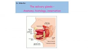
Compound tubuloacinar glands: Mandibular gland Parotid gland

Composition of compound glands: Parenchyma composed of lobes and lobules, ducts The duct system of the compound glands: Acinus Intercalated duct Striated (salivary duct) Interlobular duct Lobar duct Main duct

Model of the gland: a bunch of grapes: berry= acinus stalk of berry: intercalated duct

Intralobular striated (salivary) duct Interlobular duct

II. Type of secretory product: 1. Serous gland: Pancreas (see the micrograph) and parotid gland are purely serous producing thin watery fluid rich in proteins (mainly enzymes) composed of acini with narrow lumen (1) secretory cells have round, basally located nuclei (2) the cytoplasm of the cells is basophilic (3) 1 3 2

2. Mucous glands: Esophageal glands (see the micrograph) are purely mucous producing thick viscous fluid, rich in mucopolysaccharides for lubrication and protection of internal body surfaces composed of acini with wide lumen secretory cells have flattened nuclei at the base their cytoplasm is very weakly stained

3. Seromucous glands: mucous acini surrounded by serous cells forming a demilune shape The submandibular gland of some species (monkey, human, cattle) is seromucous. Red arrows point at mucous cells, blue arrows point at the demilune –shaped group of serous cells (Demilune of Gianuzzi)

Seromucous gland, haematein-eosin staining Seromucous gland, alciane blue staining

III. Modes of secretion: 1. • • • Merocrine secretion: The secretory process is an exocytosis The secretory cell remains completely intact Most of the glands secrete in a merocrine manner Exocrine pancreas Submandibular gland

2. Apocrine secretion: • the secretum is gathered at the apical portion of the cell • the secretum leaves the cell membrane-bounded (pinched off) • the cell remains alive, but a part of it goes with the secretory droplet Examples: sweat and mammary glands

Membrane-bound secretory droplets of a sweat gland

3. Holocrine secretion: • the secretory cell gradually fills up with secretum • the cell organelles degenerate • the cell dies, its membrane breaks and the secretum empties The sebaceous gland is a holocrine gland. Dead cells are replaced by the mitotic divison of basal cells

Absorptive epithelium: its main function is to absorb. Morphology of these epithelial cells exhibit cuticular border (intestine) or brush border (kidney tubules). Please note: at fine structural level both are microvilli ! Intestinal epithelium: the arrow shows the cuticular border EM micrograph of the apical surface with microvilli

Pigmented epithelium: Epithelial cells contain melamosomes: brown color EM Pigmented epithelium in the eye at LM level shows brown pigmentation. LM At EM level the melanosomes appear as electron dense bodies in the cytoplasm.

Sensory epithelia: Main function is sensation Types: primary, secondary sensory epithelium true nerve cells Primary sensory epithelium: olfactory epithelium

Secondary type of sensory epithelia: Example: sensory cells of the taste buds
 Gianuzzi demilune
 Epithelial tissue pogil
 Epithelial tissue pogil
 Parathyroid gland image
 Parotid duct
 Stomach lining epithelium
 Pineal gland pituitary gland
 Pituitary gland and pineal gland spiritual
 Histology of pituitary gland
 Gland histology
 Intercalated duct histology
 Simple branched alveolar gland
 Thyroid follicle histology
 Single cell gland
 Dataxin
 Sisd simd misd mimd examples
 Waiting line management
 Gianuzzi demilune
 Alectinib
 Voluntary muscles
 Lichen sclerosus vulvare
 Dental papilla gives rise to
 Stratified epithelial tissue
 Characteristics of glandular epithelium
 What kind of tissue
 Animal tissue
 Epithelial tissue with goblet cells
 Simple columnar epithelium
