Gut formation With lateral folding mesoderm is recruited


 Ectoderm Mesoderm Endoderm Trilaminar embryonic disc Figure 28.")
 (b) Figure 28. 11 b")
 Tail fold Head fold (c) Yolk sac Figure 28. 11")
 Figure 28. 11 d")


")


- Slides: 28
Gut formation
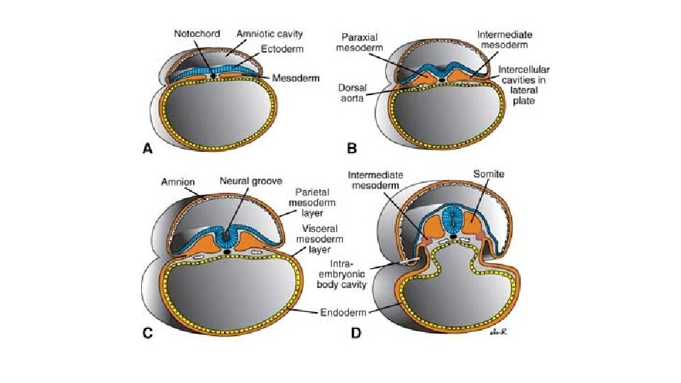
With lateral folding, mesoderm is recruited to gut wall Carlson fig 6 -20 Langman’s fig 6 -18 • Lateral folding of the embryo completes the gut tube • Mesodermal layer of the gut tube is called splanchnic (visceral) mesoderm - derived from lateral plate mesoderm • Somatic mesoderm lines body cavity
Gut tube proper Foregut: pharynx esophagus stomach proximal duodenum Midgut: distal duodenum to right 2/3 of transverse colon Hindgut: left 1/3 of transverse colon to upper part of anal canal. (These three regions are defined by their blood supply) Derivatives of gut tube thyroid parathyroid glands tympanic cavity trachea, bronchi, lungs liver, gallbladder pancreas urinary bladder
Specialization of Endoderm • Embryonic folding begins with lateral folds • Next, head and tail folds appear • Endoderm tube forms epithelial lining of the GI tract • Organs of the GI tract become apparent, and oral and anal openings perforate • Mucosal lining of respiratory tract forms from pharyngeal endoderm (foregut)
Tail Head Amnion Yolk sac (a) Ectoderm Mesoderm Endoderm Trilaminar embryonic disc Figure 28. 11 a
Lateral fold Future gut (digestive tube) (b) Figure 28. 11 b
Somites (seen through ectoderm) Tail fold Head fold (c) Yolk sac Figure 28. 11 c
Hindgut Yolk sac Neural tube Notochord Primitive gut Foregut (d) Figure 28. 11 d
Pharynx Parathyroid glands and thymus Thyroid gland Esophagus Trachea Connection to yolk sac Right and left lungs Stomach Liver Umbilical cord Pancreas Gallbladder Small intestine Allantois 5 -week embryo Large intestine Figure 28. 12
4 th week 5 th week Langman’s fig 14 -14 Langman’s fig 14 -4 Celiac artery supplies the foregut Superior mesenteric artery supplies the midgut Inferior mesenteric artery supplies the hindgut The figure on the right also shows the mesenteries; note that the liver and stomach have dorsal and ventral mesenteries whereas the rest of the gut has only a dorsal mesentery.
Regional patterning of the gut tube Gut = bilayered tube (endoderm surrounded by mesoderm) Regional gut tube patterning and organogenesis require inductive signals from other nearby structures
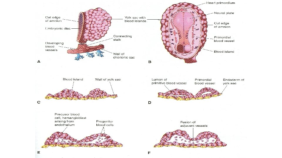
Early Development of Cardiovascular System • Starts at the beginning of the 3 rd week • Vasculogenesis and angiogenesis begins in the extraembryonic mesoderm of the yolk sac, connecting stalk and chorion • Embryonic blood vessels begin to develop about 2 days later
Early Development of Cardiovascular System • The urgent need for blood vessels to bring nourishment and oxygen to the embryo from mother causes the early formation of the cardiovascular system • A primordial uteroplacental circulation develops during the 3 rd week • Until then the embryonic nutrition is obtained from maternal blood by diffusion
Vasculogenesis & angiogenesis Formation of embryonic vascular system involves 2 processes: • Vasculogenesis • Angiogenesis
Vasculogenesis • Mesenchymal cells differentiate into endothelial precursors called Angioblast • Angioblast aggregate to form isolated angiogenic cell clusters or blood islands • Small cavities appear within the blood islands • Angioblasts flatten to form endothelial cells
Vasculogenesis • Endothelial cells arrange themselves around the cavities in blood island to form the endothelium • These endothelium lined cavities soon fuse to form networks of endothelial channels (called Vasculogenesis).
Angiogenesis • Vessels sprout into adjacent areas by endothelial budding and fuse with other vessels (called Angiogenesis).
Development of Blood Cells • Blood cells develop from the endothelial cells of vessels called hemangioblasts • Develop at the end of 3 rd week on the yolk sac and allantois • Hematogenesis does not begin until 5 th week • It occurs first in liver and later in spleen, bone marrow & lymph nodes
Development of Blood Cells • Fetal and adult erythrocytes are derived from different hematopoietic progenitor cells (hemangioblasts) • Mesenchymal cells surrounding the primordial endothelial blood vessels differentiate into the muscular and connective tissue elements of the vessels
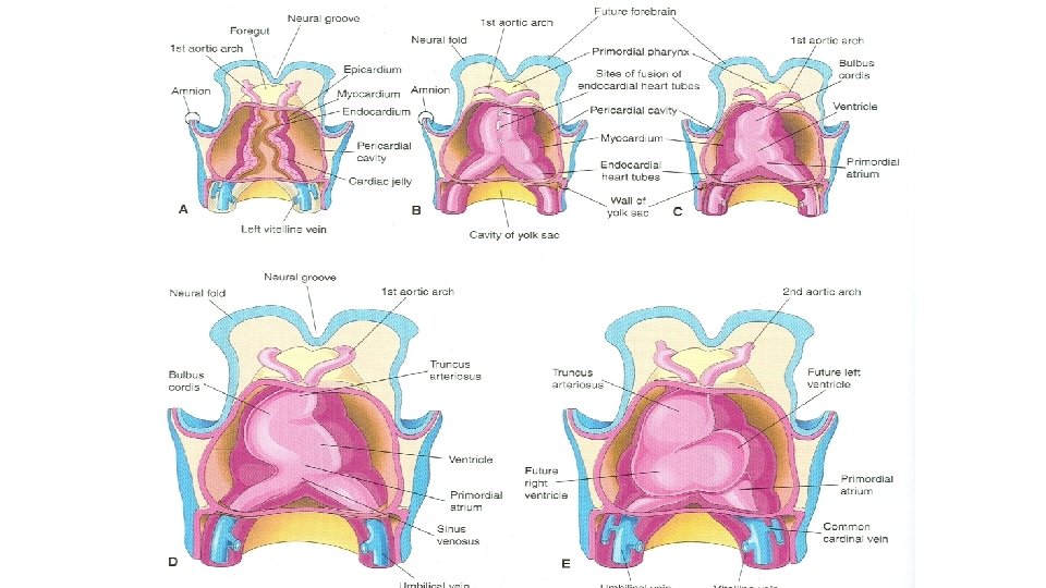
Primordial Cardiovascular System • Heart & great vessels develop from mesenchymal cells in the cardiogenic area • Paired longitudinal endothelial lined channels or endocardial heart tubes develop during the 3 rd week • These tubes fuse to form the heart tube
Primordial Cardiovascular System • The tubular heart joins with blood vessels in the embryo, connecting stalk, chorion and yolk sac to form a primordial cardiovascular system • Heart begins to beat on 21 -22 days and blood circulates • CVS is the first organ system to reach a functional state
Further Development of Chorionic Villi • Primary chorionic villi becomes secondary chorionic villi as they acquire mesenchymal cores • Before the end of third week capillaries develop in the secondary chorionic villi • Now it is called tertiary chorionic villi
Further Development of Chorionic Villi • Cytotrophoblastic extensions from these stem villi join to form a cytotrophoblastic shell that anchors the chorionic sac to the endometrium • The rapid development of chorionic villi during the third week greatly increases the surface area of chorion • This causes exchange of oxygen and nutrients between the maternal and embryonic circulations