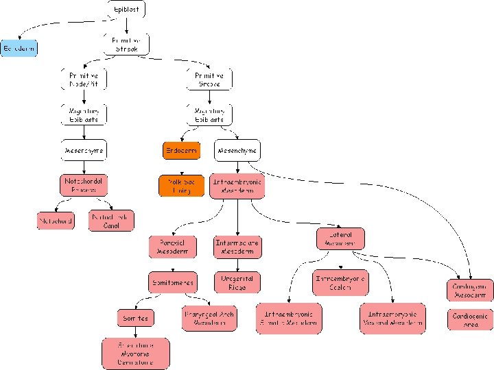
Neurulation Differentiation of Mesodermal layer Paraxial mesoderm Intermediate


 Paraxial mesoderm • Sclerotome- vertebrae, portions of neurocranium, axial skeleton • Myotome-")


 Myf 5 Pax 3 Myo")


 Intermediate mesoderm • Connective tissue of gonads, • Mesonephric and Metanephric nephrons, •")
 Septum transversum Associated with development of Heart , liver , Diaphragm • Epicardium,")
 Lateral plate mesoderm Somatopleuric layer – • Appendicular skeleton, • Connective tissue of")


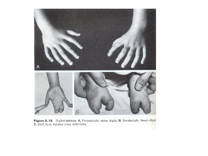
 Polydactyly Ectrodactyly")
")
 Development of splanchnopleuric mesoderm • Differentiation of mesenchyme around esophagus • Mesenchyme around")

 Angiogenic layer • • Endocardium of heart, Endothelium of blood vessels , Choroid")


- Slides: 44
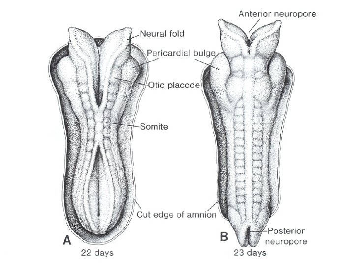
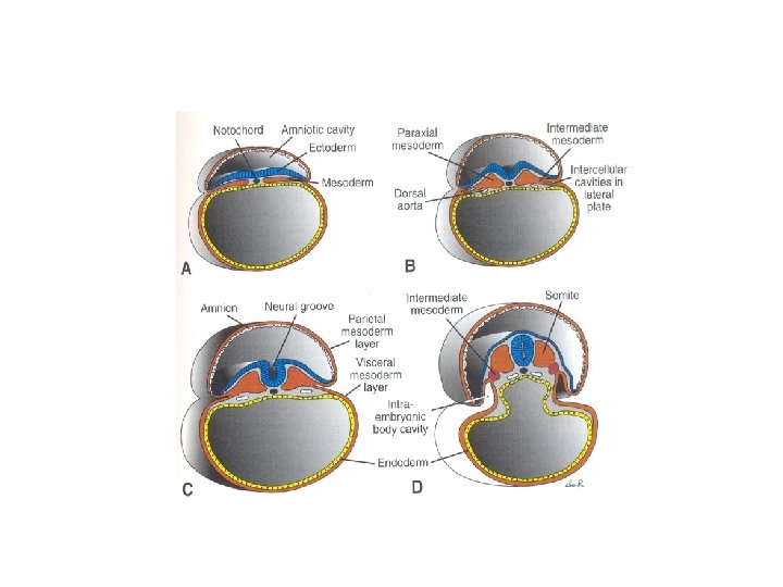
Neurulation
Differentiation of Mesodermal layer • Paraxial mesoderm • Intermediate mesoderm • Lateral plate mesoderm
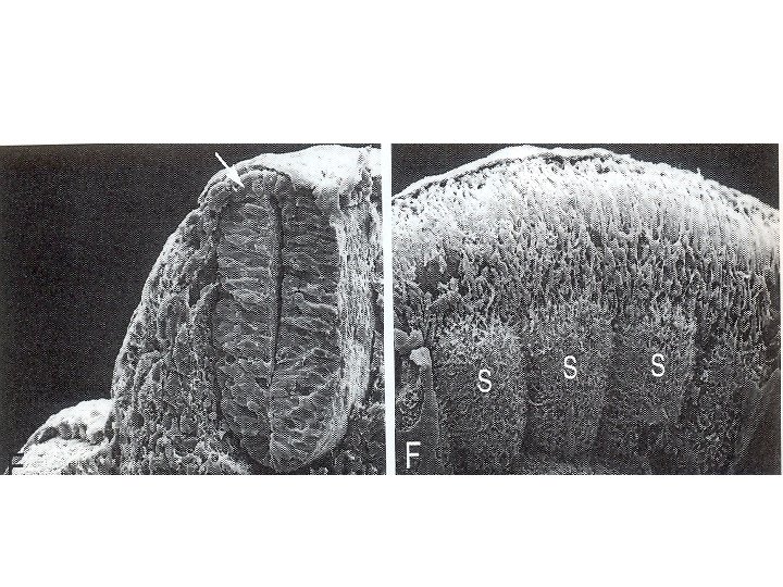
• Electron micrograph showing three parts of secondary mesoderm
Derivatives • • • Paraxial Mesoderm Intermediate Mesoderm Septum transversum Lateral plate Mesoderm Angiogenic layer
Derivatives I) Paraxial mesoderm • Sclerotome- vertebrae, portions of neurocranium, axial skeleton • Myotome- all voluntary muscles of head, . Trunk, and limbs • Dermatome- dermis of skin over dorsal regions
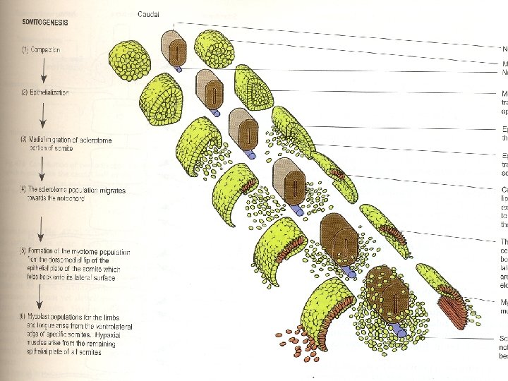
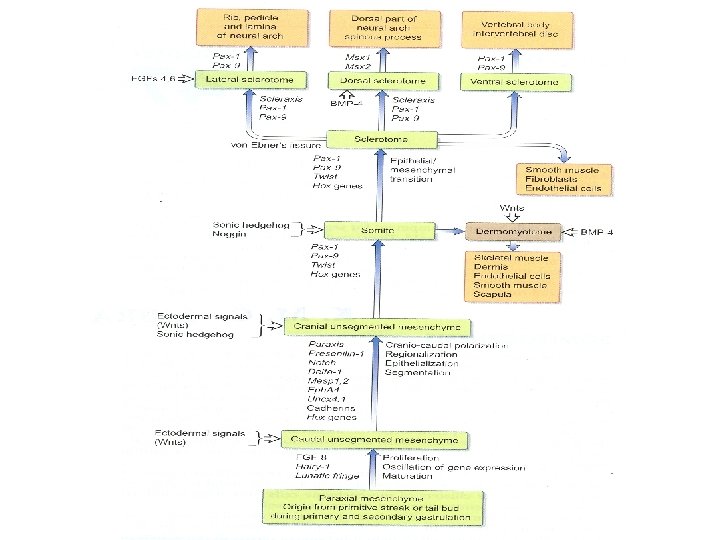
Development of sclerotome
Somitogenesis stages Compaction Epitheliasation Medial migration of sclerotome part of somites
Development of vertebra
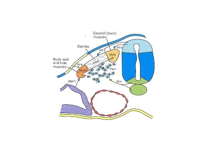
Genes in somite development • • Sonic Hedgehog (shh) Myf 5 Pax 3 Myo D Wnt Nt-3 Bmp-4 Pax -1
Derivatives of somites • Tongue muscles by occipital Myotomes • 1 st occipital myotome disappears • Vertebra • Ribs • Joints, Ligaments, Cartilages
Abnormalaties • Hemivertebra • Mal development of occipito cervical junction Arnold Chiari Syndrome - Medulla and Tonsils project through Foramen Magnum • Inappropriate fusion of lower cervical vertebra causes –Klippel Feil Syndrome
Hemi vertebra
Hemi vertebra may also leads to scoliosis
Spina bifida
Arnold chiari malformation
Klippel-Feil syndrome • Low posterior hair line • Short neck • Limitation of head and neck • Scoliosis &kyphosis
Diastematomyelia
Diastematomyelia Clinical features: - Patients may have cutaneous abnormalities - a dimple, pigmented naevi or - patch of hair along their back at the level of attachment of cord
II) Intermediate mesoderm • Connective tissue of gonads, • Mesonephric and Metanephric nephrons, • Smooth muscle and connective tissue of reproductive organs • It is not before somitogenesis Development is closely related to progress and differentiation of somites Abnormalities can cause extropy of urinary bladder
III) Septum transversum Associated with development of Heart , liver , Diaphragm • Epicardium, • Fibrous pericardium, • Portions of diaphragm, • Falciform ligament, • Sinusoids of liver , • Mesentery of esophagus. Abnormalities can cause Diaphragmatic Hernias
IV) Lateral plate mesoderm Somatopleuric layer – • Appendicular skeleton, • Connective tissue of limbs and trunk (including cartilage, tendons and ligaments) • Mesenchyme of external genitalia, • Dermis of ventral body wall and limbs.
Splanchnopleuric layer – • Smooth muscle and connective tissue of respiratory tract , • Intestinal tract, • Associated glands, • Blood vessels
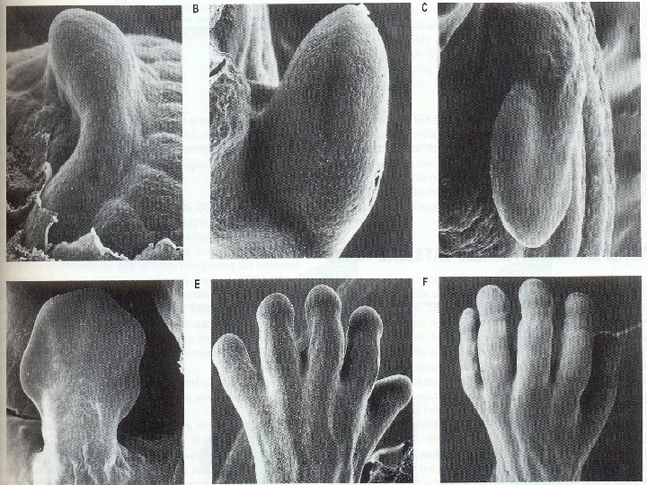
Development of limb budssomatopleuric mesoderm
• Cranio caudal axis of limb position is regulated by Homeo box gene (HOX) • Initiation of limb bud is by fibro blast growth factor -8 (FGF-8) • Out growth of limb by AER • Progressive zone • Limb patterning is regulated by ZPA along with AP axis of the limb
Effects of genes on the development of the limb buds • • AER --influences limb out growth AER– removal- Amelia Insert of AER – 2 axis of development Replacement with any other mesenchyme – no development of limbs • Replacement with lower limb mesenchyme leads to lower limb development & vice versa • Progressive zone is very specific it includes 8 stages of upper limb development
Hox genes specify each finger d-11 d-12 d-13 d-10 d-9
• • • Meromelia Amelia Phocomelia Micromelia Thalidomide Syndrome(1952 -1962 ) Polydactyly Ectrodactyly Cleft hand foot Congenital absence or deficiency of the radius
Thalidomide Syndrome(19521962 )
V) Development of splanchnopleuric mesoderm • Differentiation of mesenchyme around esophagus • Mesenchyme around trachea • Formation of lobes, their number, degree of maturity of lungs
Abnormalities – • Excessive laxity in effected air ways • Williams- Campbell Syndrome (bronchomalacia from 2 nd to 8 th generation of bronchi ) Clinical features – - Cough & tachypnoea, - Associated with tracheo- esophageal fistula - Bronchiactasis
VI) Angiogenic layer • • Endocardium of heart, Endothelium of blood vessels , Choroid plexus, Sinusoids of liver and spleen , Blood cells, Microglia, Macrophages
• Vascular system is first system to start development • Rapid vasculariziatrion and remodeling • Direction of blood flow is reversed a number of times
Theories associated with its origin • Blood islands in yolk sac endoderm • Vasculogenesis in the body of embryo is seen after the formation of extra embryonic blood vessels , hence it was believed that all blood vessels were derived from yolk sac • Recent evidences have shown that angioblastic cells are highly invasive and migratory in all directions its origin is also from endothelium of somites • Genes involved are Lmo 2 and GATA.
• Angioblast cells do not migrate into neural epithelium but form plexus of capillaries around brain Ultimate position of blood vessels is patterned by - mesenchymal population of head neural crest cells , - somatopleuric mesenchyme in limbs and - splanchnopleuric mesenchyme around viscera
References • Human embryology Inderbir singh • Langman’s embryology eigth edition • Essentials of Human Embryology A K Datta • Gray’s Anatomy 39 edition, Henry Grey