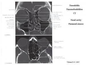
Paranasal Air Sinuses Diverticula grow from nasal cavity



- Slides: 18
Paranasal Air Sinuses • Diverticula grow from nasal cavity walls into surrounding bones • Openings of outgrowths persist as orifices of adult sinuses • At Birth- no frontal or sphenoidal sinuses • Maxillary-3 -4 mm • Ant & post Ethmoidal- Few cells
Paranasal Air Sinuses • Ethmoidal- small uptil 2 yrs grows rapidly 6 -8 yrs • Maxillary- grows slowly till puberty, fully developed in adulthood • Frontal grows from ant ethmoidal-2 yrs rapid dev. 7 yrs • Sphenidal-grows from post. Eth. 2 yrs
Pharyngeal arches & primordial Pharynx
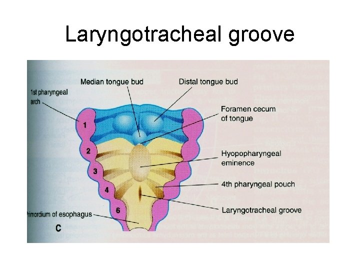
Laryngotracheal groove
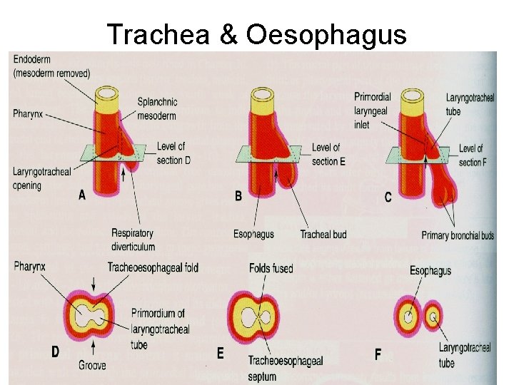
Trachea & Oesophagus
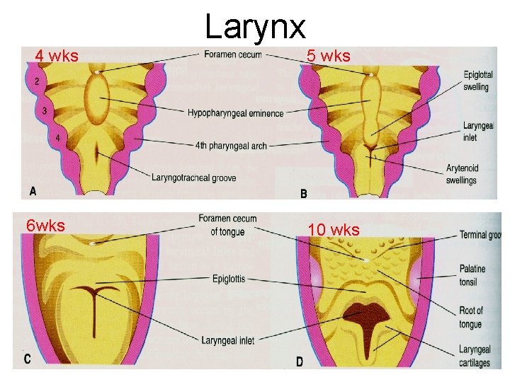
Larynx 4 wks 6 wks 5 wks 10 wks
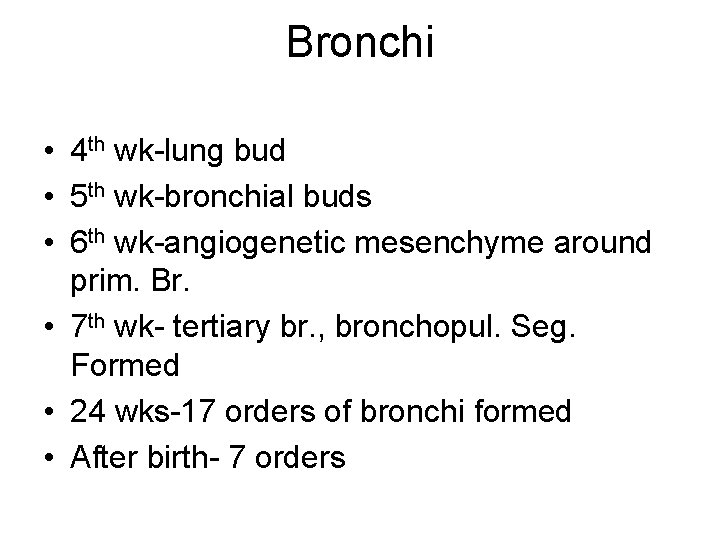
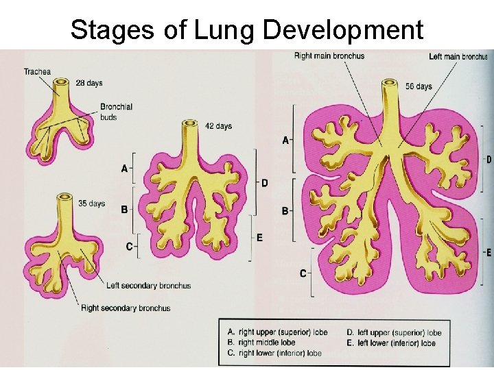
Bronchi • 4 th wk-lung bud • 5 th wk-bronchial buds • 6 th wk-angiogenetic mesenchyme around prim. Br. • 7 th wk- tertiary br. , bronchopul. Seg. Formed • 24 wks-17 orders of bronchi formed • After birth- 7 orders
Stages of Lung Development
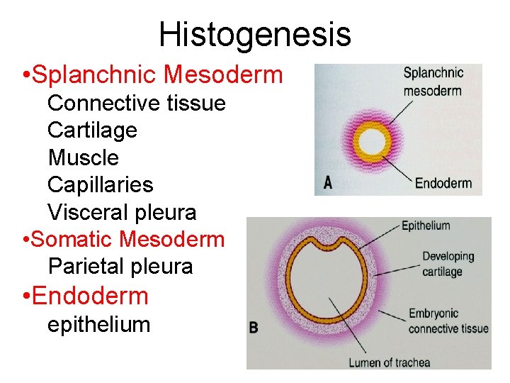
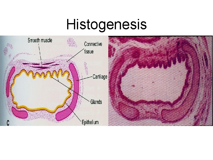
Histogenesis • Splanchnic Mesoderm Connective tissue Cartilage Muscle Capillaries Visceral pleura • Somatic Mesoderm Parietal pleura • Endoderm epithelium
Histogenesis
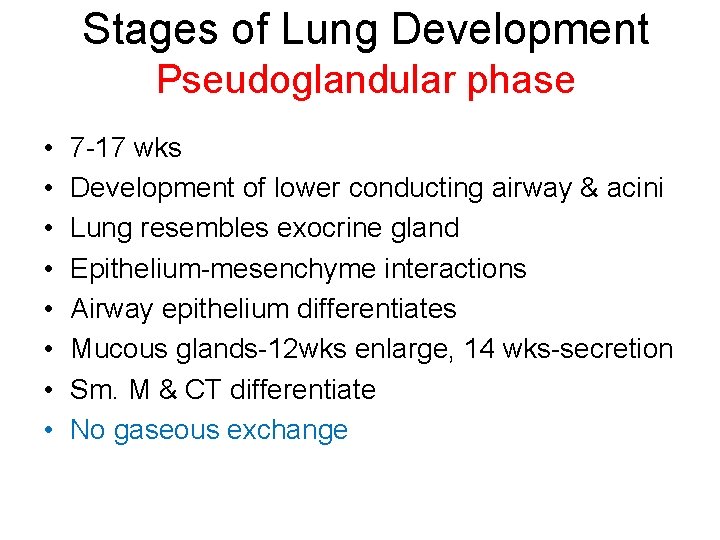
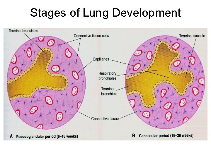
Stages of Lung Development Pseudoglandular phase • • 7 -17 wks Development of lower conducting airway & acini Lung resembles exocrine gland Epithelium-mesenchyme interactions Airway epithelium differentiates Mucous glands-12 wks enlarge, 14 wks-secretion Sm. M & CT differentiate No gaseous exchange
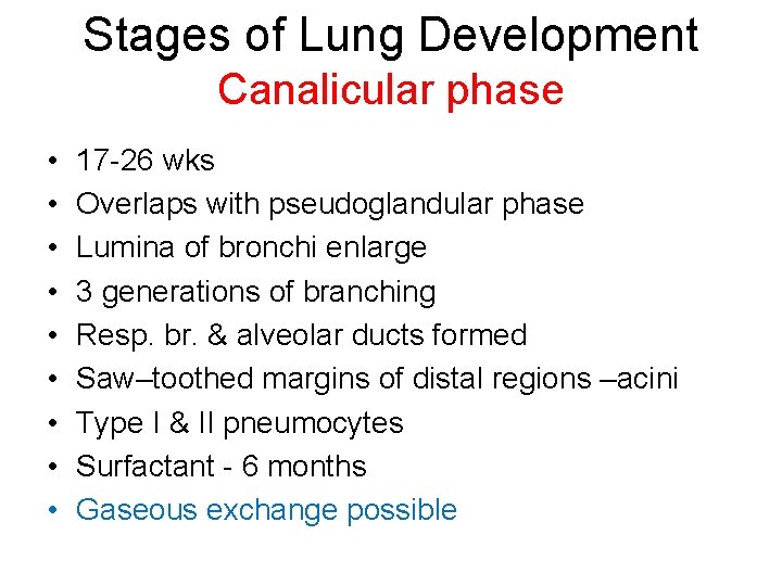
Stages of Lung Development Canalicular phase • • • 17 -26 wks Overlaps with pseudoglandular phase Lumina of bronchi enlarge 3 generations of branching Resp. br. & alveolar ducts formed Saw–toothed margins of distal regions –acini Type I & II pneumocytes Surfactant - 6 months Gaseous exchange possible
Stages of Lung Development
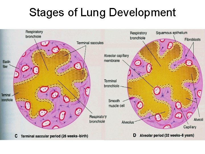
Stages of Lung Development Sacular / terminal sac phase • • 24 wks – birth More terminal sacs with thin epithelium Type I & II alveolar cells – surfactant increases Interstitial tissue decreases Cap. plexus–double cap. layer between alveoli Elastin deposited beneath ep. Lymphatics develop Fetus can survive if born
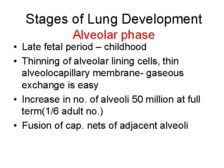
Stages of Lung Development Alveolar phase • Late fetal period – childhood • Thinning of alveolar lining cells, thin alveolocapillary membrane- gaseous exchange is easy • Increase in no. of alveoli 50 million at full term(1/6 adult no. ) • Fusion of cap. nets of adjacent alveoli
Stages of Lung Development
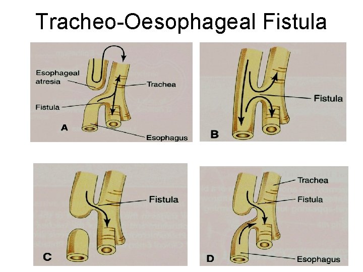
Tracheo-Oesophageal Fistula
Congenital anomalies of lungs • • Lobe of Azygous V Congenital Pul. Cysts Lung Agenesis Lung Hypoplasia Accessory Lung Abnormal fissures Abnormal lobes