GRANULOMATOUS INFLAMMATION Dr Maha Arafah Associate Professor and


 and list the cells found in granuloma along")
 and list the cells found in granuloma along")
 and list the cells found in granuloma along")

 and list the cells found in granuloma along")


 granulomatous bacterial infection that commonly affects the face and")

 2) 3) 4) 5) 6) 7) The")

 Cat-scratch disease")


 bacterial (e.")


- Slides: 41
GRANULOMATOUS INFLAMMATION Dr. Maha Arafah Associate Professor and Consultant Pathologist Office phone number: 011 - 4670360 2018
OBJECTIVES AND KEY PRINCIPLES TO BE TAUGHT: Upon completion of this lecture, the student should: � Define Granulomatous inflammation. � Recognize the morphology of granulomas (tubercles) and list the cells found in granuloma along with their appearance. � Understands the pathogenesis of granuloma formation. � Identify the two types of granulomas, which differ in their pathogenesis. � � � Foreign body granulomas Immune granulomas List the common causes of Granulomatous Inflammation.
� Define Granulomatous inflammation. Inflammation Acute inflammation Neutrophils Chronic inflammation Macrophage, Lymphocytes & Plasma cells
� Define Granulomatous inflammation. GRANULOMATOUS INFLAMMATION A form of chronic inflammation characterized by the formation of granulomas.
� Define Granulomatous inflammation. = Granuloma
� Define Granulomatous inflammation. Why is it important? Granulomas are encountered in certain specific pathologic states. Recognition of the granulomatous pattern is important because of the limited number of conditions (some life-threatening) that cause it
Recognize the morphology of granulomas (tubercles) and list the cells found in granuloma along with their appearance Granuloma = Nodular collection of epithelioid macrophages surrounded by a rim of lymphocytes Epitheloid macrophages: squamous cell-like appearance
Recognize the morphology of granulomas (tubercles) and list the cells found in granuloma along with their appearance microscopic aggregation of activated macrophages Langhans-type giant cell Granuloma Lymphocytes
Recognize the morphology of granulomas (tubercles) and list the cells found in granuloma along with their appearance Langhans Giant Cell Granuloma Lymphocytic Rim Caseous Necrosis Epithelioid Macrophage
Recognize the morphology of granulomas Section of a lymph node with caseation necrosis
Recognize the morphology of granulomas (tubercles) and list the cells found in granuloma along with their appearance The nuclei arranged either peripherally (Langhans-type giant cell) or haphazardly (foreign body-type giant cell).
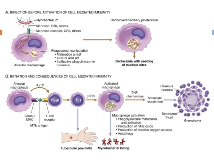
� Understands the pathogenesis of granuloma formation Granulomatous Inflammation Pathogenesis Neutrophils ordinarily remove agents that incite an acute inflammatory response. However, there are circumstances in which reactive neutrophils cannot digest the substances that provoke acute inflammation.
� Understands the pathogenesis of granuloma Granulomatous Inflammation formation Mechanism Ø What is the initiating event in granuloma formation? deposition of a indigestible antigenic material IFN-γ released by the CD 4+ T cells of the TH 1 subset is crucial in activating macrophages. Type IV hypersensitvity
� Understands the pathogenesis of granuloma formation IL-2, and IFN-γ,
� Understands the pathogenesis of granuloma formation Epithelioid cell granulomas 1. 2. When macrophages have successfully phagocytosed the injurious agent but it survives inside them. When an active T lymphocyte-mediated cellular immune response occurs. Lymphokines produced by activated T lymphocytes inhibit migration of macrophages and cause them to aggregate in the area of injury and form granulomas.
� Identify the two types of granulomas, which differ in their pathogenesis There are two types of granulomas Foreign body granuloma are incited by relatively inert foreign bodies. Typically, foreign body granulomas form when material such suture are large enough to preclude phagocytosis by a single macrophage These material do not incite any specific inflammatory immune response. The foreign material can usually be identified in the center of the granuloma, by polarized light (appears refractile). Immune granuloma are caused by insoluble particles, typically microbes, that are capable of inducing a cellmediated immune response.
� Identify the two types of granulomas, which differ in their pathogenesis Foreign body granuloma
� List the common causes of Granulomatous Inflammation Causes Non-immune granuloma � Foreign body Immune granuloma: � � Suture Bacteria � material � talc (associated with intravenous drug abuse) � � Graft � � � Parasites � � � Sarcoidosis Crohn’s disease � � Schistosomiasis Leishmaniasis Fungi � unknown Tuberculosis Leprosy Actinomycosis Cat-scratch disease Histoplasmosis Blastomycosis Metal/Dust � Berylliosis
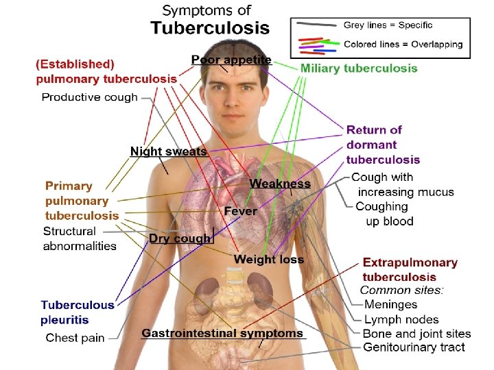
� List the common causes of Granulomatous Inflammation Tuberculosis: Mycobacterum tuberculosis Mycobacteria – ‘fungus like. . slender rods acid fast bacilli [AFB] (i. e. , they have a high content of complex lipids that readily bind the Ziehl-Neelsen [carbol fuchsin] stain and subsequently resist decolorization).
� List the common causes of Granulomatous Inflammation Pathogenesis of TB Cord factor is a glycolipid molecule found in the cell wall of Mycobacterium tuberculosis and similar species. It protects M. tuberculosis from the defenses of the host and prevents fusion between phagosomal vesicles Cord factor presence increases the production of the cytokines interleukin-12 (IL-12), IL-1β, IL -6 and TNF which are all pro-inflammatory cytokines important for granuloma formation
� List the common causes of Granulomatous Inflammation Tuberculosis
� List the common causes of Granulomatous Inflammation
� List the common causes of Granulomatous Inflammation Epitheliod histiocytes Lymphocytes Multinucleated cell Necrosis
� List the common causes of Granulomatous Inflammation Sputum , TB bacilli
Schistosomia sis
Leishmaniasis
Lepros y
Leprosy
Actinomycosis is a long-term (chronic) granulomatous bacterial infection that commonly affects the face and neck Examination of drained fluid under a microscope shows "sulfur granules" in the fluid. They are yellowish granules made of clumped organisms. filamentous, grampositive, non–acid-fast, anaerobic-tomicroaerophilic bacteria
Sarcoidosis Non-caseating granuloma
Match A and B B A 1) 2) 3) 4) 5) 6) 7) The most important cell in a. granulomatous inflammation A cytokines that is important b. in activating macrophages and c. transforming them into epithelioid cells d. Multinucleated cell in TB Antigen presenting cells e. pathogenesis of immune type granulomatous inflammation f. Microscopic finding of TB g. Found in the cell wall of TB IFN-γ Langhans cells Epitheliod histiocyes Cord factor Langerhan’s cells Type IV hypersensitivity reaction Caseating granuloma
Langerhan’s` cells Antigen presenting cells
� Which of the following diseases does not cause granulomatous inflammation a) Cat-scratch disease b) Actinomycosis c) Sarcoidosis d) Leishmaniasis e) Staphylococcus infection
What are the causes of caseous necrosis? Caseous necrosis is caused by tuberculosis, leprosy, and fungal infections.
How does caseous necrosis differ from coagulative necrosis under the microscope? In caseous necrosis, there is total loss of tissue structure, whereas in coagulative necrosis, cell outlines are retained.
What is the origin of epithelioid cells? They are transformed macrophages. Do you know the difference between granulation tissue and granulomatous inflammation? Granulation tissue contains new small blood vessels, fibroblasts, and mononuclear cells in an edematous extracellular matrix; it is part of the repair response. A granuloma is a circumscribed collection of epithelioid cells, usually surrounded by lymphocytes; it is a form of chronic inflammation.
What are the causes of granulomatous inflammation? Causes are � (1) bacterial (e. g. , Mycobacterium tuberculosis, M. leprae, Treponema pallidum) � (2) parasitic (e. g. , schistosomiasis) � (3) fungal (e. g. , histoplasmosis, blastomycosis) � (4) inorganic dusts (e. g. , silicosis, berylliosis) � (5) foreign body � (6) unknown (e. g. , sarcoidosis).
How are giant cells formed in granulomas? Giant cells are formed by fusion of macrophages. What are the other cells in a granuloma? Lymphocytes, mainly CD 4+, that caused the granulomatous reaction are present. Healing granulomas are surrounded by fibroblasts. In TB do granulomas in different organs look different? No, all granulomas look similar.
TAKE HOME MESSAGES: � Granulomatous inflammation is a distinctive pattern of chronic inflammation characterized by aggregates epithelioid macrophages Damaging stimuli which provoke a granulomatous inflammatory response include: Microorganisms which are of low inherent pathogenicity but which excite an immune response. � Granulomata are produced in response to: � ○ ○ ○ Bacterial infection parasitic infection: e. g. Schistosoma infection Certain fungi cannot be dealt with adequately by neutrophils, and thus excite granulomatous reactions. Non-living foreign material deposited in tissues, e. g. keratin from ruptured epidermal cyst. Unknown factors, e. g. in the disease 'sarcoidosis' and Crohn's diseas