Neoplasia Lecture 3 CARCINOGENESIS Dr Maha Arafah Dr


 Receptor family n")


 which is inhibitor of ABL")


 Progression of cells through cell")

")
 gene : First tumor supressor gene discovered n")

 or")


 …” Philadelphia chromosome”")

- Slides: 68
Neoplasia Lecture 3 CARCINOGENESIS Dr. Maha Arafah Dr. Abdulmalik Alsheikh, MD, FRCPC Foundation block 2012 Pathology
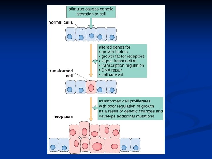
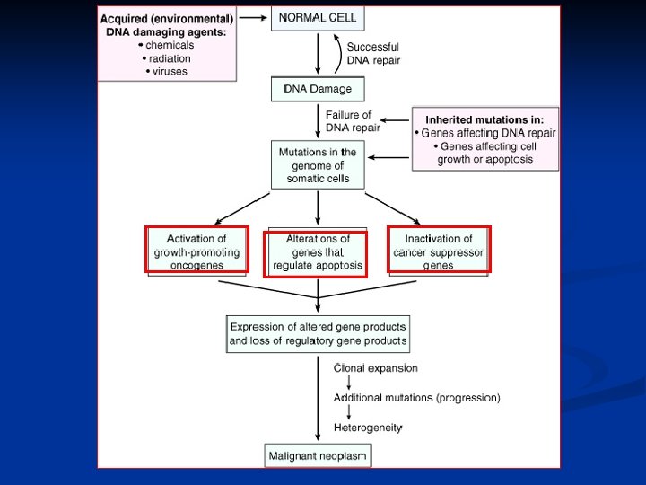
CARCINOGENESIS Carcinogenesis is a multistep process at both the phenotypic and the genetic levels. n It starts with a genetic damage: n n Environmental n Chemical n Radiation n Viral n Inhereted
Carcinogenesis Genetic damage lead to “ mutation” n single cell which has the genetic damage undergoes neoplastic proliferation ( clonal expansion) forming the tumor mass n
Carcinogenesis Where are the targets of the genetic damage? ? n Four regulatory genes are the main targets: n n Growth promoting protooncogenes n Protooncogene > mutation > oncogene Growth inhibiting (supressors) genes n Genes regulating apoptosis n DNA repair genes n
Carcinogenesis n Main changes in the cell physiology that lead to formation of the malignant phenotype: Self-sufficiency in growth signals n Insensitivity to growth-inhibitory signals n Evasion of apoptosis n Limitless replicative potential n Sustained angiogenesis n Ability to invade and metastsize n
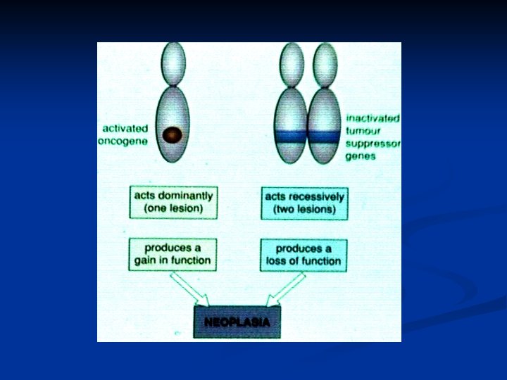
Carcinogenesis A - Self-sufficiency in Growth signals: n n Oncogene: Gene that promote autonomous cell growth in cancer cells They are derived by mutations in protooncogenes They are characterized by the ability to promote cell growth in the absence of normal growth-promoting signals Oncoproteins : are the products
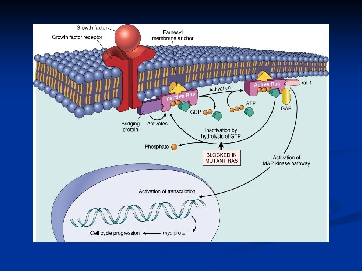
Carcinogenesis n Remember the cell cycle !! Binding of a growth factor to its receptor on the cell membrane n Activation of the growth factor receptor leading to activation of signal-transducing proteins n Transmission of the signal to the nucleus n Induction of the DNA transcription n Entry in the cell cycle and cell division n
Carcinogenesis n HOW CANCER CELLS ACQUIRE SELFSUFFICIENCY IN GROWTH SIGNALS? ?
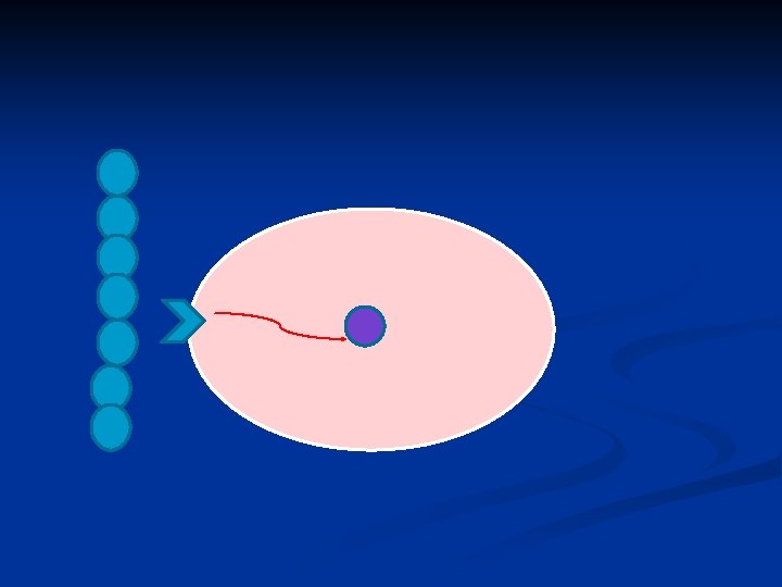
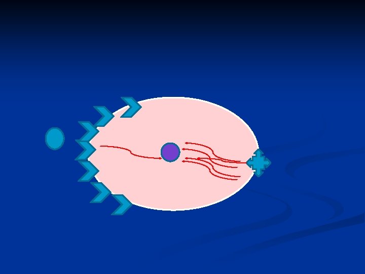
Carcinogenesis 1 - Growth factors: n Cancer cells are capable to synthesize the same growth factors to which they are responsive n E. g. Sarcomas ---- > TGF-a Glioblastoma-----> PDGF
Carcinogenesis 2 -Growth factors receptors: n n Receptors --- mutation ----continous signals to cells and uncontroled growth Receptors --- overexpression ---cells become very sensitive ----hyperresponsive to normal levels of growth factors
Carcinogenesis n Example : n Epidermal Growth Factor ( EGF ) Receptor family n HER 2 n n n Amplified in breast cancers and other tumors High levels of HER 2 in breast cancer indicate poor prognosis Anti- HER 2 antibodies are used in treatment
Carcinogenesis 3 - Signal-transducing proteins : n They receive signals from activated growth factors receptors and transmitte them to the nucleus. Examples : RAS n ABL n
Carcinogenesis n RAS : 30% of all human tumors contain mutated RAS gene. E. g : colon. Pancreas cancers n Mutations of the RAS gene is the most common oncogene abnormality in human tumors n Mutations in RAS --- cells continue to proliferate n
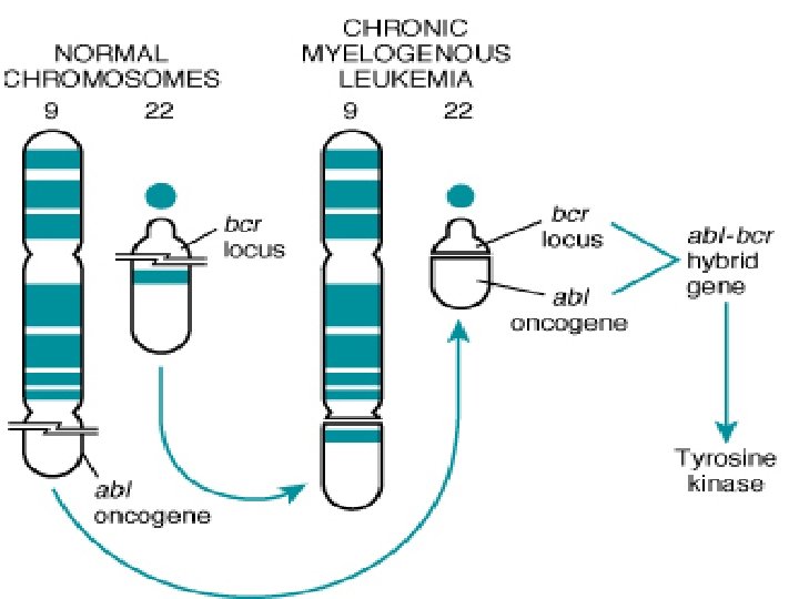
Carcinogenesis n ABL gene ABL protooncogene has a tyrosine kinase activity n Its activity is controlled by negative regulatory mechanism n E. g. : chronic myeloid leukemia ( CML ) : n n t( 9, 22) ---ABL gene transferred from ch. 9 to ch. 22 n Fusion with BCR ---> BCR-ABL n BCR-ABL has tyrosine kinase acttivity ---( oncogenec)
Carcinogenesis n CML patients are treated with ( Gleevec) which is inhibitor of ABL kinase
Carcinogenesis 4 - Nuclear transcription factors : Mutations may affect genes that regulate transcription of DNA growth autonomy n E. g. MYC n n MYC protooncogene produce MYC protein when cell receives growth signals n MYC protein binds to DNA leading to activation of growth-related genes
Carcinogenesis Normally … MYC decrease when cell cycle begins …but. . in tumors there is sustained expression of MYC continuous proliferation n E. g. Burkitt Lymphoma ; MYC is dysregulated due to t( 8, 14) n
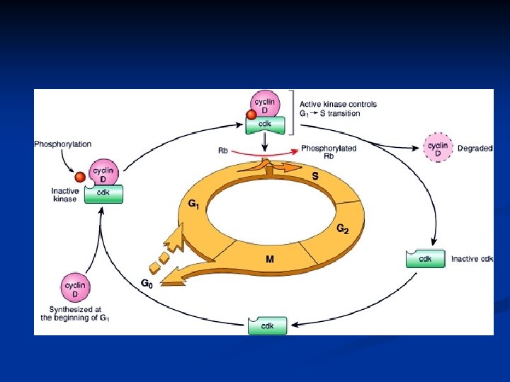
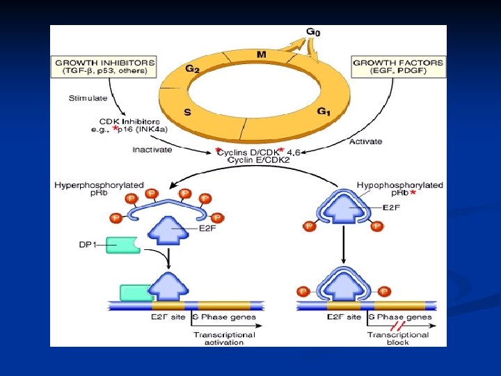
Carcinogenesis 5 - Cyclins and cyclins- dependent kinases (CDKs) Progression of cells through cell cycles is regulated by CDKs after they are activated by binding with cyclins n Mutations that dysregulate cyclins and CDKs will lead to cell proliferation …e. g. n n Cyclin D genes are overexpressed in breast, esophagus and liver cancers. n CDK 4 is amplified in melanoma and sarcomas
Carcinogenesis n Main changes in the cell physiology that lead to formation of the malignant phenotype: A- Self-sufficiency in growth signals B- Insensitivity to growth-inhibitory signals C- Evasion of apoptosis D- Limitless replicative potential E- Sustained angiogenesis F- Ability to invade and metastsize
Carcinogenesis 2. Insensitivity to growth-inhibitory signals n Tumor suppressor genes control ( apply brakes) cells proliferation n If mutation caused disruption to them cell becomes insensitive to growth inhibition uncontrolled proliferation n Examples: RB, TGF-b, APC, P 53
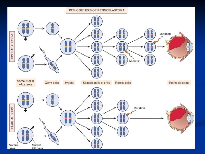
Carcinogenesis n RB ( retinoblastoma ) gene : First tumor supressor gene discovered n It was discovered initially in retinoblastomas n Found in other tumors, e. g. breast ca n RB gene is a DNA-binding protein n RB is located on chromosome 13 n
Carcinogenesis RB gene exists in “ active “ and “ inactive” forms n If active will stop the advancing from G 1 to S phase in cell cycle n If cell is stimulated by growth factors inactivation of RB gene brake is released cells start cell cycle …G 1 S M …then RB gene is activated again n
Carcinogenesis Retinoblastoma is an uncommon childhood tumor n Retinoblastoma is either sporadic (60%) or familial ( 40% ) n Two mutations required to produce retinoblastoma n Both normal copies of the gene should be lost to produce retinoblastoma n
Carcinogenesis n Transforming Growth Factor- b pathway: TGF-b is an inhibitor of proliferation n It regulate RB pathway n Inactivation of TGF-b lead to cell proliferation n Mutations in TGF-b pathway are present in : 100% of pancreatic cancers 83% of colon cancers
Carcinogenesis n Adenomatous Polyposis Coli – b Catenin pathway: APC is tumor supressor gene n APC gene loss is very common in colon cancers n It has anti-proliferative action through inhibition of b-Catenin which activate cell proliferation n Individuals with mutant APC develop thousands of colonic polyps n
Adenomatous Polyposis Coli
Carcinogenesis One or more of the polyps will progress to colonic carcinoma n APC mutations are seen in 70% to 80% of sporadic colon cancers n
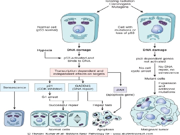
Carcinogenesis n P 53 It has multiple functions n Mainly : n n Tumor suppressor gene ( anti-proliferative ) n Regulates apoptosis
Carcinogenesis P 53 senses DNA damage n Causes G 1 arrest to give chance for DNA repair n Induce DNA repair genes n If a cell with damaged DNA cannot be repaired, it will be directed by P 53 to undergo apoptosis n
Carcinogenesis With loss of P 53, DNA damage goes unrepaired n Mutations will be fixed in the dividing cells, leading to malignant transformation n
Carcinogenesis P 53 is called the “ guardian of the genome” n 70% of human cancers have a defect in P 53 n It has been reported with almost all types of cancers : e. g. lung, colon, breast n In most cases, mutations are acquired, but can be inhereted, e. g : Li-Fraumeni syndrome n
Carcinogenesis n Main changes in the cell physiology that lead to formation of the malignant phenotype: A- Self-sufficiency in growth signals B- Insensitivity to growth-inhibitory signals C- Evasion of apoptosis D- Limitless replicative potential E- Sustained angiogenesis F- Ability to invade and metastsize
Carcinogenesis n Evasion of apoptosis: Mutations in the genes regulating apoptosis are factors in malignant transformation n Cell survival is controlled by genes that promote and inhibit apoptosis n
Evasion of apoptosis n n Reduced CD 95 level inactivate death – induced signaling cascade that cleaves DNA to cause death tumor cells are less susceptible to apoptosis DNA damage induced apoptosis (with the action of P 53 ) can be blocked in tumors
Carcinogenesis n Main changes in the cell physiology that lead to formation of the malignant phenotype: A- Self-sufficiency in growth signals B- Insensitivity to growth-inhibitory signals C- Evasion of apoptosis D- Limitless replicative potential E- Sustained angiogenesis F- Ability to invade and metastsize
n Limitless replicative potential: n n n Normally there is progressive shortening of telomeres at the ends of chromosomes Telomerase is active in normal stem cells but absent in somatic cells In tumor cells : activation of the enzyme telomerase, which can maintain normal telomere length
Carcinogenesis n Main changes in the cell physiology that lead to formation of the malignant phenotype: A- Self-sufficiency in growth signals B- Insensitivity to growth-inhibitory signals C- Evasion of apoptosis D- Limitless replicative potential E- Sustained angiogenesis F- Ability to invade and metastsize
Carcinogenesis n Sustained angiogenesis n Neovascularization has two main effects: n Perfusion supplies oxygen and nutrients n Newly formed endothelial cells stimulate the growth of adjacent tumor cells by secreting growth factors, e. g : PDGF, IL-1 n Angiogenesis is required for metastasis
n How do tumors develop a blood supply? Tumor-associated angiogenic factors n These factors may be produced by tumor cells or by inflammatory cells infiltrating the tumor e. g. macrophages n Important factors : n n Vascular endothelial growth factor( VEGF ) n Fibroblast growth factor
Carcinogenesis n Main changes in the cell physiology that lead to formation of the malignant phenotype: A- Self-sufficiency in growth signals B- Insensitivity to growth-inhibitory signals C- Evasion of apoptosis D- Limitless replicative potential E- Sustained angiogenesis F- Ability to invade and metastsize
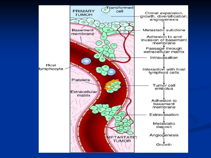
Carcinogenesis n Ability to invade and metastsize: n Two phases : n Invasion of extracellular matrix n Vascular dissimenation and homing of tumor cells
Carcinogenesis n Invasion of ECM: Malignant cells first breach the underlying basement membrane n Traverse the interstitial tissue n Penetrate the vascular basement membrane n Gain access to the circulation n v Invasion of the ECM has four steps:
1. Detachment of tumor cells from each other
2. Attachments of tumor cells to matrix components
3. Degradation of ECM by collagenase enzyme
4. Migration of tumor cells
Carcinogenesis n Vascular dissemination and homing of tumor cells: May form emboli n Most travel as single cells n Adhesion to vascular endothelium n extravasation n
Carcinogenesis n Main changes in the cell physiology that lead to formation of the malignant phenotype: A- Self-sufficiency in growth signals B- Insensitivity to growth-inhibitory signals C- Evasion of apoptosis D- Limitless replicative potential E- Sustained angiogenesis F- Ability to invade and metastsize
Genomic Instability Enabler of malignancy n Due to defect in DNA repair genes n Examples: n Hereditary Nonpolyposis colon carcinoma(HNPCC) n Xeroderma pigmentosum n Familial breast cancer n
Genomic Instability n Familial breast cancer: Due to mutations in BRCA 1 and BRCA 2 genes n These genes regulate DNA repair n Account for 80% of familial breast cancer n They are also involved in other malignancies n
Molecular Basis of multistep Carcinogenesis Cancer results from accumulation of multiple mutations n All cancers have multiple genetic alterations, involving activation of several oncogenes and loss of two or more tumor suppressor genes n
Molecular Basis of multistep Carcinogenesis
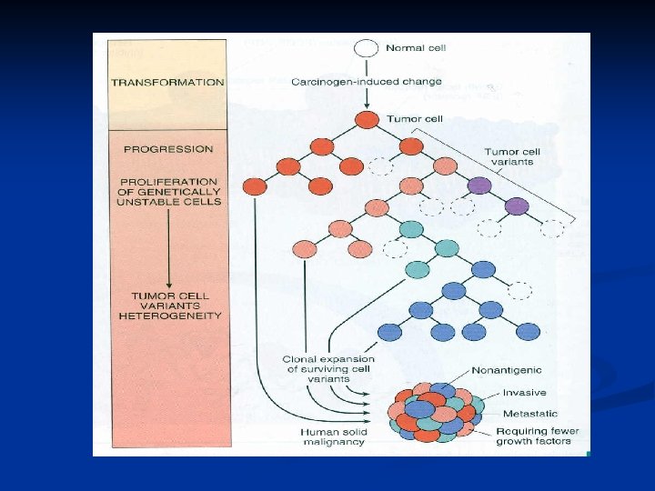
Tumor progression Many tumors become more aggressive and acquire greater malignant potential…this is called “ tumor progression” …not increase in size!! n By the time, the tumor become clinically evident, their constituent cells are extremely heterogeneous n
Karyotypic Changes in Tumors n Translocations: In CML : t(9, 22) …” Philadelphia chromosome” n In Burkitt Lymphoma : t(8, 14) n In Follicular Lymphoma : t(14, 18) n Deletions n Gene amplification: n n Breast cancer : HER-2
Translocations Gene amplification