
Pathology of infectious diseases LEC 4 Learning objectives
Pathology of infectious diseases LEC. 4 ﺍﻳﻤﺎﻥ ﺳﻌﻮﺩ ﺧﻠﻴﻔﺔ. ﺩ
Learning objectives: You should: - recognize parasitic infection and its causative microorgans, mode of transmission, types and clinical manifestation. know fungal infection and its classification. Understand HIV infection, its pathogenesis, clinical phases and associated opportunistic infections and neoplasms.
Parasites: PROTOZOA ( single cell) Plasmodium (malaria) � Leishmania � Entamoeba � Trypanosoma � Toxoplasma � Giardia � META-ZOA (HELMINTHS) Echinococcus Granulosus � Schistosomiasis. � ECTO-PARASITES i. e. ARTHROPODS
Mucocutaneous leishmaniasis: Caused by leishmania Brasiliensis. � The lesions are at the mucocutaneous � junction e. g nose, upper lip. The lesion doesn’t heal spontaneously, but it � progresses and erodes the skin, mucous membrane and even the cartilage causing a gross disfigurement. Mic. : granuloma formation. �

Mucocutaneous leishmania

Visceral leishmaniasis: The disease is called Kal-azar which means the (black � fever). Caused by leishamania Donovani. � The m. o disseminate throughout the lymphoreticular system. � Clinically : hepatosplenomegaly, fever, LAP. � Mic. : Biopsy from the lesion shows L-D bodies, (leishmania � donovani) Outcome: it is fatal and may lead to associated bacterial � infection.
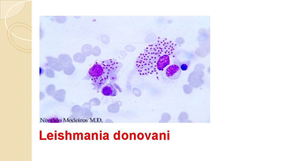
Leishmania donovani
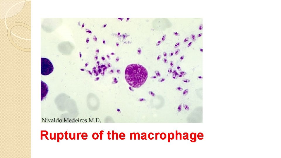
Rupture of the macrophage
Amoebiasis: Cause: Entamoeba histolytica. � Site: � 1 - The colon which manifest as amoebic dysentery. 2 Extraintestinal which manifest itself as liver and lung abscesses.
Intestinal amoebiasis Mode of transmission: contaminated food and water that � contain the E. histolytica cyst. Morphologically: the characteristic lesion in the colon is the � flask shape ulcer, caused by the trophozoite. Mic. : mucosal necrosis, invasion of the submucosa &spread � laterally. Diagnosis: GSE (general stool exam)……to see the � trophozoite in case of active disease, or the cyst in healthy carrier. Clinically: fever &diarrhea. � Complications: perforation &peritonitis. �
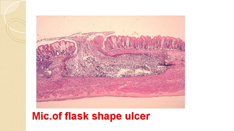
Mic. of flask shape ulcer
Hepatic Amoebisis: Passage of the trophozoite from the colon to the � liver via the portal vein will cause Amoebic liver abscess. Grossly: � The abscess is pale , shaggy with necrotic rim and contains red-brown thick fluid composed of dead hepatocytes+ blood+ fibrin…. . giving the characteristic Anchovy sauce Clinically : Fever hepatosplenomegaly, high � WBC titer.
Complications: Rupture of the abscess to the: Peritoneum. � Pleura. � Pericardium. � Diaphragm lung abscess. �
Giardiasis: Caused by Giardia Lamblia. � Route of infection: Contaminated food and water by giardia � cyst. Site: Duodenum & small intestine. � The cyst will hatch into a trophozoite that adheres to the � mucosal surface without penetrating it. � - Villous atrophy -chronic inflammatory cells infiltrating the area (lymphocytes and plasma cells) -trophozoite can be detected near the mucosal surface.
Clinically: most patients are Asymptomatic, 10% develop chronic diarrhea. Diagnosis: GSE to detect the cyst. Intestinal biopsy To detect the trophozoite
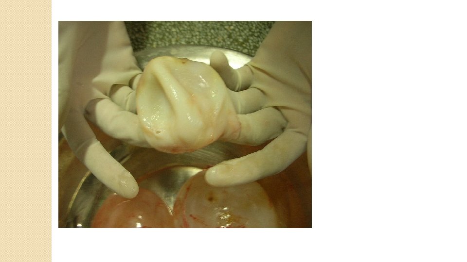
Hydatid cyst: Caused: by Echinococcus Granulosus larvae. � Man is considered as the intermediate host , dog is the � final host having the adult worm & shed the eggs onto the grass or green plants, the eggs will be ingested by man which will hatch and pass from the intestine through the portal system to the liver and other organs.
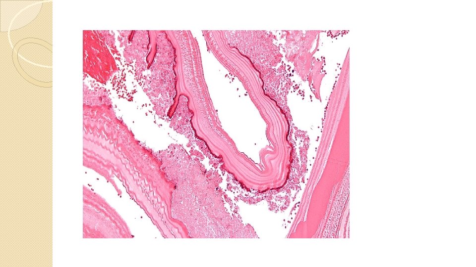
Morphology: The cyst is composed of: � 1 - Outer fibrous capsule. 2 - Acellular white laminated layer. 3 - Thin cellular (germinal) layer which give rise to scolices. Rupture of the cyst will release the scolices which will � form new cyst from each scolex. Effects of the cyst: Organ compression. � Rupture of the cyst which will cause sever allergic � (Anaphylactic ) reaction.
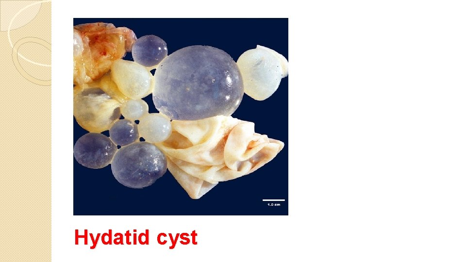
Hydatid cyst
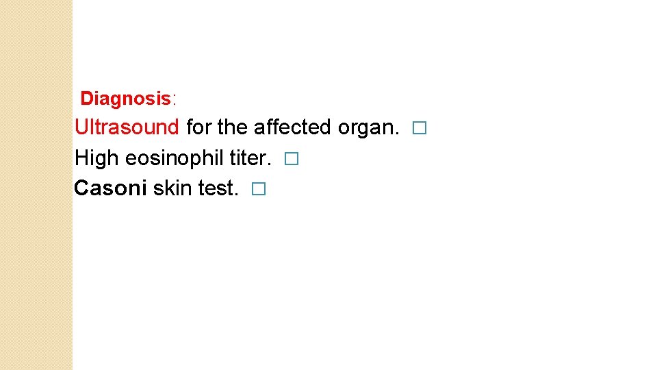
Diagnosis: Ultrasound for the affected organ. � High eosinophil titer. � Casoni skin test. �
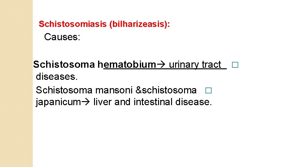
Schistosomiasis (bilharizeasis): Causes: Schistosoma hematobium urinary tract � diseases. Schistosoma mansoni &schistosoma � japanicum liver and intestinal disease.
Route of infection: The bilharizeal ovum pass in the urine or feces � contamination of water hatch the cercaria which will enter the skin and pass to the blood stream reaching either the urinary bladder or intestine and liver. In the affected organs, the parasite is attached to the � endothelium of the blood vessels in which it will shed the eggs that pass with the stool or urine.
Clinicopathology: The first lesion occurs at the site of entry of the ovum � through the skin rash which is called swimmer itch. Constitutional signs and symptoms including fever, � hepatosplenomegaly, diarrhea, cough…. etc. at the site of egg shedding (urinary bladder, intestine, � liver…) there is inflammatory reaction e. g cystitis…. associated with hematuria, colitis …. associated with diarrhea.
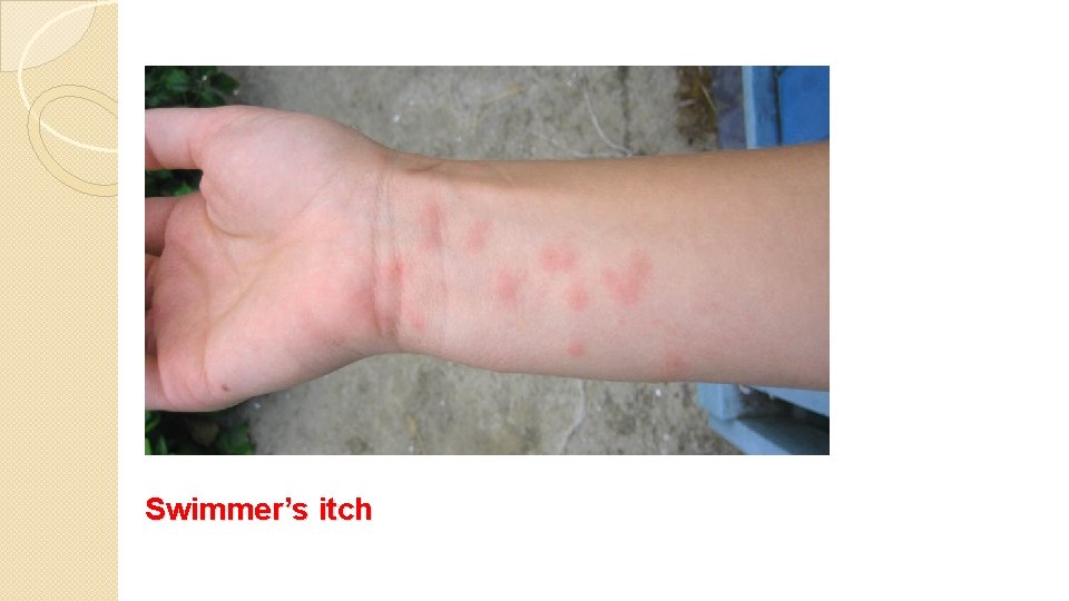
Swimmer’s itch
At the liver there will be a granuloma formation � around the implanted eggs which will heal by fibrosis , this will cause obstruction of the portal system causing the shift of blood to the systemic circulation ending in esophageal varices hematamesis (vomiting of blood).
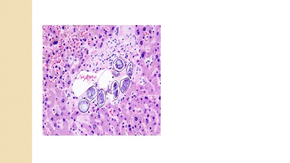
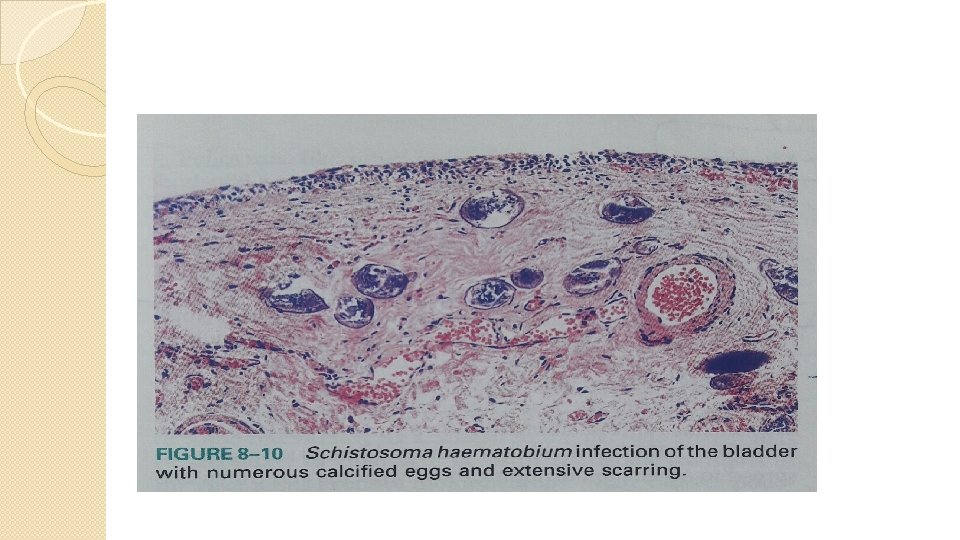
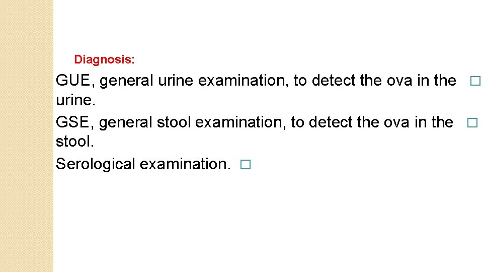
Diagnosis: GUE, general urine examination, to detect the ova in the � urine. GSE, general stool examination, to detect the ova in the � stool. Serological examination. �
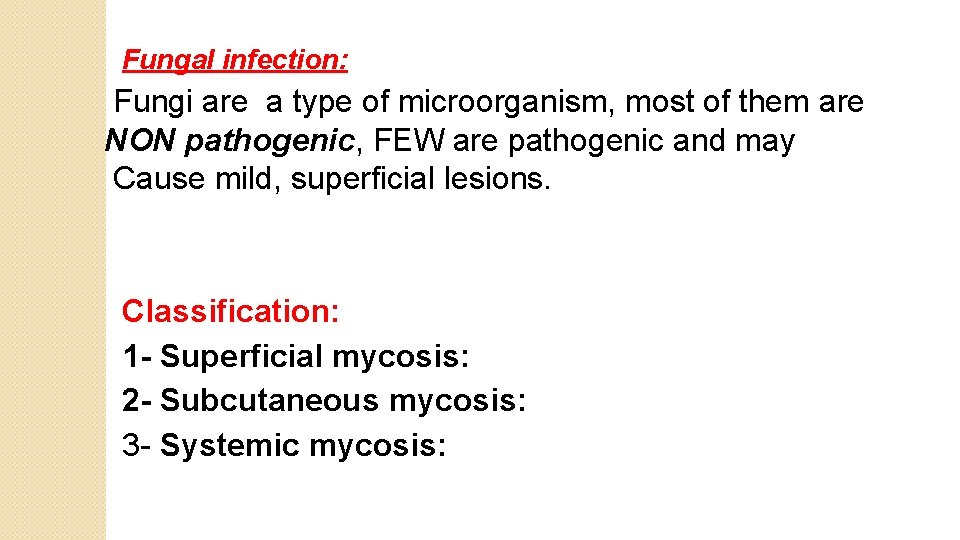
Fungal infection: Fungi are a type of microorganism, most of them are NON pathogenic, FEW are pathogenic and may Cause mild, superficial lesions. Classification: 1 - Superficial mycosis: 2 - Subcutaneous mycosis: 3 - Systemic mycosis:
1 - Superficial mycosis: The fungus infiltrate the cornified layer of the skin causing sever tissue destruction to the epidermis &skin appendages. e. g Tinea, Candida. 2 - Subcutaneous mycosis: Following inoculation of the fungus by a traumatic implantation. The host reaction is either: Suppuration (acute reaction). � Epitheloid granuloma (chronic). e. g mycetoma. �
3 - Systemic mycosis: Most of them will cause respiratory diseases; the mode of transmission is by Inhalation leads to pulmonary lesion, which will disseminate through the lymphatic system and may cause visceral involvement. e. g Histoplasmosis , mucormycosis , aspergillosis.
Candidiasis: Candida albicans: It is a commensal microorganism normally present in the mouth, intestine, moist skin. Any clinical condition which cause decrease in the immunity will activate the Candida &change it into pathogenic fungus. e. g on such conditions, diabetes mellitus , leukemia, pregnancy, patient on immunosuppressive treatment.
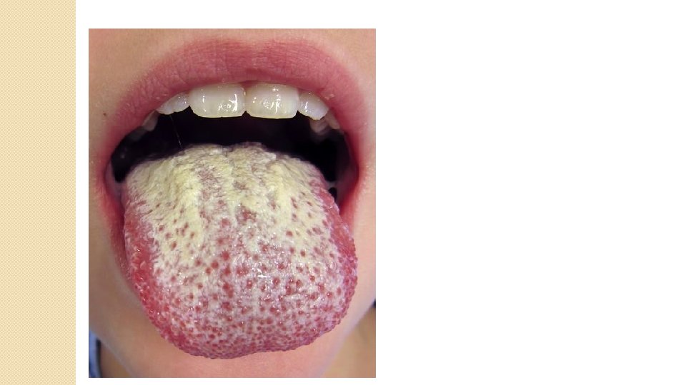
Types of Candida infection: 1) Mucosal infection: Seen as white dirty looking pseudomembrane on the oral cavity composed of matted organisms &inflammatory debri. In the vagina, it is usually associated with intense itching &curdlike discharge. In the esophagus , the white plaques &pseudomembrane on the esophageal mucosa cause dysphagia &retrosternal pain
2)Mucocutaneous infection: The lesion here is more destructive involving the perineum (diaper rash), nail folds &hair follicles, mammary folds, webs of the fingers. The lesion will show hyperplastic epithelium &chronic granulomatous inflammation underneath it.
3) Systemic or invasive candidiasis : Blood borne dissemination of the organsms to various tissues Seen in cases with decrease immunity. The organs involved are: heart valves (in prosthetic valves or intravenous drug abusers), lung (candida pneumonia), liver (hepatic abscess) and kidney (renal abscess), brain (meningitis).
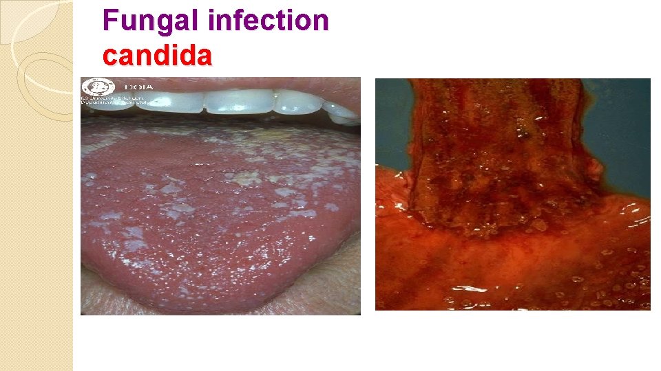
Fungal infection candida
Aspergillosis: It causes respiratory & systemic infections, which occur almost exclusively in Immunocompromised individuals. Pulmonary lesions include: 1 - Bronchopneumonia. 2 - Abscess. 3 - Small infarcts caused by invasion of the small blood vessels by the fungus leading to its obstruction.
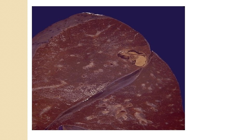
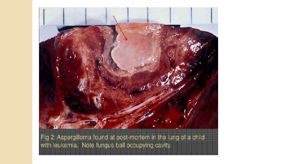
4 - Fungus ball formation which is called (Aspergilloma), it is a mass of aspergillous fungus contained in a new or a pre-existing cavity (lung cyst or post tuberculous cavity), act as ball valves occluding the cavity predisposing to infection &hemoptysis 5 - Chronic granulomatous inflammation. 6 - Allergic bronchopulmonary aspergillosis in asthma with exacerbation of symptoms due to hypersensitivity reaction against the fungi.
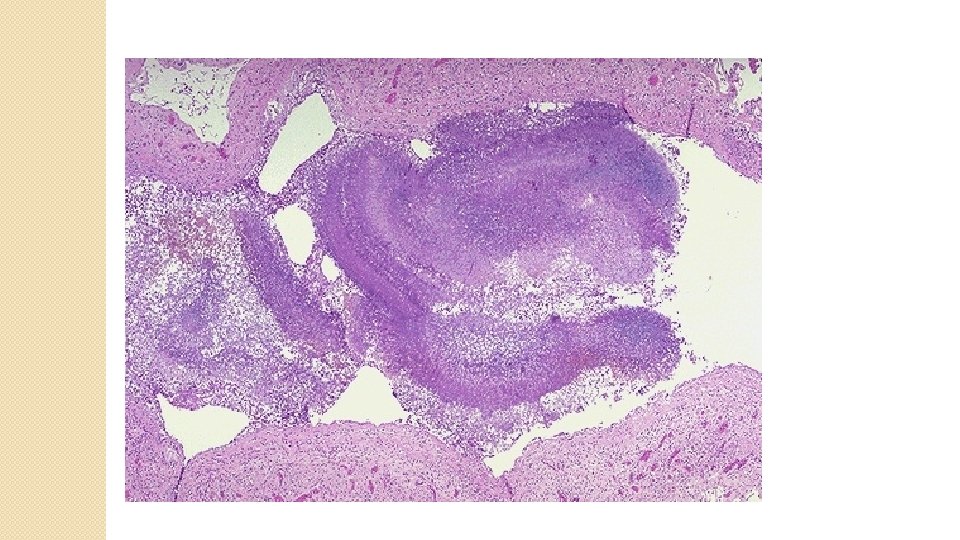
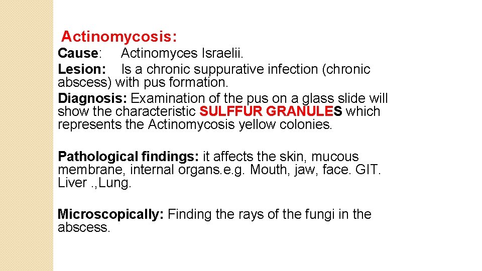
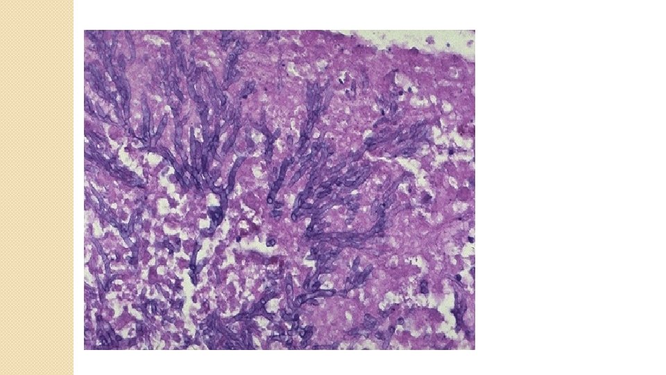
Actinomycosis: Cause: Actinomyces Israelii. Lesion: Is a chronic suppurative infection (chronic abscess) with pus formation. Diagnosis: Examination of the pus on a glass slide will show the characteristic SULFFUR GRANULES which represents the Actinomycosis yellow colonies. Pathological findings: it affects the skin, mucous membrane, internal organs. e. g. Mouth, jaw, face. GIT. Liver. , Lung. Microscopically: Finding the rays of the fungi in the abscess.
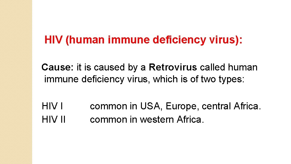
HIV (human immune deficiency virus): Cause: it is caused by a Retrovirus called human immune deficiency virus, which is of two types: HIV I common in USA, Europe, central Africa. HIV II common in western Africa.
Structure of the virus: The virus is composed of: 1 - Two RNA strands. 2 - Reverse transcriptase enzyme. 3 - An outer envelope from the host cell membrane
Transmission: Sexually transmitted (homosexual or bisexual males � &heterosexual contacts of high risk groups). Parenteral route (I. V drug abusers, hemophiliacs, blood � transfusion) From mother to baby (through the placenta, intrapartum � during delivery or ingestion of infected breast milk).
Pathogenesis: At the contact surface which is usually a mucosal surface, the macrophages will get the virus, then they will secrete certain mediators result in accumulation of CD 4 +ve T- lymphocytes, then the virus will get inside the CD 4+ cells and start to use its reverse transcriptase enzyme to form proviral DNA & get incorporated into the host DNA result in formation of complete viral particles that bud from the cell membrane & will direct the cell for producing more HIV particles.
Outcome: 1 - Decrease CD 4+ helper T lymphocytes. 2 - Increase CD 8+ cytotoxic T lymphocytes. 3 - Impaired cell mediated immunity. 4 - Impaired humoral immunity. 5 - Abnormal macrophage function, decrease in chemotactic substances. If the virus remains inside the cell then this is called LATENT infection. If the virus replicate and release a new viruses cell rupture and death i. e loss of the CD 4 + cells , this is called PRODUCTIVE infection.
Natural history of HIV infection: Three phases: Early acute phase � Middle chronic phase � Final crisis phase ◦
AIDS: (acquired immune deficiency syndrome) It consists of groups of clinical signs and symptoms which will appear when the lymphocyte count drops to < 200 / microlit. In which the characteristic opportunistic infections and neoplasms of AIDS will appear.
Opportunistic infections: Protozoal infection (Pneumocystis carinii, toxoplasmosis) � Viral infection (cytomegalovirus, herpes simplex, varicella � zoster Bacterial infection (mycobacterium T. B, nocardiosis � &salmonella) Fungal infection (Candida, Histoplasma, Cryptococcus) � GIT protozoal infections. �
Malignant neoplasms: Patients with AIDs have increased risk of malignancy because of: - Profound defects in T cell immunity. - Dysregulated B cell &monocyte functions. - Multiple infections with known (e. g herpesvirus type 8, EBV, human papilloma virus) &unknown viruses.
these neoplasms include: Kaposi sarcoma: it is a vascular tumor appears as � purple nodule over the skin. Malignant lymphoma, mainly of the non Hodgkin type � (diffuse large cell type of B cell origin) Primary lymphoma of the brain. � Invasive cancer of uterine cervix due to increase � prevalence of human papilloma virus.
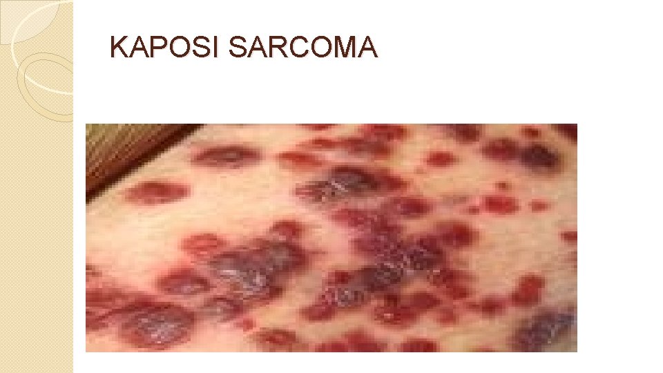
KAPOSI SARCOMA

Thanks for attention
- Slides: 57