Glycolysis 5903 Pathway overview 1 Add phosphoryl groups


 Pyruvate decarboxylase requires thiamine pyrophosphate TPP as")


![An increase in flux, DJ causes [A] to increase and when [A] increases the](https://slidetodoc.com/presentation_image_h/19f704ee207886cd177e2228b396279c/image-30.jpg "An increase in flux, DJ causes [A] to increase and when [A] increases the")
 is a measure of the sensitivity of a reactions fractional change in flux")


- Slides: 41
Glycolysis 5/9/03
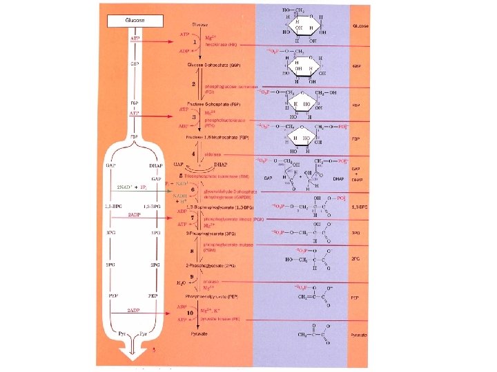
Pathway overview 1. Add phosphoryl groups to activate glucose. 2. Convert the phosphorylated intermediates into high energy phosphate compounds. 3. Couple the transfer of the phosphate to ADP to form ATP. Stage I A preparatory stage in which glucose is phosphorylated and cleaved to yield two molecules of glyceraldehyde-3 phosphate - uses two ATPs Stage II glyceraldehyde-3 -phosphate is converted to pyruvate with the concomitant generation of four ATPs-net profit is 2 ATPs per glucose. Glucose + 2 NAD+ + 2 ADP +2 Pi 2 NADH + 2 pyruvate + 2 ATP + 2 H 2 O + 4 H+
Front half of glycolysis
Phosphoglycerate Kinase: First ATP generation step + ADP 1, 3 BPG + ATP 3 PG
GAP + Pi + NAD+ 1, 3 -BPG + NADH + 6. 7 k. J • mol-1 1, 3 BPG + ADP 3 PG + ATP -18. 8 k. J • mol-1 GAP+Pi+NAD+ +ADP 3 PG+NADH+ATP -12. 1 k. J • mol-1
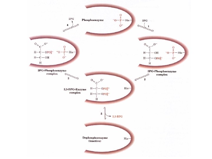
Phosphoglycerate mutase 2 PG 3 PG 2, 3 BPG
Phosphoglycerate mutase requires a phosphorylated form of the enzyme to be active. Only 2, 3 BPG can phosphorylate the unphosphorylated enzyme. Phospho Histidine residue
Glycolysis influences oxygen transport
Oxygen saturation curves in erythrocytes
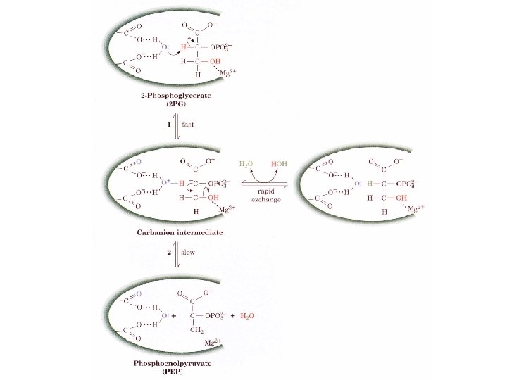
Enolase generation of a second “high energy” intermediate + H 2 O 2 Phosphoglycerate Phosphoenol pyruvate
Pyruvate kinase: Second ATP generation step
The second half of glycolysis
The metabolic fate of pyruvate
The need to regenerate NAD+ from NADH A. Homolactic fermentation: conversion of pyruvate to lactate LDH Pyruvate NADH L-Lactate NAD+ Mammals have two different types of enzymes: Isozymes M type for muscle H type for heart
Lactate dehydrogenase is a tetramer H 4 has a low Km for pyruvate and is allosterically inhibited by high concentrations of pyruvate. M 4 has a higher Km for pyruvate and is not allosterically regulated Although all five types can exist, H 4, H 3 M, H 2 M 2 HM 3, M 4 The M predominates in anaerobic muscle tissues which favor the formation of lactate while the H 4 form predominates in aerobic tissues like heart where the formation of pyruvate from lactate is preferred
Pro-R hydride is transferred from C 4 of NADH to C 2 of pyruvate with the concomitant transfer of a proton from His 195 All muscle lactate is transferred to the liver where it is turned back to glucose
Alcoholic fermentation A two step process: 1) Pyruvate decarboxylase requires thiamine pyrophosphate TPP as a cofactor. 2) Alcohol dehydrogenase requires Zn+2 as a cofactor
Thiamine pyrophosphate The build up of negative charges seen in decarboxylation reactions on the carbonyl atom in the transition state is unstable and TPP helps stabilize the negative charge
Reaction mechanism of pyruvate decarboxylation 1. Nucleophilic attack by the ylid from of TPP on the carbonyl 2. Departure of CO 2 and resonance-stabilization of the carbanion. 3. Protonation of the carbanion 4. Elimination of TPP ylid to form acetaldehyde
Long distance hydrogen bonding and general acid catalysis from Glu 51 with the aminopyrimidine ring leads to the formation of the ylid form of TPP.
Deficiencies of TPP lead to Beriberi Vitamin B 1 Beriberi was prevalent in the rice consuming countries of the Orient where polished rice is preferred. TPP is found in the brown outer layers of rice. Neurological atrophy, cardiac failure, endema nowadays found in alcoholics who would rather drink than eat.
Alcohol dehydrogenase
Energetics of Fermentation DG ' Glucose 2 lactate + 2 H+ -196 k. J • mol-1 of glucose Glucose 2 CO 2 + 2 ethanol -235 k. J • mol-1 of glucose Formation of 2 ATP +61 k. J • mol-1 of glucose equals 31% and 26% efficient for energy conservation Under physiological conditions this efficiency approaches 50%
Glycolysis is for rapid ATP production Glycolysis is about 100 times faster than oxidativephosphorylation in the mitochondria Fast twitch muscles - short blasts of energy and are nearly devoid of mitochondria use exclusively glycolysis for ATP Slow twitch muscles are dark red, rich in mitos obtain ATP from OX-phos. , i. e. flight muscles of migratory birds and the muscles of long distance runners
Control of Metabolic Flux At equilibrium DJ = 0 and far from equilibrium DJ=vf The flux throughout the pathway is constant at steady state conditions and control of flux requires: 1) The flux-generating step varies with the organisms metabolic needs 2). The change in flux is felt throughout the pathway
A diagrammatic representation of substrate cycling and control of flux
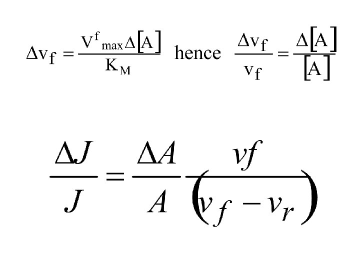
An increase in flux, DJ causes [A] to increase and when [A] increases the is a Dvf So DJ = Dvf Plugging in the Michaelis -Menton equation
vf/(vf-vr) is a measure of the sensitivity of a reactions fractional change in flux to its fractional change in substrate concentration 1. In an irreversible reaction, vr approaches 0 and vf/(vf-vr) approaches 1. The reaction is therefore requires a nearly fractional increase in its substrate concentration order to respond to a fractional increase in flux 2. As a reaction approaches equilibrium, vr approaches vf and vf/(vf-vr) approaches infinity. The reaction’s response to a fractional increase in flux therefore requires a much smaller fractional increase in its substrate concentrations.
Flux is controlled at the rate limiting step Usually the product is removed much faster than it is formed so that the rate-determining step is far from equilibrium. Because of the fractional change in the flux DJ/J when vf>>vr is directly proportional to the change is substrate concentrations other mechanisms are needed to achieve factors of over 100 as seen in glycolysis. • Allosteric regulation • Covalent modification • Substrate cycling • Genetic control
Covalent modification-Protein phosphorylation
Three steps to elucidate common controlling mechanisms in a pathway 1. Identify the rate determining steps: Those with a large negative DG and measure flux through the pathway and each step with inhibitors. 2. Identify In vitro allosteric modifiers of the pathway study each enzymes kinetics, mechanisms and inhibition patterns. 3. Measure in vivo levels of modulators under conditions consistent with a proposed control mechanism
Free energy changes in glycolysis Reaction enzyme DG ´ DG 1 Hexokinase -20. 9 -27. 2 2 PGI +2. 2 -1. 4 3 PFK -17. 2 -25. 9 4 Aldolase +22. 8 -5. 9 5 TIM +7. 9 +4. 4 6+7 8 GAPDH+PGK PGM -16. 7 +4. 7 -1. 1 -0. 6 9 Enolase -3. 2 -2. 4 10 PK -23. 3 -13. 9
Only three enzymes function with large negative DG’s Hexokinase, Phosphofructokinase and pyruvate kinase The other enzymes operate near equilibrium and their rates are faster than the flux through the pathway. Specific effectors of Glycolysis Enzymes Inhibitors Hexokinase PFK G 6 P ATP, citrate, PEP Activators none ADP, AMP, c. AMP FBP, F 2, 6 BP, F 6 P NH 4, Pi Pyruvate kinase ATP none
PFK: the major flux controlling enzyme of glycolysis in muscle
PFK activity as a function of G 6 P
AMP concentrations not ATP control glycolysis ATP concentrations only vary about 10% from resting to active cells. [ATP] is buffered by creatine phosphate and adenylate kinase. 2 ADP ATP + AMP A 10% decrease in ATP produces a four fold increase in AMP because ATP = 50 AMP in muscle. AMP activates PFK by the action of adenylate kinase.
Substrate cycling Fructose-6 phosphate +ATP Fructose 1, 6 -bisphosphate Fructose-6 phosphate + Pi The net result is the breakdown of ATP. Two different enzymes control this pathway PFK and Fructose 1, 6 bisphosphatase. If these both were not controlled a futile cycle would occur. Specific effectors of Glycolysis Enzymes Inhibitors Activators Phosphatase AMP ATP, citrate