Epilepsy Surgery Stephan Eisenschenk MD Department of Neurology

- Slides: 52
Epilepsy Surgery Stephan Eisenschenk, MD Department of Neurology
Candidates for Epilepsy Surgery Persistent seizures despite appropriate pharmacological treatment (usually at least two drugs at limits of tolerability) Impairment of quality of life due to ongoing seizures S-Slide 2 Stephan Eisenschenk, MD Department of Neurology
Problems of intractable epilepsy • Potential for injury • Socially disabling – Employment (and schooling) – Driving – Living independently – Stigmatization Stephan Eisenschenk, MD Department of Neurology
Epilepsy Surgery • As many as 100, 000 patients with epilepsy in the United States are potential surgical candidates • ~ 5, 000 new potential cases occur each year • 1, 500 epilepsy surgeries are performed each year Stephan Eisenschenk, MD Department of Neurology
When should surgery be considered? • After adequate trials of 2 -3 good antiepileptic medications • The type of epilepsy and initial testing may influence this decision Stephan Eisenschenk, MD Department of Neurology
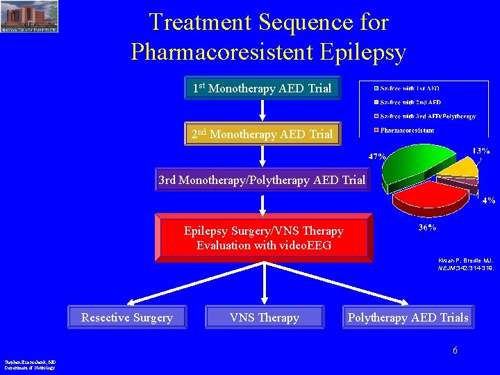
Treatment Sequence for Pharmacoresistent Epilepsy 1 st Monotherapy AED Trial 2 nd Monotherapy AED Trial 3 rd Monotherapy/Polytherapy AED Trial Epilepsy Surgery/VNS Therapy Evaluation with video. EEG Kwan P, Brodie MJ. NEJM; 342: 314 -319. Resective Surgery VNS Therapy Polytherapy AED Trials 6 Stephan Eisenschenk, MD Department of Neurology
Presurgical Evaluation History and exam MRI scan – Mesial Temporal Sclerosis (MTS), tumor, vascular malformation, dysplasia Video/EEG monitoring with scalp EEG – interictal epileptiform discharges – ictal • Seizure semiology • Ictal EEG discharge – Additional electrodes S-Slide 7 Stephan Eisenschenk, MD Department of Neurology
Presurgical Evaluation Functional Imaging – PET • hypometabolism interictally – SPECT • hypoperfusion interictally • hyperperfusion ictally • subtraction and co-registration with MRI S-Slide 8 Stephan Eisenschenk, MD Department of Neurology
Presurgical Evaluation Neuropsychological testing – Pre-operative baseline – Aid in localization – Predicting risk of cognitive decline with surgery Wada (intracarotid amobarbital) test – language • lateralization – Memory • prediction of postoperative decline Stephan Eisenschenk, MD Department of Neurology
Inpatient Video EEG Monitoring • Goal: to capture the patient’s typical seizures • Medications are tapered or withdrawn • Sleep deprivation, hyperventilation, photic stimulation and possibly other provocative maneuvers are tried Stephan Eisenschenk, MD Department of Neurology
Inpatient Video EEG Monitoring • Assess the interictal EEG using a longer recording • Confirm the diagnosis of epilepsy • Confirm the type of epilepsy – Generalized vs. partial – Temporal vs. extratemporal • Localize the site of seizure onset Stephan Eisenschenk, MD Department of Neurology
MRI head- seizure protocol • Coronal T 2 • High resolution 3 T MRI (coronal and axial) • Thin coronal cuts through the temporal lobes Stephan Eisenschenk, MD Department of Neurology
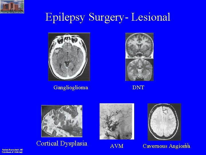
Epilepsy Surgery- Lesional Ganglioma Cortical Dysplasia Stephan Eisenschenk, MD Department of Neurology DNT AVM 13 Cavernous Angioma
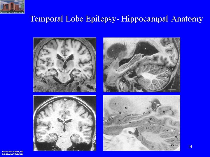
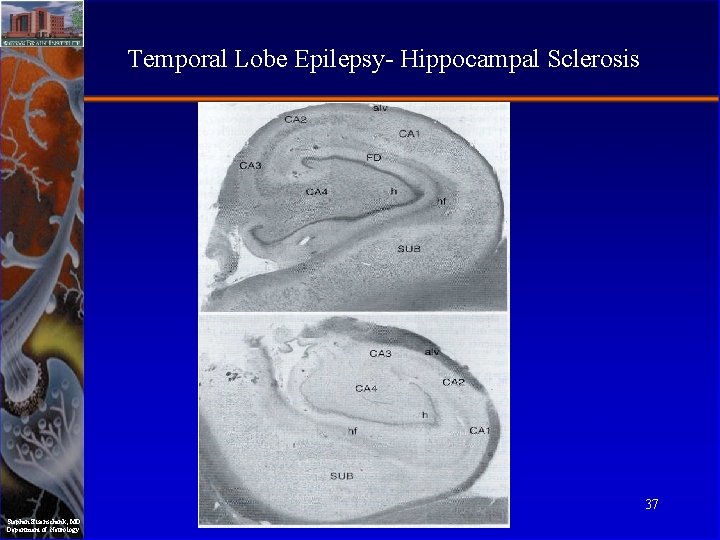
Temporal Lobe Epilepsy- Hippocampal Anatomy 14 Stephan Eisenschenk, MD Department of Neurology
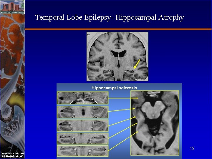
Temporal Lobe Epilepsy- Hippocampal Atrophy 15 Stephan Eisenschenk, MD Department of Neurology
Concordance • • Interictal EEG findings Ictal semiology Ictal EEG onset MRI • Tests should show concordant findings • If a contradictory or unclear picture emerges, additional testing is warranted Stephan Eisenschenk, MD Department of Neurology
If not concordant • MEG • Metabolic studies – PET – Ictal SPECT (SISCOM) Stephan Eisenschenk, MD Department of Neurology
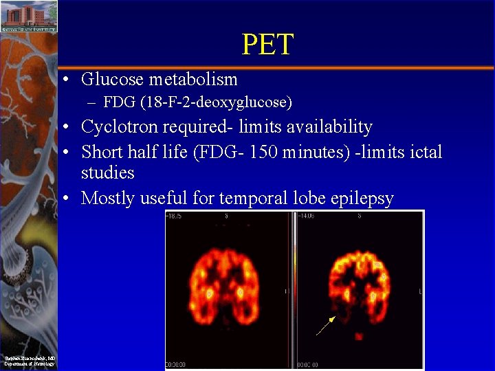
PET • Glucose metabolism – FDG (18 -F-2 -deoxyglucose) • Cyclotron required- limits availability • Short half life (FDG- 150 minutes) -limits ictal studies • Mostly useful for temporal lobe epilepsy Stephan Eisenschenk, MD Department of Neurology
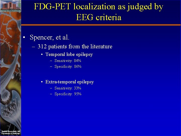
FDG-PET localization as judged by EEG criteria • Spencer, et al. – 312 patients from the literature • Temporal lobe epilepsy – Sensitivity: 84% – Specificity: 86% • Extra-temporal epilepsy – Sensitivity: 33% – Specificity: 95% Stephan Eisenschenk, MD Department of Neurology
Ictal SPECT • SISCOM (subtraction ictal SPECT with coregistration onto MRI) – Ictal injection at the time of the seizure with scan – Interictal injection with scan – Co-registration onto MRI Stephan Eisenschenk, MD Department of Neurology
Ictal SPECT • Good confirmatory test- added utility in – Non-lesional (MRI negative) temporal and extra -temporal epilepsies – Limiting resection in cases of diffuse hemispheric abnormalities • Due to regulations regarding administration of radiopharmaceuticals not currently feasible in many states (including Texas) Stephan Eisenschenk, MD Department of Neurology
Criteria for “skip” vs Phase 2 – CONCORDANT DATA = ‘skip’ to resection • EEG/ MRI/ Neuropsych – 2 concordant tests and no discordant test – usually EEG + MRI – If data NOT Concordant • Unilateral seizure activity = subdural grids • Bilateral temporal lobe seizure activity = bilateral depths Stephan Eisenschenk, MD Department of Neurology
The WADA Test • Helps determine which side of the brain controls language function and how each side of the brain controls memory function – Language is usually controlled on the L, Memory can be controlled by both sides of the brain (the test tells physicians which side has the better memory function) • Need this information to help the patient /family make an informed decision based on possible risk factors for specific neurologic deficits such as memory problems or aphasia Stephan Eisenschenk, MD Department of Neurology
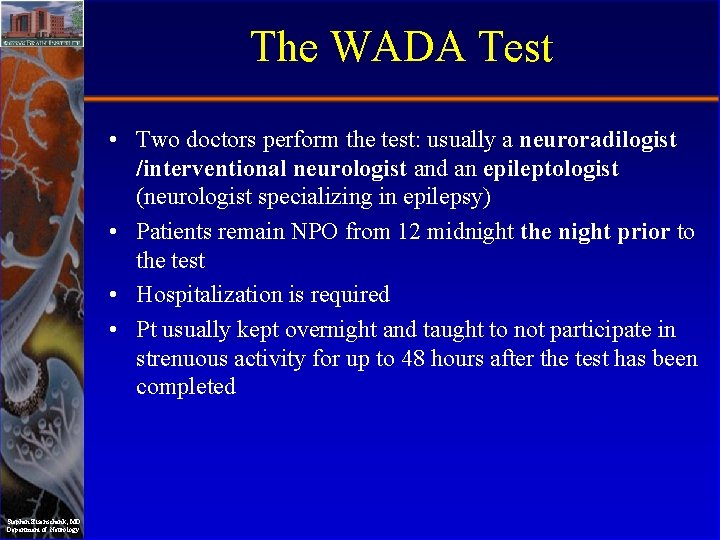
The WADA Test • Two doctors perform the test: usually a neuroradilogist /interventional neurologist and an epileptologist (neurologist specializing in epilepsy) • Patients remain NPO from 12 midnight the night prior to the test • Hospitalization is required • Pt usually kept overnight and taught to not participate in strenuous activity for up to 48 hours after the test has been completed Stephan Eisenschenk, MD Department of Neurology
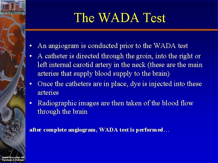
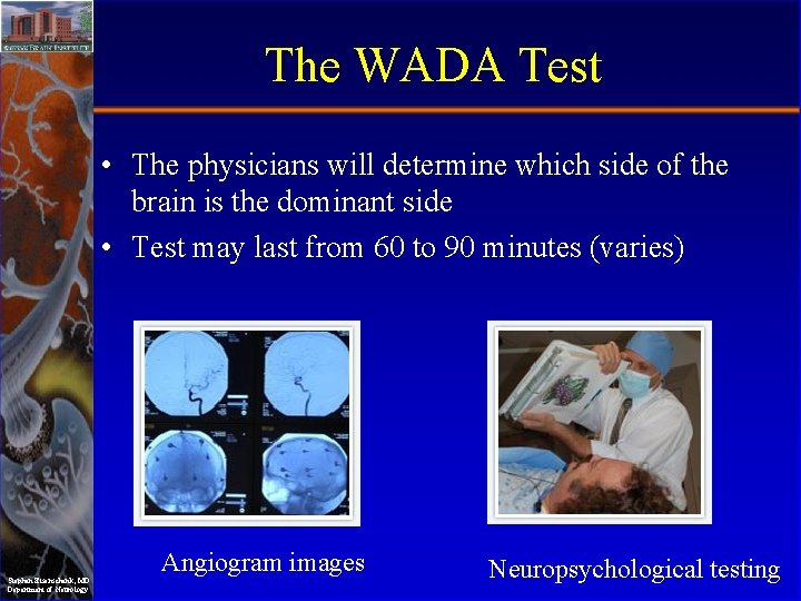
The WADA Test • An angiogram is conducted prior to the WADA test • A catheter is directed through the groin, into the right or left internal carotid artery in the neck (these are the main arteries that supply blood supply to the brain) • Once the catheters are in place, dye is injected into these arteries • Radiographic images are then taken of the blood flow through the brain after complete angiogram, WADA test is performed… Stephan Eisenschenk, MD Department of Neurology
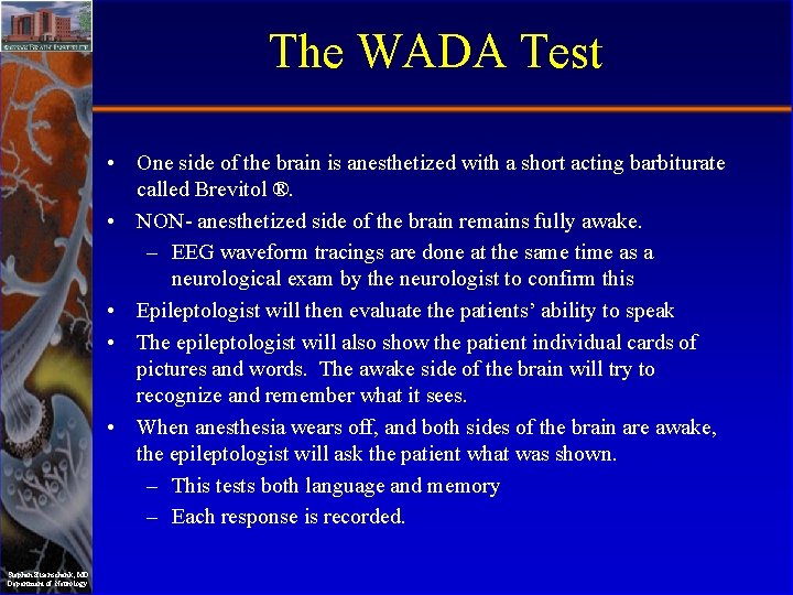
The WADA Test • One side of the brain is anesthetized with a short acting barbiturate called Brevitol ®. • NON- anesthetized side of the brain remains fully awake. – EEG waveform tracings are done at the same time as a neurological exam by the neurologist to confirm this • Epileptologist will then evaluate the patients’ ability to speak • The epileptologist will also show the patient individual cards of pictures and words. The awake side of the brain will try to recognize and remember what it sees. • When anesthesia wears off, and both sides of the brain are awake, the epileptologist will ask the patient what was shown. – This tests both language and memory – Each response is recorded. Stephan Eisenschenk, MD Department of Neurology
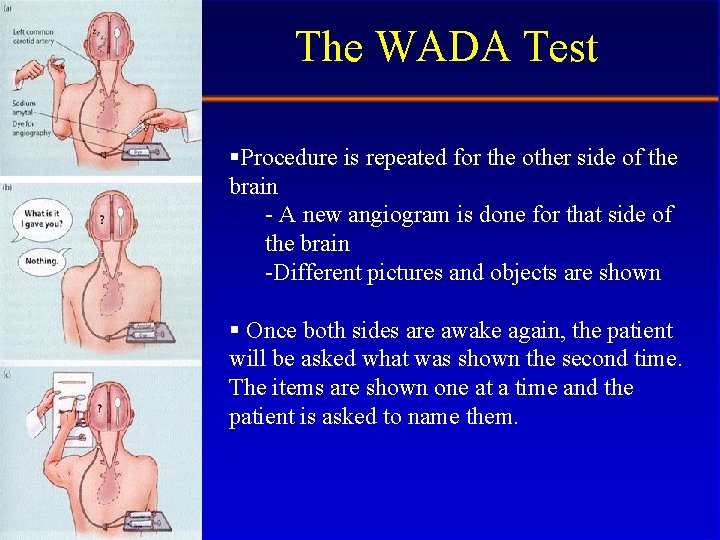
The WADA Test §Procedure is repeated for the other side of the brain - A new angiogram is done for that side of the brain -Different pictures and objects are shown § Once both sides are awake again, the patient will be asked what was shown the second time. The items are shown one at a time and the patient is asked to name them. Stephan Eisenschenk, MD Department of Neurology
The WADA Test • The physicians will determine which side of the brain is the dominant side • Test may last from 60 to 90 minutes (varies) Stephan Eisenschenk, MD Department of Neurology Angiogram images Neuropsychological testing
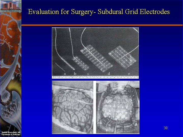
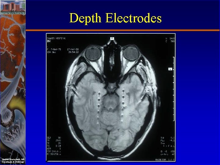
Presurgical Evaluation Intracranial EEG when needed – Grids and strips, most commonly subdural – Parenchymal “depth” electrodes, especially for recording from hipppocampus – Identification of ictal onset – Brain mapping • cortical stimulation • SSEPs • Functional MRI S-Slide 29 Stephan Eisenschenk, MD Department of Neurology
Evaluation for Surgery- Subdural Grid Electrodes 30 Stephan Eisenschenk, MD Department of Neurology
Depth Electrodes Stephan Eisenschenk, MD Department of Neurology
Types of Surgical Procedures Resective Surgery: single seizure focus in non-eloquent region. Palliative Surgery: – For drop attacks: corpus callosotomy – For Rasmussen’s encephalitis or hemimegalencephaly: hemispherectomy S-Slide 32 Stephan Eisenschenk, MD Department of Neurology
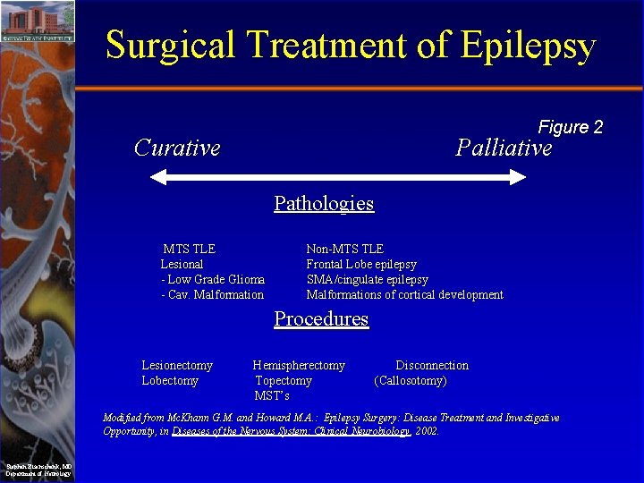
Surgical Treatment of Epilepsy Figure 2 Curative Palliative Pathologies MTS TLE Lesional - Low Grade Glioma - Cav. Malformation Non-MTS TLE Frontal Lobe epilepsy SMA/cingulate epilepsy Malformations of cortical development Procedures Lesionectomy Lobectomy Hemispherectomy Topectomy MST’s Disconnection (Callosotomy) Modified from Mc. Khann G. M. and Howard M. A. : Epilepsy Surgery: Disease Treatment and Investigative Opportunity, in Diseases of the Nervous System: Clinical Neurobiology , 2002. Stephan Eisenschenk, MD Department of Neurology
Consideration of intracranial monitoring • Options – Subdural strips – Subdural grids – Depth electrodes • Record additional seizures with the intracranial electrodes in place to better localize seizure onset Stephan Eisenschenk, MD Department of Neurology
Cortical stimulation • Can be done at the time of surgery or through implanted intracranial electrodes to localize functional cortex including – Language cortex – Motor cortex – Sensory cortex Stephan Eisenschenk, MD Department of Neurology
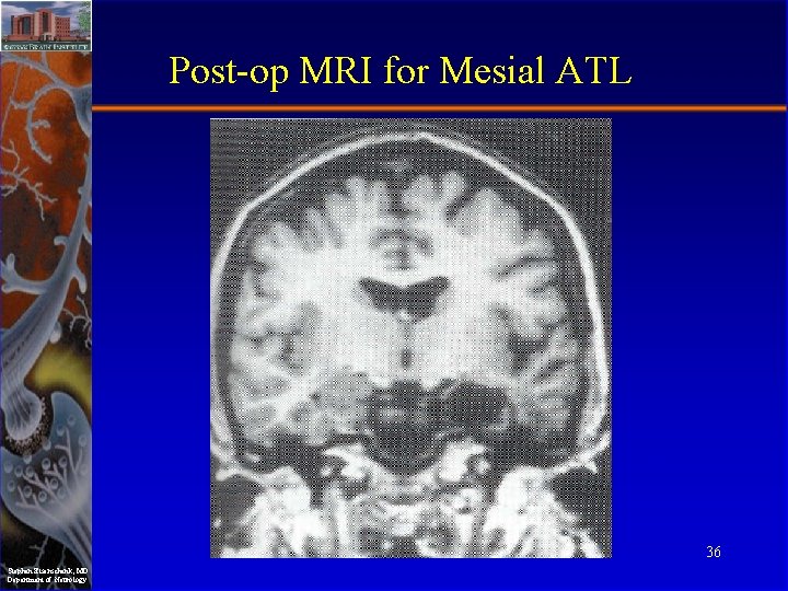
Post-op MRI for Mesial ATL 36 Stephan Eisenschenk, MD Department of Neurology
Temporal Lobe Epilepsy- Hippocampal Sclerosis 37 Stephan Eisenschenk, MD Department of Neurology
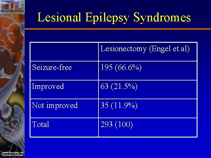
Lesional Epilepsy Syndromes • Predictive of – An unfavorable response to AED medication – A favorable response to surgery • Operative success – Lesional pathology – Location of lesion – Completeness of resection Stephan Eisenschenk, MD Department of Neurology
Lesional Epilepsy Syndromes Lesionectomy (Engel et al) Stephan Eisenschenk, MD Department of Neurology Seizure-free 195 (66. 6%) Improved 63 (21. 5%) Not improved 35 (11. 9%) Total 293 (100)
Lesional Epilepsy Syndromes. Tumors • Commonly low grade gliomas, DNETs, gangliomas • With full resection 43 -90% seizure free depending on the study Stephan Eisenschenk, MD Department of Neurology
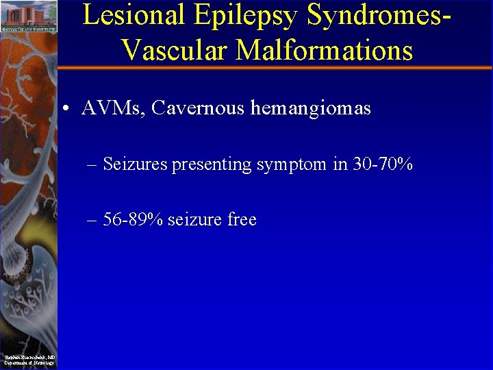
Lesional Epilepsy Syndromes. Vascular Malformations • AVMs, Cavernous hemangiomas – Seizures presenting symptom in 30 -70% – 56 -89% seizure free Stephan Eisenschenk, MD Department of Neurology
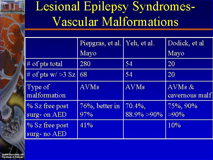
Lesional Epilepsy Syndromes. Vascular Malformations # of pts total Stephan Eisenschenk, MD Department of Neurology Piepgras, et al. Yeh, et al. Mayo 280 54 Dodick, et al Mayo 20 # of pts w/ >3 Sz 68 54 20 Type of malformation AVMs & cavernous malf % Sz free post surg- on AED 76%, better in 70. 4%, 75%, 90% 97% 88. 9% >90% % Sz free post surg- no AED 41% 10%
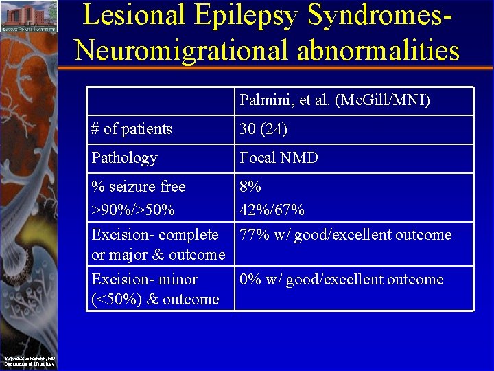
Lesional Epilepsy Syndromes. Neuromigrational abnormalities Palmini, et al. (Mc. Gill/MNI) # of patients 30 (24) Pathology Focal NMD % seizure free 8% >90%/>50% 42%/67% Excision- complete 77% w/ good/excellent outcome or major & outcome Excision- minor (<50%) & outcome Stephan Eisenschenk, MD Department of Neurology 0% w/ good/excellent outcome
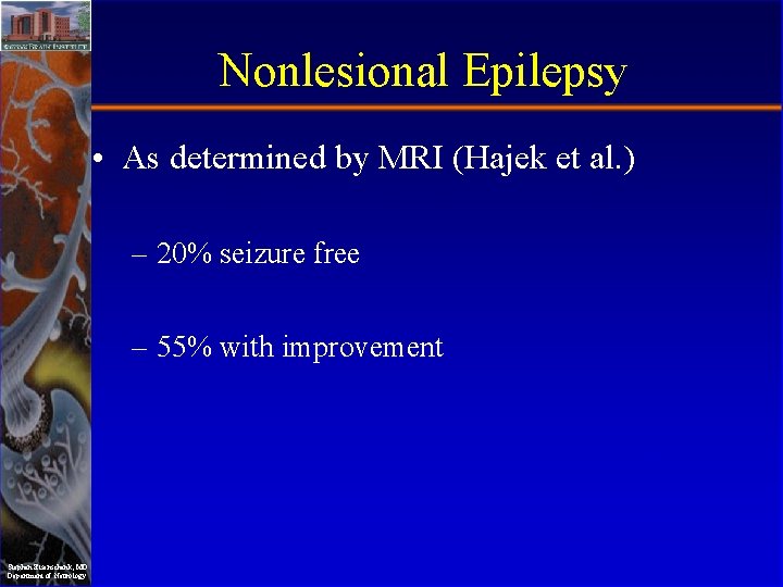
Nonlesional Epilepsy • As determined by MRI (Hajek et al. ) – 20% seizure free – 55% with improvement Stephan Eisenschenk, MD Department of Neurology
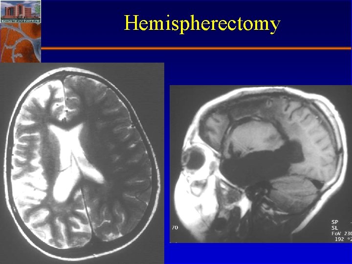
Hemispherectomy • Current practice is to perform a modified radical hemispherectomy leaving the frontal and occipital poles in place though disconnected • Reserved for certain catastrophic childhood epilepsies – Rasmussen’s encephalitis – Hemimegalencephaly Stephan Eisenschenk, MD Department of Neurology
Hemispherectomy Stephan Eisenschenk, MD Department of Neurology
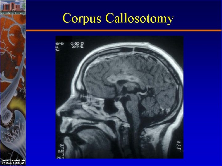
Corpus Callosotomy • Useful as a palliative procedure in patients with symptomatic generalized epilepsy associated with certain seizure types – Tonic – Atonic • Almost always done as a staged procedure – Anterior 2/3’s corpus callosotomy first – Completion of corpus callosotomy if initial surgery ineffective or not effective enough Stephan Eisenschenk, MD Department of Neurology
Corpus Callosotomy Stephan Eisenschenk, MD Department of Neurology
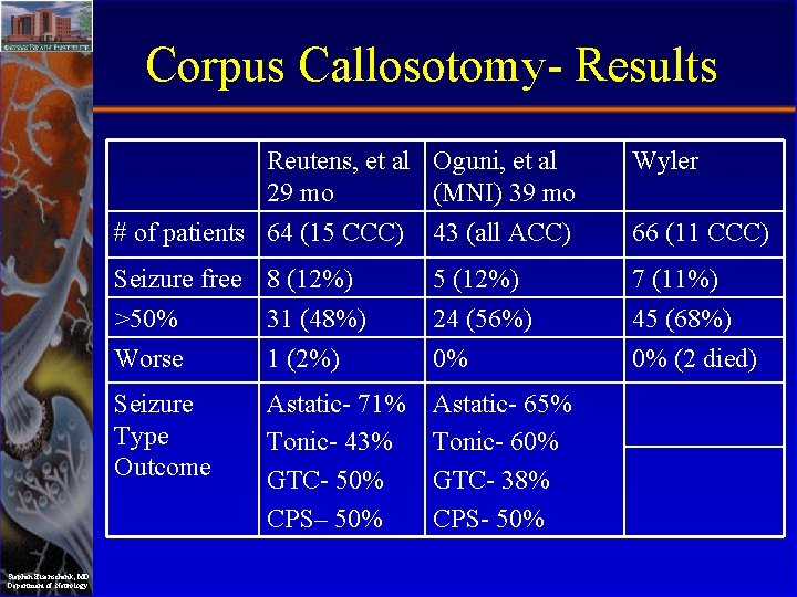
Corpus Callosotomy- Results Reutens, et al Oguni, et al 29 mo (MNI) 39 mo Stephan Eisenschenk, MD Department of Neurology Wyler # of patients 64 (15 CCC) 43 (all ACC) 66 (11 CCC) Seizure free 8 (12%) 5 (12%) 7 (11%) >50% Worse 31 (48%) 1 (2%) 24 (56%) 0% 45 (68%) 0% (2 died) Seizure Type Outcome Astatic- 71% Tonic- 43% GTC- 50% CPS– 50% Astatic- 65% Tonic- 60% GTC- 38% CPS- 50%
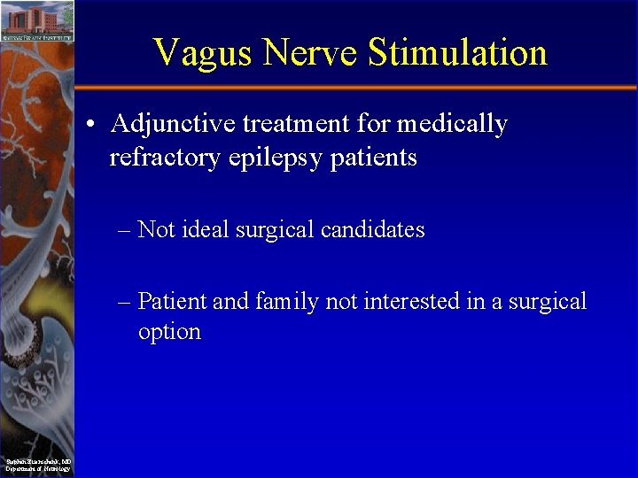
Vagus Nerve Stimulation • Adjunctive treatment for medically refractory epilepsy patients – Not ideal surgical candidates – Patient and family not interested in a surgical option Stephan Eisenschenk, MD Department of Neurology
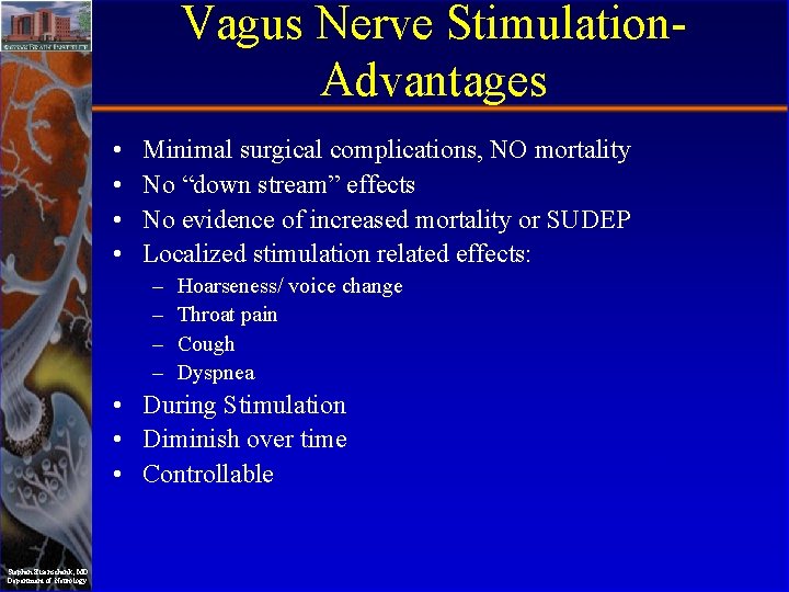
Vagus Nerve Stimulation. Advantages • • Minimal surgical complications, NO mortality No “down stream” effects No evidence of increased mortality or SUDEP Localized stimulation related effects: – – Hoarseness/ voice change Throat pain Cough Dyspnea • During Stimulation • Diminish over time • Controllable Stephan Eisenschenk, MD Department of Neurology
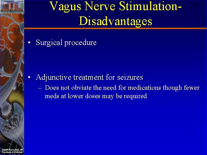
Vagus Nerve Stimulation. Disadvantages • Surgical procedure • Adjunctive treatment for seizures – Does not obviate the need for medications though fewer meds at lower doses may be required Stephan Eisenschenk, MD Department of Neurology