Degenerative Disease of the Spine Khalid A Al


- Slides: 34

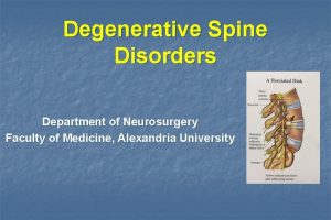
Degenerative Disease of the Spine Khalid A. Al. Saleh, FRCSC Assistant Professor Dept. of Orthopedic Surgery

Introduction • Degeneration: – “deterioration of a tissue or an organ in which its function is diminished or its structure is impaired” • Other terms: – “Spondylosis” – “Degenerative disc disease” – “Facet osteoarthrosis”

Etiology • Multi-factorial – Genetic predisposition – Age-related – Some environmental factors: • • • Smoking Obesity Previous injury, fracture or subluxation Deformity Operating heavy machinery, such as a tractor

Anatomy • Anterior elements: – Vertebral body – Inter-vertebral disc • Degeneration occurs at the disc • Posterior elements – Pedicles, laminae, spinous process, transverse process, facet joints (2 in each level) • Osteoarthrosis occurs at the facet joints

Anatomy, cont. • Neurologic elements: – Spinal cord – Nerve roots – Cauda equina

Cervical and Thoracic Spine Anatomy

Lumbar Spine Anatomy

Pathology: The inter-vertebral disc • The first component of the 3 joint complex present in each vertebral segment from C 2 to S 1 – It is primarily loaded in FLEXION • Composed of annulus fibrosus and nucleus pulposus – Degeneration of the nucleus causes loss of cellular material and loss of hydration • Movement is impaired-painful- and could become unstable

The inter-vertebral disc, cont. • Disc degeneration will also cause – Loss of disc height→ • Abnormal loading of facet joints • Stenosis in the inter-vertebral foramen – Bulging of the disc into the spinal canal • Contributing to spinal stenosis – Herniation of the nucleus into spinal canal • Causing radiculopathy (e. g. sciatica in the lumbar spine)

Pathology: The facet joints • Scientific name: “zygapophysial joints” – Synovial joints – 2 in each segment • Together with the disc, form the 3 joint complex • Are primarily loaded in EXTENSION – Pattern of degeneration similar to other synovial joints • Loss of hyaline cartilage, formation of osteophytes, laxity in the joint capsule

The facet joints, cont. • Facet degeneration will cause: – Hypertrophy, osteophyte formation • Contributing to spinal stenosis or foraminal stenosis – Laxity in the joint capsule • Leading to instability (degenerative spondylolisthesis)

Presentation • Falls into 2 catagories: – Mechanical pain: due to joint degeneration or instability • “Axial pain” in the neck or back • Activity related-not present at rest – Neurologic symptoms: due to neurologic impingement • Spinal cord – Presents as myelopathy, spinal cord injury • Cauda equina & Nerve roots – Presents as radiculopathy (e. g. sciatica) or neurogenic claudication

Presentation, cont. • Mechanical pain – Associated with movement • Sitting, bending forward (flexion): – originating from the disc » “discogenic pain” • Standing, bending backward (extension) : – originating from the facet joints » “Facet syndrome” – Instability-e. g. spondylolisthesis- also causes mechanical pain

Presentation, cont. • Neurologic symptoms – Spinal cord • Myelopathy: – Loss of motor power and balance – Loss of dexterity » Objects slipping from hands – UMN deficit (rigidity, hyper-reflexia, positive Babinski. . ) – Slowly progressive “step-wise” deterioration. • Spinal cord injury – With Spinal stenosis, there is a higher risk of spinal cord injury – Complete or incomplete

Presentation, cont. • Cauda equina & Nerve roots – Radiculopathy • LMN deficit • Commonest is sciatica, but cervical root impingement causes similar complaints in the upper limb – Neurogenic claudication • Pain in both legs caused by walking • Must be differentiated from vascular claudication

Vascular vs. Neurogenic claudication

Break for 5 minutes

The Cervical spine: introduction • Degenerative changes typically occur in C 3 -C 7 • Presents with axial pain, myelopathy, radiculopathy • Physical examination: – Stiffness (loss of ROM) – Neurologic exam • • Weakness Loss of sensation Hyper-reflexia, hypertonia Special tests: Spurling’s sign

The Cervical spine: Management • Conservative treatment – First line of treatment for axial neck pain and mild neurologic symptoms (e. g. mild radiculopathy without any motor deficit) • Physiotherapy: – Focus on ROM and muscle strengthening • Non-steroidal anti-inflammatory medications (NSAID) – E. g. Diclofenac, ibuprofen, naproxen • Neuropathic medication: for radiculopathy pain – E. g. Gabapentin or pregabalin

The Cervical spine: Management • Surgical management – Indicated for: • Spinal stenosis causing myelopathy • Disc herniation causing severe radiculopathy and weakness • Failure of conservative treatment of axial neck pain or mild radiculopathy – Procedures: • Anterior discectomy and fusion • Posterior laminectomy

Anterior Discectomy and fusion

The Lumbar spine • Degenerative changes typically occur in L 3 -S 1 • Presents with axial pain, Sciatica, neurogenic claudication • Physical examination: – Stiffness (loss of ROM) – Neurologic exam • • Weakness Loss of sensation Hypo-reflexia, hypo-tonia Special tests: SLRT

The Lumbar spine: management • Axial low back pain – Conservative treatment if first-line and mainstay of treatment • Physiotherapy: core muscle strengthening, posture training • NSAID – Surgical treatment indicated for: • Instability or deformity e. g. high-grade spondylolisthesis • Failure of conservative treatment

Lumbar spondylosis

The Lumbar spine: management • Spinal stenosis – Conservative treatment is first line of treatment • Activity modification, analgesics, epidural corticosteroid injections – Surgical treatment • Indicated for – Motor weakness e. g. drop foot – failure of –minimum- 6 months of conservative treatment • Spinal decompression (laminectomy) is the commonest procedure

Spinal Stenosis

The Lumbar spine: management • Disc herniation – Conservative treatment is first line of treatment for mild sciatica without motor deficit • Short (2 -3 day) period of rest, NSAID, physiotherapy, epidural cortico-steroid injection • 95% of sciatica resolves within the first 3 months without surgery – Surgical treatment: • Indicated for cauda-equina syndrome, motor deficit, failure of 3 months of conservative treatment • Procedure: Discectomy (only the herniated part)

Disc Herniation

The Lumbar spine: management • Degenerative Spondylolisthesis – Typically at L 4 -5 – Causes spinal stenosis – Conservative treatment first, – Surgery if Grade 3 or more or failed conservative managment. • Other spondylolisthesis types: – Isthmic: • Usually at L 5 -S 1, • Has par inter-articularis defect

Spondylolisthesis, foramenal stenosis

Spinal Fusion

The Lumbar spine: management • Degenerative scoliosis – Combination of elements from the prior conditions • Deformity, Instability • Spinal stenosis, Disc herniation – Also treated conservatively first, unless severe neurologic deficit or instability present – Usually requires multi-level instrumentation, fusion and decompression

Degenerative scoliosis

Thanks, Questions?
 Degenerative myelopathy wayne
 Fascetaria
 Communicable disease and non communicable disease
 911 in roman
 When islam started
 Khalid alsadhan
 King khalid hospital
 Fariza khalid
 Dr sana khalid
 Khalid mustafa
 Fariza khalid
 Khalid chaouch
 Dr khalid waheed
 Khalid bakri
 Khalid karaoui
 Dr sana khalid
 Nationality vs citizenship
 Khalid al habib
 Cipralex side effects
 Dr. khalid bazaid
 Complete the tag question your name is ali khalid
 Khalid hossain books
 Jurnal jamal
 Khalid farhan course google drive
 Nauman khalid md
 Dr arzoo khalid
 Dr samra khalid
 Genial tubercle
 Spine meninges
 Shilla procedure
 Spine blood spatter definition
 Histoire des arts frida kahlo
 Genial tubercle
 Psis level with s2
 Extreme fabric connect
