Copper Dysfunction in Alzheimers Disease Rosanna Squitti Department


")


 a Cu(I) (Multhaup G et al.")


 . 62 Cu: ceruloplasmin ratio AD: 79 Ctrl: 76")


 the")


")


- Slides: 34
Copper Dysfunction in Alzheimer’s Disease Rosanna Squitti, Department of Neuroscience, Fatebenefratelli Foundation, AFa. R Division Rome, Italy
Signs of Alzheimer’s disease • In a schematic representation, five primary functional and anatomical features characterize the AD brain: • (a) loss of neurons and synapses in the cerebral cortex • (b) density and distribution of extracellular amyloid plaques • (c) presence of intracellular neurofibrillary tangles containing hyperphosphorylated Tau • (d) increased oxidative damage of lipids, proteins and nucleic acids • (e) loss of biometal homeostasis
Copper and AD (> 1394)
Evidence sustaining the role of copper dysfunction in Alzheimer’s disease • • • Biochemical in vitro Clinical Epidemiological Meta-analyses Genetics
Copper in physiology: body distribution
Squitti et al. , Neurobiol Aging 2014
Copper dysfunction in Alzheimer’s disease: Biochemical evidence in vitro
The Cu. BD sequence APP reduces Cu (II) a Cu(I) (Multhaup G et al. , Science 1996) H 2 O 2 production by A Cu 2 (Opazo et al. , J Biol Chem. 2002) Aβ deposits dissolution by biometal depletion (Cherny et al. , J Biol Chem 1999) APP/A metalloprotein controlling [Cu]in (White AR et al. , Brain Res 1999) (Barnham et al. J Biol Chem 2003)
Model of copper toxicity Physiological Aβ oxidation Haber–Weiss Fenton reactions copper Rogue soluble Aβ copper H 2 O 2 Diffuse amyloid oxidation Oxidative stress toxicity plaques (modified by Bush AI Trends Neurosci 2003)
What happens in living patients? Does a copper dysfunction occur in AD patients?
Copper dysfunction in Alzheimer’s disease: Clinical evidence
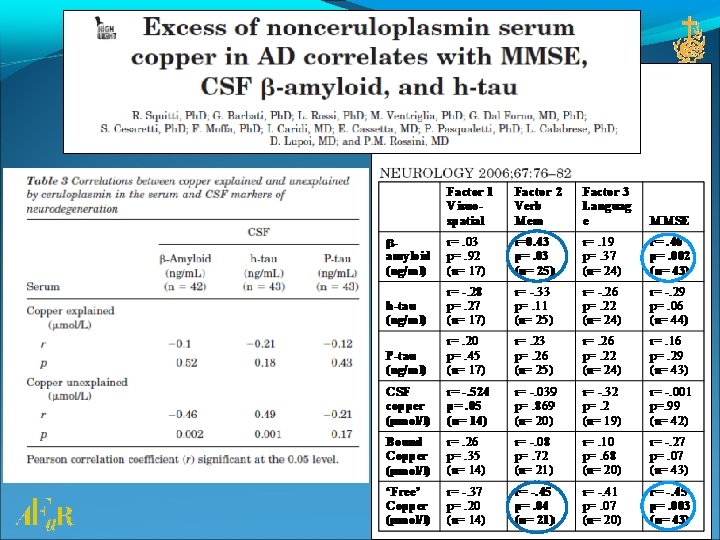
AD copper subtype: 12 years of evidence 2002 Squitti et al. , Elevation of serum copper levels in Alzheimer's disease - Neurology 2003 Squitti et al. , Elevation of serum copper levels discriminates Alzheimer's disease from vascular dementia - Neurology 2005 Squitti et al. , Excess of serum copper not related to ceruloplasmin in Alzheimer disease - Neurology 2006 Squitti et al. , Excess of nonceruloplasmin serum copper in AD correlates with MMSE, CSF [beta]-amyloid, and h-tau - Neurology 2007 Babiloni et al. , Free copper and resting temporal EEG rhythms correlate across healthy, mild cognitive impairment, and Alzheimer's disease subjects - Clin Neurophysiol 2007 Squitti et al. , 'Free' copper in serum of Alzheimer's disease patients correlates with markers of liver function - J Neural Transm 2008 Squitti et al. , Ceruloplasmin fragmentation is implicated in 'free' copper deregulation of Alzheimer's disease - Prion 2009 Squitti et al. , Longitudinal prognostic value of serum "free" copper in patients with Alzheimer disease - Neurology 2014 Squitti R et al, Value of serum non-ceruloplasmin copper for prediction of MCI conversion to AD. Ann Neurol
Cu (deviation from controls’ mean) . 62 Cu: ceruloplasmin ratio AD: 79 Ctrl: 76 AD: 47 VAD: 24 Ctrl: 44. 60. 58. 56. 54. 52. 50. 48. 46 C AD VAD
Non-Cp Cu AD Prognosis Non-Cp Cu in MCI 81 AD 1 year follow-up (Squitti et al. , Neurology 2009) MCI: 83 Ctrl: 100 AD: 105 (Squitti et al. , J Alzh Dis 2010)
Non-Cp Cu MCI conversion Faster rate of conversion to AD: Among the MCI, the 20% more rapid to convert to AD employs about 4 years if they have normal levels of Non-Cp. Cu and less than 1. 5 years if they have levels higher than 1. 6 µmol/L Increased risk for AD: After 4 years, the probability of conversion to AD is less than 20% in MCI with low Non-Cp-Cu and almost 50% in the MCI with higher than 1. 6 µmol/L (Squitti et al. , Annals of Neurology 2014)
Copper dysfunction in Alzheimer’s disease: epidemiologic data
Dietary Supplements and Mortality Rate in Older Women (The Iowa Women’s Health Study) the use of vitamin and mineral supplements in relation to total mortality in 38 772 older women: Copper was associated with a 18% increased of mortality (Mursu et al, Arch Intern Med. ) Relationship between Mortality from Alzheimer's Disease and Soil Metal Concentration in Mainland China. Shen Positive J Alzheimers Dis. 2014 (Morris et al Arch Neurol 2006)
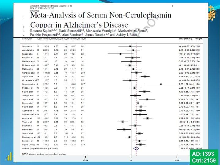
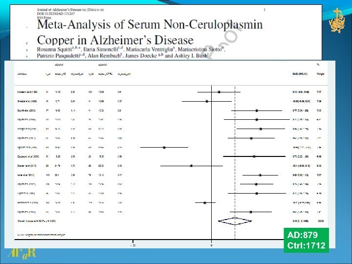
Copper dysfunction in Alzheimer’s disease: Meta-analytic data
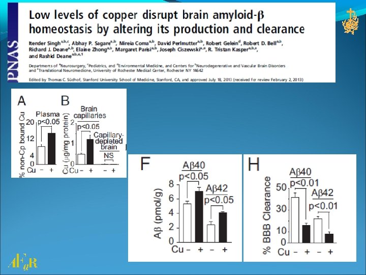
25% Labile copper increased in AD in Brodman area (James et al Free Radic Biol Med 2012) Non-Cp-Cu in the AD cascade
Copper dysfunction in Alzheimer’s disease: genetics
ATP 7 B (60% of LOAD)
The copper gene ATP 7 B associates with AD 2013 Squitti R, et al. , ATP 7 B Variants as Modulators of Copper Dyshomeostasis in Alzheimer's Disease - Neuromolecular Med. 2013 Bucossi et al. , Intronic rs 2147363 variant in ATP 7 B transcription factor-binding site associated with Alzheimer’s disease - J Alzheimers Dis. 2013 Squitti R et al. , Linkage disequilibrium and haplotype analysis of the ATP 7 B gene in Alzheimer's disease - Rejuvenation Res. 2012 Squitti R. Copper dysfunction in Alzheimer's disease: from metaanalysis of biochemical studies to new insight into genetics - J Trace Elem Med Biol 2012 Bucossi et al. , Association of K 832 R and R 952 K SNPs of Wilson's disease gene with Alzheimer's disease - J Alzheimers Dis. 2012 Squitti R, Polimanti R. Copper hypothesis in the missing hereditability of sporadic Alzheimer's disease: ATP 7 B gene as potential harbor of rare variants - J Alzheimers Dis 2011 Bucossi et al. , Association between the c. 2495 A>G ATP 7 B Polymorphism and Sporadic Alzheimer's Disease - Int J Alzheimers Dis.
ATP 7 B informative SNPs in AD risk rs 732774 rs 1061472 Block 1 rs 732774, rs 1061472 Transmembrane domains rs 2147363 rs 1801243 Block 2 rs 2147363, rs 1801243 Copper domains AD: 285 Ctrl: 230 (Squitti et al. , 2013 Rejuvenation Res. 16: 3 -10)
ATP 7 B loci in transmembrane domains associate with an increased risk for AD Block 1 2 SNP ID rs 732774 Best genetic model recessive rs 1061472 Log-additive rs 2147363 Log-additive rs 1801243 Log-additive OR adjusted (95%CI) P value 2. 31 < 0. 001 (1. 41 -3. 77) 1. 73 0. 002 (1. 23 -2. 43) 1. 58 0. 010 (1. 11 -2. 25) 1. 52 0. 010 (1. 10 -2. 09) (Squitti et al. , 2013 Rejuvenation Res. 16: 3 -10)
ATP 7 B SNPs modulate Non-Cp copper in AD patients AD: 399 Ctrl: 303 [Non-Cp copper] in AD and rs 732774 AD carriers of GG genotype have higher levels of Non-Cp copper than carriers of AA+AG genotypes (p value = 0. 005) (Squitti et al. , Neuromolecular Med. 2013)
Copper dysfunction in Alzheimer’s disease: 1 patent, 1 CE-certified blood test for Non-Cp-Cu (C 4 D)
kit to measure Non-Cp copper Ref Patent: PCT/EP 2012/072063 switch-off coumarin fluorescent probe for detecting Non-Cp copper
Conclusions Serum Non-Cp-Cu increases HIGHER than normal values are present in about 50% of amnestic MCI subjects and 60% of AD patients. Non-Cp-Cu correlates with: • the major deficits of AD • markers of AD (beta-amyloid and Tau) in the CSF • Copper in CSF • Clinical worsening • MCI state • MCI conversion to AD • Copper dysfunction has a genetic basis on ATP 7 B gene variants
Acknowledgements Patrizio Pasqualetti, Ph. D Mariacarla Ventriglia, Ph. D Emanuele Cassetta, MD Mariacristina Siotto, Ph. D Serena Bucossi, Ph. D Paolo Maria Rossini, MD Luisa Benussi, Ph. D Roberta Ghidoni, Ph. D Filomena Moffa, Ph. D Giuliano Binetti, MD Renato Polimanti, Ph. D Stefania Mariani, Ph. D Fabrizio Vernieri, MD Ilaria Simonelli, Ph. D