Chronic Rhinosinusitis CRS Nasal polyps Fungal RS Dr
 Nasal polyps & Fungal RS Dr. Islam Alzayadneh MD Otolaryngology, Head")


")


 • This > Reduces nasal symptoms. > Improve quality of")


. Compare it with the inferior turbinate appearance (IT)")


. • FESS is")

 • Allergic fungal RS")

 • hyphal elements detected by fungal stains. • Culture :")


- Slides: 48
Chronic Rhinosinusitis (CRS) Nasal polyps & Fungal RS Dr. Islam Alzayadneh MD Otolaryngology, Head and Neck Surgeon Mutah University School of Medicine-Special Surgery Department Undergraduate Seminars 2019
Classification • Rhinosinusitis task force of the American academy : �Acute 7 days – 4 weeks. �Subacute 4 weeks – 12 weeks. �Recurrent ARS. ≥ 4 Episodes of ARS / Y. �Chronic RS. ≥ 12 weeks / Y. �Acute exacerbations of CRS. • European position paper on RS 2007 �Acute rhinosinusitis ≤ 4 weeks. �Chronic rhinosinusitis ≥ 12 weeks.
Other classification of Chronic Rhinosinusitis • Symptoms for > 12 weeks • Two main subtypes: • CRS without nasal polyps (CRSs. NP) • CRS with nasal polyps (CRSw. NP) • Strongly associated with asthma and aspirin tolerance Meltzer et al. JACI 2004; 114: 155
Epidemiology • Chronic sinusitis is one of the more prevalent chronic illnesses in the United States, affecting persons of all age groups. • The overall prevalence of CRS in the United States is 146 per 1000 population. • For unknown reasons, the incidence of this disease appears to be increasing yearly. This results in a conservative estimate of 18 -22 million physician visits in the United States each year and a direct treatment cost of $3. 4 -5 billion annually. • Chronic sinusitis is the fifth most common disease treated with antibiotics. Up to 64% of patients with AIDS develop chronic sinusitis
Predisposing factors to CRS Host factors Systemic. Environmental factors Microorganisms AR. Immunodeficiency. Viral, bacterial and fungal. Genetic/congenital Cystic fibrosis. Local. Anatomic obstruction. GERD/PLR. Pollutants Cigarette. Medications:
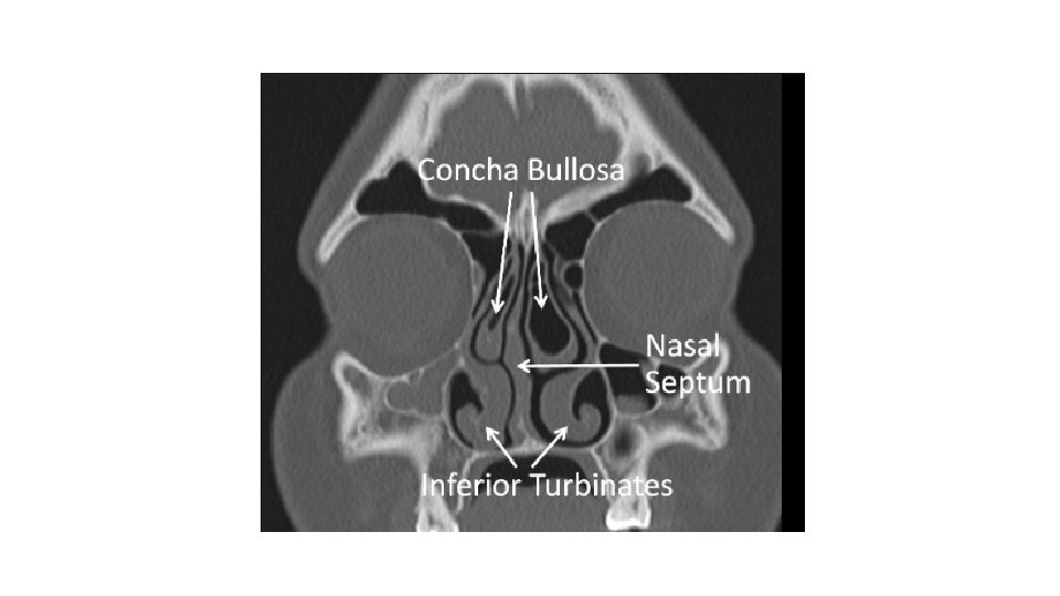
Predisposing factors to CRS • The following conditions and risk factors predispose patients to the development of chronic sinusitis: 1. Anatomic abnormalities of the ostiomeatal complex (e. g. , septal deviation, concha bullosa, deviation of uncinate process, Haller cells) 2. Allergic rhinitis 3. Aspirin sensitivity 4. Asthma 5. Nasal polyps 6. Nonallergic rhinitis (e. g. , vasomotor rhinitis, rhinitis Medicamentosa, cocaine abuse) 7. Defects in mucociliary clearance 8. Nasotracheal intubation 9. Nasogastric intubation 10. Hormonal (e. g. , puberty, pregnancy, oral contraception)
Predisposing factors to CRS 11. Obstruction by tumor 12. Immunologic disorders (e. g. , common variable immunodeficiency, immunoglobulin A [Ig. A] deficiency, Ig. G subclass deficiency, AIDS) 13. Cystic fibrosis 14. Primary ciliary dyskinesia, Kartagener syndrome 15. Wegener granulomatosis 16. Repeated viral upper respiratory tract infections 17. Smoking 18. Environmental irritants and pollutants 19. Gastroesophageal reflux disease (GERD). The reflux of gastric contents may play a contributing role in some cases of CRS; this relationship still needs to be better defined 20. Periodontitis/significant dental disease 21. Systemic diseases (ie, granulomatosis with polyangiitis (Wegener granulomatosis), Churg-Strauss vasculitis, sarcoidosis
Physiology of the sinuses • Functions of the sinuses • Normal cellular component , mucosa • Cilia ( PH , mechanical , thermal) • Mucus secreting goblet cells • Basal cells
Pathophysiology of CRS
Pathophysiology of CRS • CRS is a proliferative process with remarkable thickening of the mucosa and lamina propria, eosinophils being the predominant infiltrative cell in CRS. • T-cell Cytokines Stimulation of various cell type Chemokines Attract eosinophils. • ↑ Levels of IL-4 & IL-5 in sinonasal tract ↑ Life & proliferation of eosinophils. • Degranulation of eosinophils Release of a number of destructive enzymes Epithelial damage Disruption of the normal barrier + M. C. clearance activity �Bacteria & fungi colonizing the sinus cavity, �Irritation of sensory nerve endings.
Approach to CRS
Clinical picture Symptoms Associated with the Diagnosis of Rhinosinusitis Major symptoms • Purulent anterior nasal discharge • Purulent posterior nasal discharge • Nasal obstruction • Facial congestion • Facial pain/pressure • Hyposmia/anosmia • Fever (acute) Minor symptoms • Headache • Otalgia/aural fullness • Halitosis • Dental pain • Cough • Fever • Fatigue
Diagnosis • In order to diagnose CRS you need one of those 2 symptoms for more than 12 weeks 1. Blockage/congestion. 2. Discharge anterior/posterior (discolored) Plus either: • Endoscopic signs of: - Polyps; - Mucopurulent discharge from middle meatus; - or Edema/mucosal obstruction primarily in middle meatus; And/or: • Com. tomography (CT) changes - Mucosal changes within ostiomeatal complex and sinuses.
Physical examination • Anterior rhinoscopy. • Endoscopy.
Plain Films • Plain radiographs have less role in CRS than in acute rhinosinusitis. • Plain films may show mucosal thickenings or sinus opacities.
CT scans • The imaging study of choice with fine coronal sections at the level of the ostiomeatal complex. • After failure of maximal medical therapy. • Before surgical procedures. • For evaluation of suspected complications. • When neoplasm is a possibility.
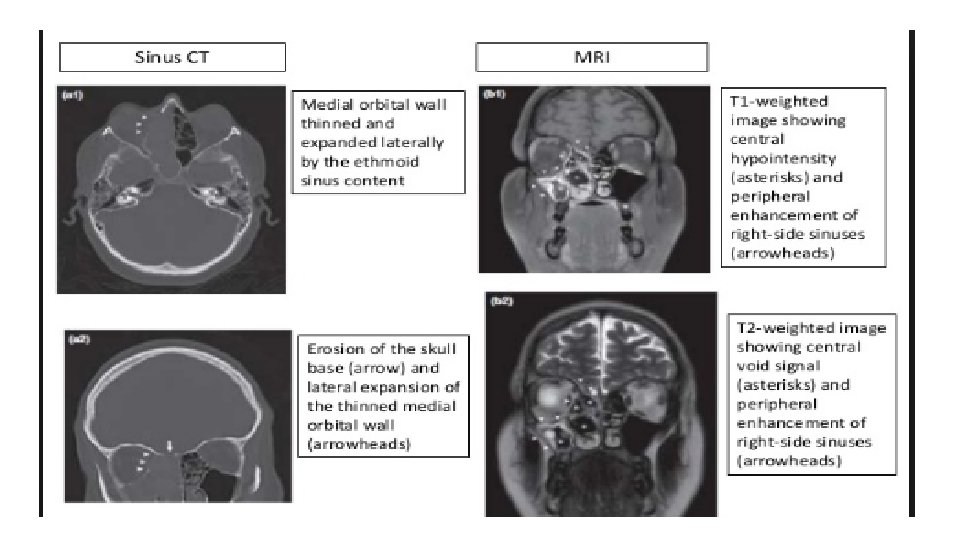
MRI • MRI is generally reserved for complex cases, (better evaluation of soft tissue) 1. Paranasal sinus tumors. 2. Cases with cranial base and orbital involvement. 3. Fungal sinusitis
Cultures and biopsies • Maxillary sinus tap, endoscopically directed middle meatal culture • Biopsy samples from the maxillary sinus mucosa of patients with chronic sinusitis show basement membrane thickening, atypical gland formation, goblet cell hyperplasia, mononuclear cell infiltration, and sub-epithelial edema. The mononuclear cell infiltrate often predominantly demonstrates neutrophils in acute disease and eosinophils in chronic disease. Rarely, squamous cell metaplasia may be seen.
Management • Assess patients with CRS or recurrent acute rhinosinusitis for multiple chronic conditions that would modify management (e. g. , asthma, cystic fibrosis, immunodeficiency, ciliary dyskinesia) • Assess for nasal polyps in patients with CRS • Recommend saline nasal irrigation, topical intranasal corticosteroids, or both for symptomatic relief of CRS According to recommendations of last update 2015 guidelines by the American Academy of Otolaryngology-Head and Neck Surgery Foundation
Nasal douching (saline irrigation) • This > Reduces nasal symptoms. > Improve quality of life. > Improve endoscopic findings. • Mechanism - Prevents crust accumulation. - Promotes mucociliary clearance
Corticosteroids • Initial oral steroid therapy followed by topical steroid therapy was found to be more effective than topical steroid therapy alone in decreasing polyp size and improving olfaction in patients with CRS with at least moderate nasal polyposis. • For maintenance therapy topical corticosteroids are the main stay. • Side effects of systemic steroids: 1. Osteoporosis. 2. Growth retardation. 3. Cataract , glaucoma • Ant leukotriene agents can be adjunctive to the effect of the steroids, especially in patients with asthma or an allergy to aspirin
Antimicrobial therapy • An adequate antibiotic trial in CRS usually consists of a minimum of 3 -4 weeks of treatment, preferably culture directed. • Empiric therapeutic regimens include the combination of a penicillin (eg, amoxicillin) plus a betalactamase inhibitor (eg, clavulanic acid), a combination of metronidazole plus a macrolide or a second- or third-generation cephalosporin, and the newer quinolones (eg, moxifloxacin). • Antibiotic therapy is often required for up to 6 weeks or longer and should not be discontinued until the patient is asymptomatic. Discontinuation of antimicrobial therapy prior to complete resolution increases the likelihood of relapse.
Decongestants • Vasoconstriction of dilated blood vessels. • Symptomatic relief nasal congestion. • No therapeutic efficacy for the treatment of sinusitis or polyps.
Surgical Treatment of Chronic Rhinosinusitis • Surgical care is used as an adjunct to medical treatment in some cases. Surgical care is usually reserved for cases that are refractory to medical treatment and for patients with anatomic obstruction. • The goal in surgical treatment is to reestablish sinus ventilation and to correct mucosal opposition in order to restore the mucociliary clearance system. Surgery strives to restore the functional integrity of the inflamed mucosal lining. • Recent advances in endoscopic technology and a better understanding of the importance of the ostiomeatal complex in the pathophysiology of sinusitis have led to the establishment of functional endoscopic sinus surgery (FESS) as the surgical procedure of choice for the treatment of chronic sinusitis
Extent of surgery Extended surgery doesn´t yield better results than limited surgery. {Surgery stops there , where pathology stops } Professor Heinz Stammberger (1946 -2018) The Father of Endoscopic Sinus Surgery
Complications of CRS • Mucoceles are chronic, slowly expanding lesions in any of the sinuses that may result in bony erosion and subsequent extension beyond the sinus. • If the Mucocele becomes secondarily infected and the contents purulent, it is described as a pyocele.
Nasal polyps Edematous pedunculated masses arising from the mucous lining of paranasal sinuses
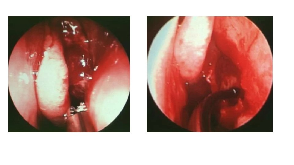
• Glistening and pale growth liken to peel grape appearance are characteristic features of nasal polyps. • It is painless and can be quite mobile on probing as compared to nasal turbinates.
Close-up view of nasal polyp (P). Compare it with the inferior turbinate appearance (IT)
Theories of pathogenesis 1. Bernstein theory : Inflammatory changes in lateral nasal wall or sinus mucosa, polyps originate from contact area. Ulceration, reepithelialisation and new gland formation. Inflammatory processes from epithelial cells, endothelium and fibroblasts. Integrity of sodium channels affected 2. Vasomotor theory: increased vascular permeability and impaired vascular regulation cause detoxification of mast-cell products (eg, histamine). The prolonged effects of these products within the polyp stroma result in marked edema (especially in the polyp pedicle) that is worsened by venous drainage obstruction. 3. Epithelial rupture theory suggests that rupture of the epithelium of the nasal mucosa is caused by increased tissue turgor in illness (eg, allergies, infections). This rupture leads to prolapse of the lamina propria mucosa, forming polyps.
Classification Multifactorial , Asthma is a common association Polyps can also be a presenting feature of fungal sinusitis or Sino nasal malignancy
Antrochoanal polyp Seen more commonly in adolescents and young children
Ethmoid polyps Usually presents in middle aged adults
Clinical presentation
Physical exam
Diagnosis
Management ethmoidal polyps • Aggressive medical management as mentioned previously in management of CRSw. NP ( combination of systemic and topical steroids, antimicrobial therapy, nasal irrigation). • For refractory symptoms despite aggressive medical therapy FESS + polypectomy. • Endoscopic sinus surgery appears to improve both olfaction and quality of life in chronic rhinosinusitis patients with nasal polyps. • Polyposis recurrence is common following treatment with medical or surgical.
Management Antrochoanal polyps • Simple polypectomy ( avulsion from the stalk). • FESS is a better technique that not only removes the polyps but also opens the clefts in the middle meatus, where they most often form, which helps decrease the recurrence rate.
Fungal sinusitis Classification
Classification of Infection • Non-invasive • Sinus fungal ball (Mycetoma) • Allergic fungal RS • Invasive • Acute fulminant invasive fungal sinusitis • Chronic invasive fungal sinusitis • Granulomatous invasive fungal sinusitis
Sinus Fungal Ball • Sequestration without invasion • Aspergillus • Maxillary sinus (70 -80% ) • concomitant in southern India. • normal immunologic status and in immunocompromised as well • Treatment : surgical (FESS) � surgical removal of the fungus ball � No medical treatment is required � recurrence or relapse is exceptional.
Allergic Fungal Rhinosinusitis (AFRS) • hyphal elements detected by fungal stains. • Culture : variety of fungal species. • vary geographically: ** In North America : Bipolaris and Alternaria and Curvularia. ** In northern India : Aspergillus flavus • Diagnosis : high Ig. E + no invasion • Treatment : 1. Surgery (FESS) 2. Post operative topical +/- systemic steroids 3. Immunotherapy
Diagnostic criteria of AFRS
Acute fulminant Invasive Fungal Rhinosinusitis • Altered host defenses : ** transplantation ** DKA ** leukemia. • Mainly due to Aspergillus (A. fumigatus or A. flavus. ), Mucormycosis • The time course is rapid, and on histopathologic inspection, the fungus can be seen invading tissue. • Invasive FRS may be present : ** Ophthalmoplegia, orbital swelling ** proptosis, and visual loss • Nasal endoscopy : necrosis of the nasal mucosa, only edema and pallor • Management : 1. Urgent Endoscopic debridement. 2. Combined with systemic antifungal therapy (Amphotericin B)
Thank you Good luck