
Approach to the Patient with Liver Disease Liver

- Slides: 106
Approach to the Patient with Liver Disease
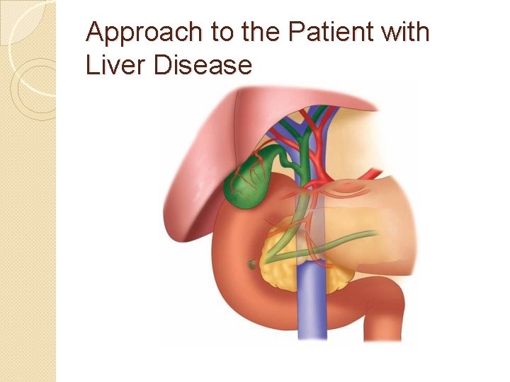
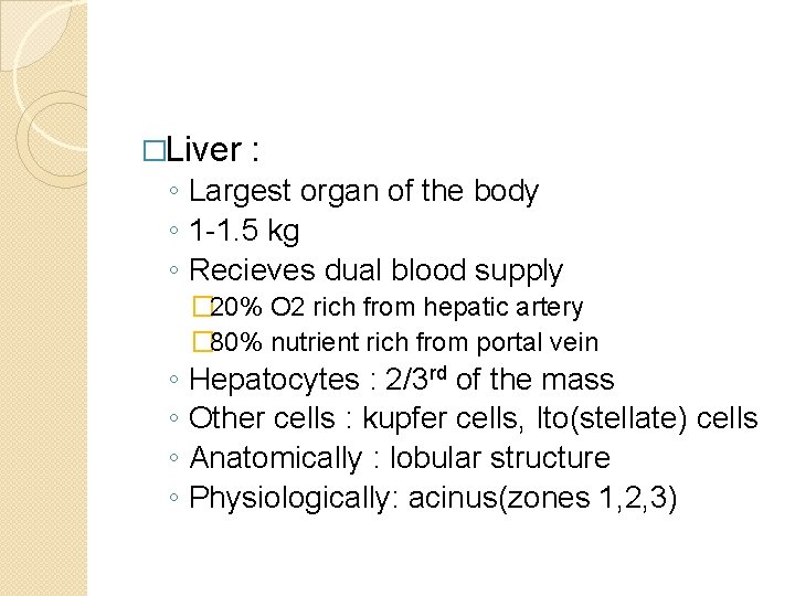
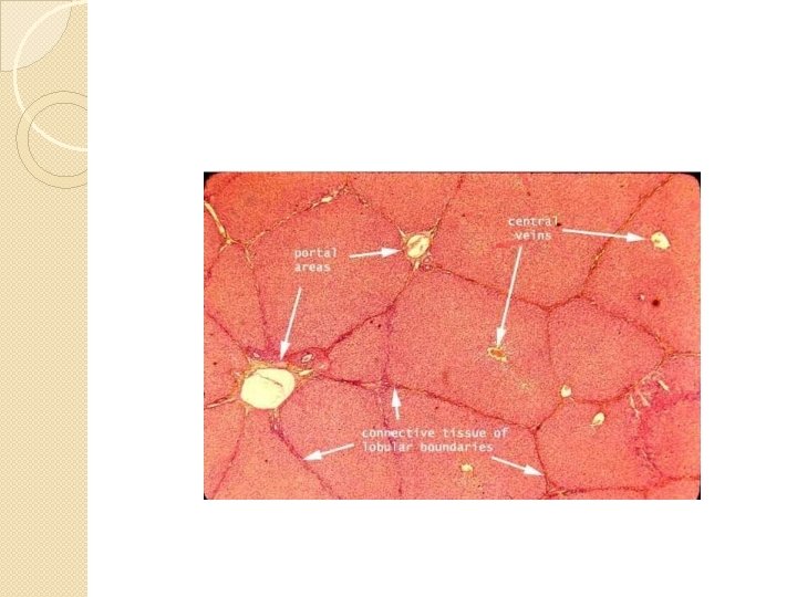
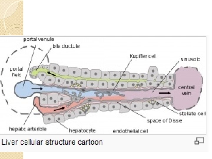
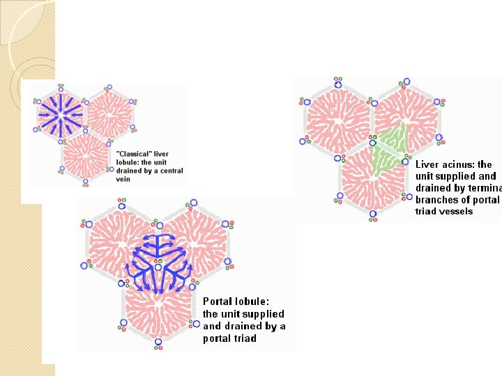
�Liver : ◦ Largest organ of the body ◦ 1 -1. 5 kg ◦ Recieves dual blood supply � 20% O 2 rich from hepatic artery � 80% nutrient rich from portal vein ◦ ◦ Hepatocytes : 2/3 rd of the mass Other cells : kupfer cells, Ito(stellate) cells Anatomically : lobular structure Physiologically: acinus(zones 1, 2, 3)
Clinical History � focus on the ◦ symptoms of liver disease—their nature, patterns of onset, and progression ◦ potential risk factors for liver disease � Non specific symptoms ◦ Fatigue: intermittent, afternoon fatigue ◦ Nausea: induced by food smell, vomiting not significant ◦ RUQ pain: distension of Glisons capsule �Severe: gall bladder ds, liver abcsess, severe venooclusive ◦ Itching : early in cholestatic ds, late with hepatocellular(cirhosis) ◦ Jaundice: most reliable marker of severity �Jaundice without darkening of urine indirect bilirubinemia due to hemolytic anemia
�Major risk factors ◦ include details of alcohol use, ◦ medications (including herbal compounds, birth control pills, and over-the-counter medications), ◦ personal habits, ◦ sexual activity, travel, ◦ exposure to jaundiced or other high-risk persons, ◦ injection drug use, ◦ recent surgery, ◦ remote or recent transfusion with blood and blood products, ◦ occupation, accidental exposure to blood or needlestick, and familial history of liver disease
Risk factors for viral hepatitis �Hepatitis B: sexual, mother to child, transfusion, IVD abuse �Hepatitis C: IVD, transfusion, sexual, mother to child �Hepatitis A: contact with jaundiced person, with day care babies
�Alcohol: ◦ > 3 drinks(33 -45 gm) per day in men and > 2 in females for long time(> 10 yrs) ◦ Abuse and dependance ◦ CAGE questions
�Familial liver diseases ◦ ◦ Wilsons Hemochromatosis @ 1 anti trypsin deficiency Familial intrahepatic cholestasis
Evaluation of liver function �To know the presence of liver disease �To identify the type of liver disease �To gauge the severity �To follow treatment response �Not specific or sensitive �No one test tells the liver function �Used as battery of tests ◦ Aminotransferases, ALP, bili, alb, PT
Diagnostic work up of patients with diseases of hepatobiliary system. Biochemical tests: q Tests for the detoxification capacity of the liver : ◦ ο Serum ammonia level ◦ ο Serum bilirubin, urine bilirubin level q Tests for synthetic function of the liver: ◦ ο Serum albumin level ◦ ο Prothrombin time q Tests indicating hepatocellular damage ◦ ο Transaminases (serum glutamic oxalate transferase (SGOT) and serum glutamic pyruvate transferase (SGPT)) q Tests which show cholestasis ◦ ο Alkaline phosphatase ◦ ο (Direct) bilirubin level
Ultrasound : help full in diagnosing the following diseases: �o Cirrhosis �o Metastasis �o Fluid filled lesions – cysts, abscess �o Cholelithiasis 3. Biopsy 4. Cholangiography: is contrast study of the biliary tree
Liver disease � 3 patterns ◦ Hepatocellular ◦ Cholestatic ◦ Mixed
Hepatitis
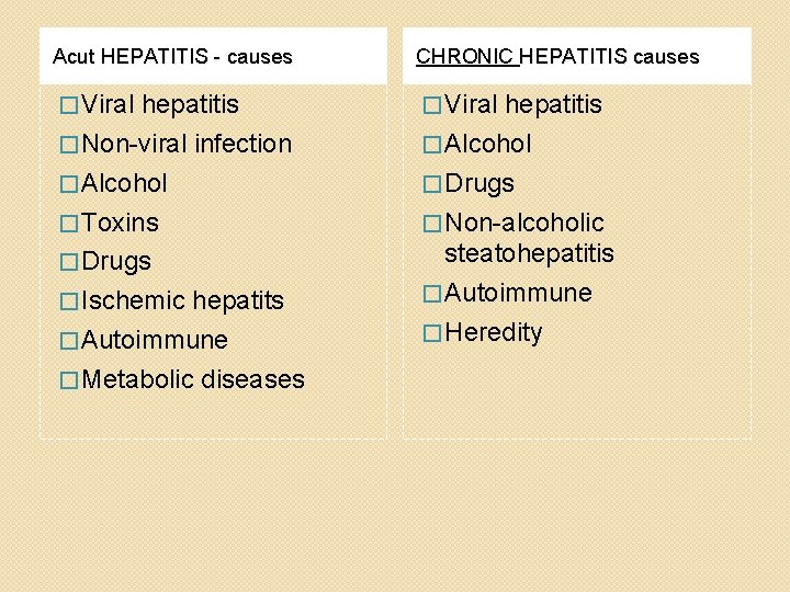
�Hepatitis is a broad category of clinico- pathologic conditions resulting from viral, toxic, pharmacologic or immune mediated damage to the liver. �Hepatocellular necrosis and inflammatory cell infiltration of the liver are common pathologic features. �There are two types of hepatitis, which are defined based on duration: ◦ I. Acute hepatitis: lasts for about 6 months or less. ◦ II. Chronic hepatitis: sustained inflammatory response for over 6 months
Acut HEPATITIS - causes CHRONIC HEPATITIS causes � Viral hepatitis � Non-viral infection � Alcohol � Drugs � Toxins � Non-alcoholic � Drugs steatohepatitis � Autoimmune � Heredity � Ischemic hepatits � Autoimmune � Metabolic diseases
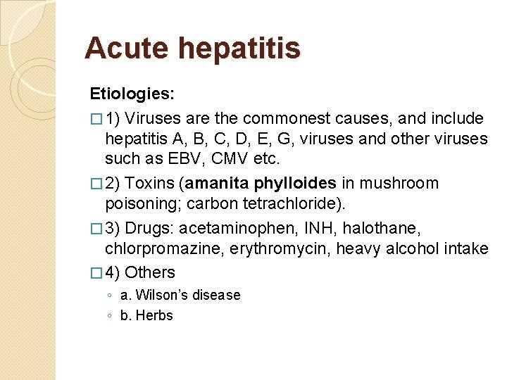
Acute hepatitis Etiologies: � 1) Viruses are the commonest causes, and include hepatitis A, B, C, D, E, G, viruses and other viruses such as EBV, CMV etc. � 2) Toxins (amanita phylloides in mushroom poisoning; carbon tetrachloride). � 3) Drugs: acetaminophen, INH, halothane, chlorpromazine, erythromycin, heavy alcohol intake � 4) Others ◦ a. Wilson’s disease ◦ b. Herbs
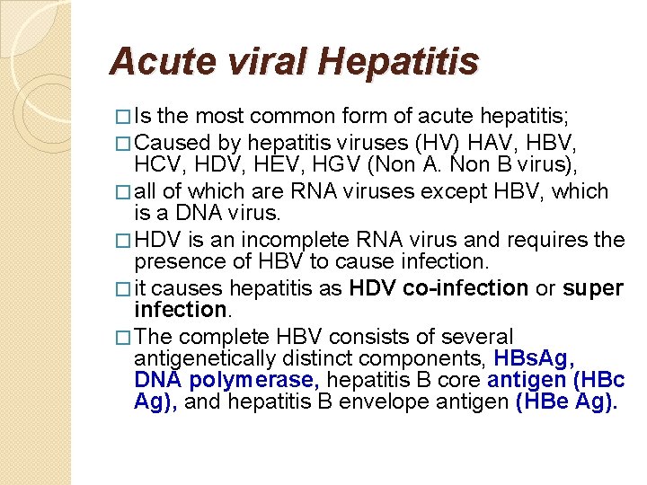
Acute viral Hepatitis � Is the most common form of acute hepatitis; � Caused by hepatitis viruses (HV) HAV, HBV, HCV, HDV, HEV, HGV (Non A. Non B virus), � all of which are RNA viruses except HBV, which is a DNA virus. � HDV is an incomplete RNA virus and requires the presence of HBV to cause infection. � it causes hepatitis as HDV co-infection or super infection. � The complete HBV consists of several antigenetically distinct components, HBs. Ag, DNA polymerase, hepatitis B core antigen (HBc Ag), and hepatitis B envelope antigen (HBe Ag).
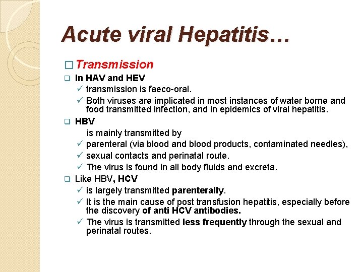
Acute viral Hepatitis… � Transmission In HAV and HEV ü transmission is faeco-oral. ü Both viruses are implicated in most instances of water borne and food transmitted infection, and in epidemics of viral hepatitis. q HBV is mainly transmitted by ü parenteral (via blood and blood products, contaminated needles), ü sexual contacts and perinatal route. ü The virus is found in all body fluids and excreta. q Like HBV, HCV ü is largely transmitted parenterally. ü It is the main cause of post transfusion hepatitis, especially before the discovery of anti HCV antibodies. ü The virus is transmitted less frequently through the sexual and perinatal routes. q
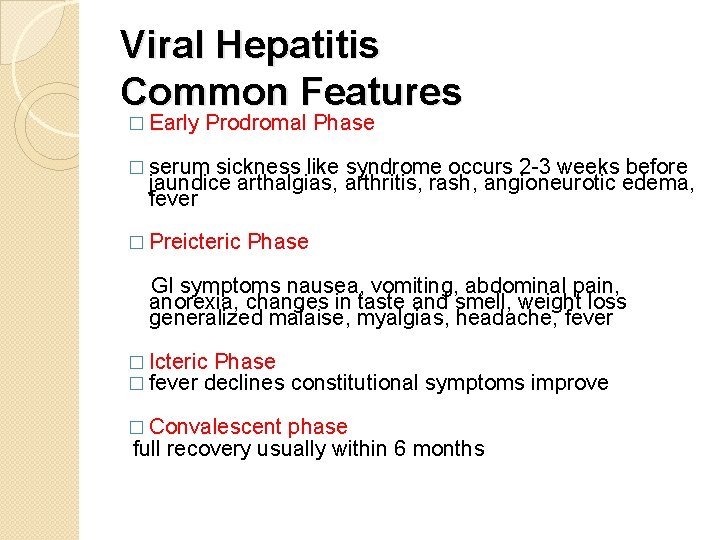
Viral Hepatitis Common Features � Early Prodromal Phase � serum sickness like syndrome occurs 2 -3 weeks before jaundice arthalgias, arthritis, rash, angioneurotic edema, fever � Preicteric Phase GI symptoms nausea, vomiting, abdominal pain, anorexia, changes in taste and smell, weight loss generalized malaise, myalgias, headache, fever � Icteric Phase � fever declines constitutional symptoms improve � Convalescent phase full recovery usually within 6 months
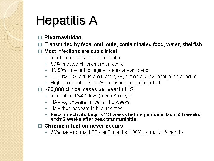
Hepatitis A Picornaviridae � Transmitted by fecal oral route, contaminated food, water, shellfish � Most infections are sub clinical � ◦ ◦ ◦ � >60, 000 clinical cases per year in U. S. ◦ ◦ � Incidence peaks in fall and winter 80% infected children are anicteric 10 -50% infected college students are anicteric 30 -50% U. S. adults are HAV Ig. G+, but only 3 -5% recall prior jaundice High attack rate: 70 -90% exposed become infected Incubation 15 -49 days (mean 30 days) HAV Ag appears in liver at 1 -2 weeks HAV then appears in bile and stool Fecal infectivity begins 2 -3 weeks before jaundice, lasts 4 -5 weeks, ends 2 weeks after peak transaminitis Chronic infection never occurs ◦ 60% have normal LFT’s at 2 months; 100% normal at 6 months
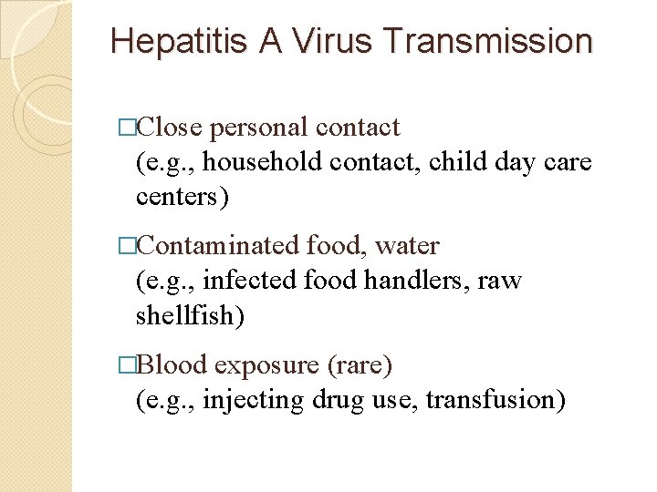
Hepatitis A Virus Transmission �Close personal contact (e. g. , household contact, child day care centers) �Contaminated food, water (e. g. , infected food handlers, raw shellfish) �Blood exposure (rare) (e. g. , injecting drug use, transfusion)
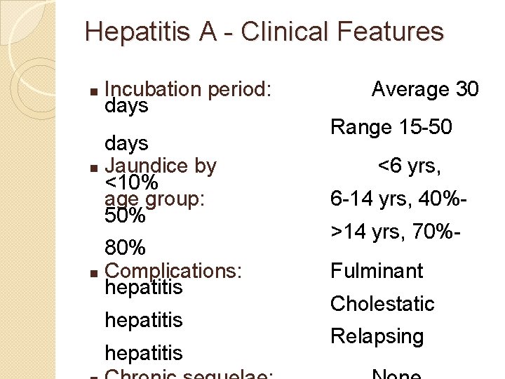
Hepatitis A - Clinical Features n Incubation period: days n Jaundice by <10% age group: 50% 80% n Complications: hepatitis Average 30 Range 15 -50 <6 yrs, 6 -14 yrs, 40%>14 yrs, 70%Fulminant Cholestatic Relapsing
Hepatitis A Atypical Features � Death is uncommon ◦ Overall mortality rate is 0. 14% ◦ If age > 40, mortality rate is 1. 1% � Prolonged cholestasis >3 months ◦ Severe pruritis, fatigue, weight loss, diarrhea ◦ May improve with steroids � Relapsing hepatitis ◦ Occurs in 6 -12% of cases, 4 -15 weeks after recovery � Extrahepatic manifestations ◦ Rash (14%), arthralgias (11%), ◦ Immune complex diseases: leukocytoclastic vasculitis, glomerulonephritis, arthritis, cryroglobulinemia ◦ Extremely rare: myocarditis, transverse myelitis, optic neuritis, polyneuritis, aplastic anemia
Laboratory Diagnosis �detection of anti HAV antibody which is Ig. M for acute infection and Ig. G later on. �The Ig. G antibody confers lifelong immunity
Hepatitis A Prevention � General prevention ◦ ◦ � HAV Immunoglobulin ◦ ◦ ◦ � Water chlorination Boil water 20 minutes Wash hands Avoid contaminated food Can prevent 85 -95% infections if given within two weeks of exposure Household and sexual contacts Day care contacts Prison contacts Common source outbreaks HAV Vaccine ◦ ◦ ◦ ◦ 90 -98% successful with one injection, 100% with two injections Protection begins after 1 -2 weeks, may last 20 years Give to all of the above Travelers to endemic areas Homosexuals, IV drug abusers Persons with HCV and HBV Military
Hepatitis B Virus �Member of hepadnavirus family with partly single-stranded DNA �Ä Dane particle is probably complete virion and contains HBs. Ag �Ä Incomplete forms (spherical and filamentous particles) also contain HBs. Ag �Ä Virion cores (HBc. Ag) are contained within the incomplete forms �Ä HBe. Ag exists in serum and on hepatocytes
Hepatitis B cont…. � Transmission route is variable ◦ HBV is found in blood and all body fluids except stool ◦ “Western” societies: percutaneous, hetero/homosexual contact is most common ◦ “Non-western” societies: perinatal transmission is most common � Epidemiology ◦ Worldwide � 2 billion people have markers of infection � 400 million have chronic infection (5%) ◦ U. S. � 1. 25 million chronic infections (50% Asian) � 200 thousand acute infections per year � 250 deaths/year from fulminant HBV � 4000 deaths/year from chronic HBV � 800 deaths/year from HBV related hepatomas
Hepatitis B Virus Modes of Transmission § Sexual - sex workers and homosexuals are particular at risk. § Parenteral - IVDA, Health Workers are at increased risk. § Perinatal - Mothers who are HBe. Ag positive are much more likely to transmit to their offspring than those who are not. Perinatal transmission is the main means of transmission in high prevalence populations
Transmission cont… �Without intervention, a mother who is positive for the hepatitis B surface antigen confers a 20% risk of passing the infection to her offspring at the time of birth. �This risk is as high as 90% if the mother is also positive for the hepatitis B e antigen. �Roughly 16 -40% of unimmunized sexual partners of individuals with hepatitis B will be infected through sexual contact. �The risk of transmission is closely related to the rate of viral replication in the infected individual at the time of exposure.
Concentration of Hepatitis B Virus in Various Body Fluids �High ; blood , serum , wound exudates �Moderate ; semen , vaginal fluid , saliva �Low/Not detectable , urine , feces , sweat , tears , breast milk
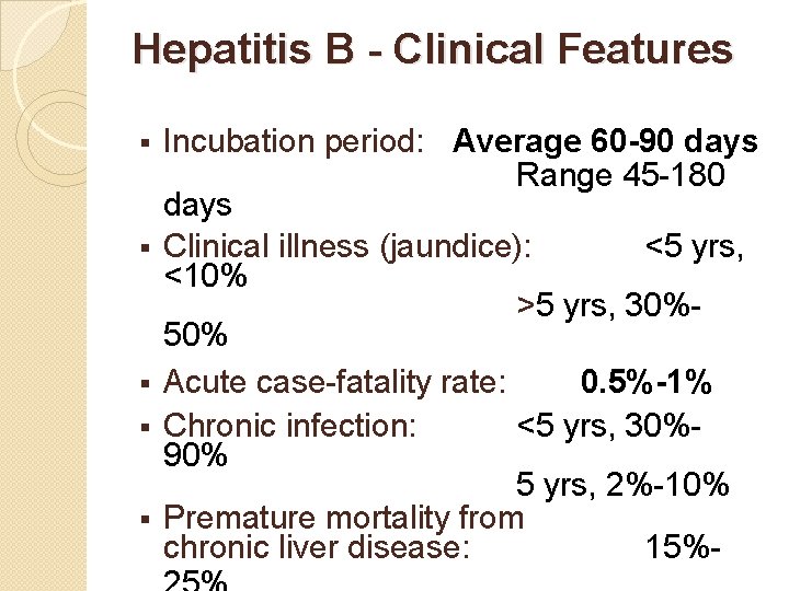
Hepatitis B - Clinical Features § § § Incubation period: Average 60 -90 days Range 45 -180 days Clinical illness (jaundice): <5 yrs, <10% >5 yrs, 30%50% Acute case-fatality rate: 0. 5%-1% Chronic infection: <5 yrs, 30%90% 5 yrs, 2%-10% Premature mortality from chronic liver disease: 15%-
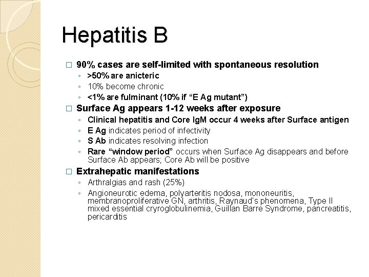
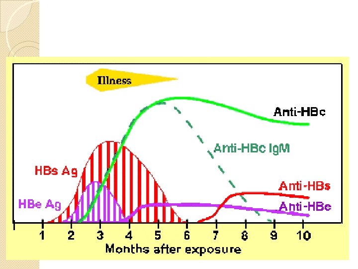
Hepatitis B � 90% cases are self-limited with spontaneous resolution ◦ >50% are anicteric ◦ 10% become chronic ◦ <1% are fulminant (10% if “E Ag mutant”) � Surface Ag appears 1 -12 weeks after exposure ◦ ◦ � Clinical hepatitis and Core Ig. M occur 4 weeks after Surface antigen E Ag indicates period of infectivity S Ab indicates resolving infection Rare “window period” occurs when Surface Ag disappears and before Surface Ab appears; Core Ab will be positive Extrahepatic manifestations ◦ Arthralgias and rash (25%) ◦ Angioneurotic edema, polyarteritis nodosa, mononeuritis, membranoproliferative GN, arthritis, Raynaud’s phenomena, Type II mixed essential cryroglobulinemia, Guillan Barre Syndrome, pancreatitis, pericarditis
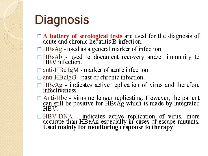
Diagnosis �A battery of serological tests are used for the diagnosis of acute and chronic hepatitis B infection. � HBs. Ag - used as a general marker of infection. � HBs. Ab - used to document recovery and/or immunity to HBV infection. � anti-HBc Ig. M - marker of acute infection. � anti-HBc. Ig. G - past or chronic infection. � HBe. Ag - indicates active replication of virus and therefore infectiveness. � Anti-Hbe - virus no longer replicating. However, the patient can still be positive for HBs. Ag which is made by integrated HBV. � HBV-DNA - indicates active replication of virus, more accurate than HBe. Ag especially in cases of escape mutants. Used mainly for monitoring response to therapy
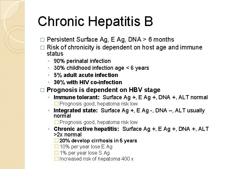
Chronic Hepatitis B Persistent Surface Ag, E Ag, DNA > 6 months � Risk of chronicity is dependent on host age and immune status � ◦ ◦ � 90% perinatal infection 30% childhood infection age < 6 years 5% adult acute infection 30% with HIV co-infection Prognosis is dependent on HBV stage ◦ Immune tolerant: Surface Ag +, E Ag +, DNA +, ALT normal � Prognosis good, hepatoma risk low ◦ Integrated state: Surface Ag +, E Ag -, DNA –, ALT usually normal � Prognosis good, hepatoma risk low ◦ Chronic active hepatitis: Surface Ag +, E Ag +, DNA +, ALT >2 x normal � 20% develop cirrhosis in 5 years � 10% per year lose E Ag � 1% per year lose S Ag � Increased risk of hepatoma 400 x
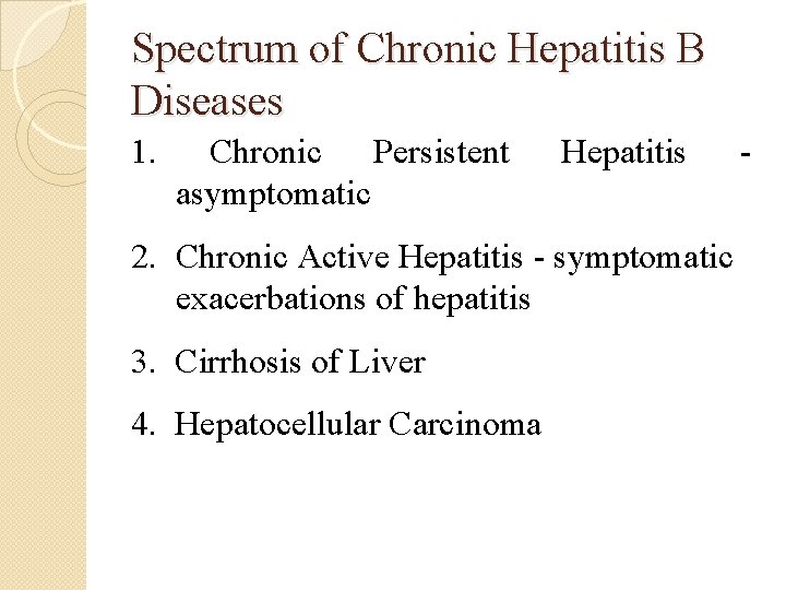
Spectrum of Chronic Hepatitis B Diseases 1. Chronic Persistent asymptomatic Hepatitis 2. Chronic Active Hepatitis - symptomatic exacerbations of hepatitis 3. Cirrhosis of Liver 4. Hepatocellular Carcinoma -
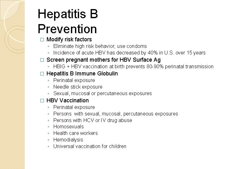
Hepatitis B Prevention � Modify risk factors ◦ Eliminate high risk behavior; use condoms ◦ Incidence of acute HBV has decreased by 40% in U. S. over 15 years � Screen pregnant mothers for HBV Surface Ag ◦ HBIG + HBV vaccination at birth prevents 80 -90% perinatal transmission � Hepatitis B Immune Globulin ◦ Perinatal exposure ◦ Needle stick exposure ◦ Sexual, mucosal or percutaneous exposures � HBV Vaccination ◦ ◦ ◦ ◦ Perinatal exposure Persons with sexual, mucosal, percutaneous exposures Persons with HCV or IV drug abuse Homosexuals Health care workers Hemodialysis Universal vaccination for children
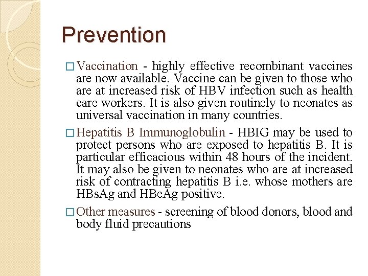
Prevention � Vaccination - highly effective recombinant vaccines are now available. Vaccine can be given to those who are at increased risk of HBV infection such as health care workers. It is also given routinely to neonates as universal vaccination in many countries. � Hepatitis B Immunoglobulin - HBIG may be used to protect persons who are exposed to hepatitis B. It is particular efficacious within 48 hours of the incident. It may also be given to neonates who are at increased risk of contracting hepatitis B i. e. whose mothers are HBs. Ag and HBe. Ag positive. � Other measures - screening of blood donors, blood and body fluid precautions
Treatment �Interferon - for HBe. Ag +ve carriers with chronic active hepatitis. Response rate is 30 to 40%. �Lamivudine - a nucleoside analogue reverse transcriptase inhibitor. Well tolerated, most patients will respond favorably. However, tendency to relapse on cessation of treatment. Another problem is the rapid emergence of drug resistance. �Successful response to treatment will result in the disappearance of HBs. Ag, HBV-DNA, and seroconversion to HBe. Ag
Treatment cont… � Who to treat? ◦ Chronic active disease > 6 months ◦ Surface Ag +, DNA +, E Ag + or – (if E Ag mutant) ◦ ALT > 100, and/or active hepatitis on biopsy � Goal of treatment ◦ Stop viral replication, HBV DNA becomes neg ◦ Convert E Ag pos to neg, E AB becomes pos ◦ Improvement in histology, prevention of progression to cirrhosis ◦ With successful treatment, loss of Surface Ag may occur in 1 -2% per year
�Alpha-interferon 2 b �Lamivudine �Adefovir �Famciclovir �Entecavir �Emtricitabine
Liver Transplant in Hepatitis B � 10% liver transplants are for HBV � Prior to effective immunoprophylaxis 80% of transplants became reinfected with very poor survival � Most centers stopped transplanting for active HBV � Reinfection rate can now be reduced effectively: ◦ HBIG for 6 -12 months � 20% reinfected ◦ Addition of Lamivudine to HBIG � 5 -10% reinfected � 2 year survival with transplant is now 70 -80%
Hepatitis C �HCV is a linear, ss RNA virus �HCV is the only member of the genus Hepacivirus in the family Flaviviridae - A core of genetic material (+ ss. RNA) - A lipid fatty envelope (cellular origin) - Two viral envelope glycoproteins (E 1 and E 2)
TRANSMISSION- primarily by: 1. Direct percutaneous exposures to infectious blood, such as injection drug use 2. The transfusion of contaminated blood products 3. Inefficiently transmitted through sexual contact; however, persons with multiple sexual partners have an increased risk 4. From mother to child in approximately 5 -6% of pregnant women who have chronic infection at the time of delivery. 5. Tattooing with contaminated needle
HCV cont… v IP: 15 -160 days (mean: 50 days); CLINICAL FEATURES : § Acute HCV infection is often silent, evident if there is circumstantial evidence for new infection, such as a recent exposure to a known HCV-infected person or clinical features of acute hepatitis with the exclusion of other causes of hepatitis. § Acute hepatitis C rarely causes fulminant hepatic failure. LONG-TERM EFFECTS : § 50 -85% with HCV develop chronic infection, § 15 - 50% of newly infected clear the virus spontaneously. § 10 -15% with chronic HCV infection develop progressive fibrosis of the liver leading to cirrhosis. § Hepatocellular carcinoma (HCC) risk is about 1 - 4% / year § Deaths from CLD: 1 -5%
Hepatitis C cont…. DIAGNOSIS: ACUTE: 1) marked elevations in ALT (>7 times) with or without symptoms of acute hepatitis; 2) negative tests for acute hepatitis A (Ig. M anti. HAV) and acute hepatitis B (Ig. M anti-HBc); and 3) a positive anti-HCV screening immunoassay 4) HCV RNA may be detected by PCR 1 -3 weeks after exposure, but viremia may be transient, i. e. , a negative qualitative HCV RNA does not preclude acute HCV infection
v. TESTING RECOMMENDATIONS FOLLOWING A KNOWN EXPOSURE: 1) nucleic acid test for HCV RNA immediately after exposure, at week 4 and at week 12; 2) anti-HCV by immediately after exposure and at week 12 3) serum ALT and AST immediately after exposure, at week 4 and 12. CHRONIC: A quantitative HCV nucleic acid test, v. If negative, the more sensitive qualitative HCV nucleic acid test (PCR) should be obtained
Hepatitis C Factors Associated with Disease Progression �Age > 40 �Male �Alcohol > 50 gm/d �Immunosuppression: HIV, transplant, etc. �Infection by blood transfusion �Co-infection with HBV �Genotype 1
TREATMENT: 1. antiviral therapy is beneficial in treating persons with acute HCV infection; 2. however, the timing and the optimal treatment regimen in this setting are uncertain. 3. In one study, 52% of symptomatic individuals with acute hepatitis C spontaneously cleared the virus within 12 weeks. 4. Of those who did not clear the virus, treatment initiated within 3 -6 months of infection resulted in a sustained virological response in 81%. 5. The combination of interferon + ribavirin is significantly effective in clearing viremia and establishing sustained viral response rates
HCV/HIV co infection �Co infection with HCV is found in at least 30% of persons infected with HIV �HIV infection leads to: ◦ an increased risk of acute hepatic failure ◦ more rapid progression of chronic hepatitis C to cirrhosis ◦ in addition, HCV increases the hepatotoxicity of HAART
Prevention of Hepatitis C § Screening of blood, organ, tissue donors § High-risk behavior modification § Blood and body fluid precautions
Hepatitis D �Defective single stranded RNA virus - requires HBV for synthesis of envelope protein (HBs. Ag encapsulates genome) �Therefore only exists as co-infection with HBV or as superinfection of chronic HBs. Ag-positive individual
Hepatitis D (Delta) Virus cont… � Epidemiology � • Spread most commonly by percutaneous or permucosal exposure � - Perinatal transmission reported but rare � • More prevalent in Mediterranean countries, Romania, some isolated � areas in the Amazon River basin � Ä Clinical Features � • Coinfection with HBV ◦ - Fulminant hepatitis more common (2 -5%) ◦ - Less commonly become HBs. Ag carriers � • Superinfection in chronic HBV ◦ Fulminant hepatitis more common (up to 20%) ◦ - More likely to develop chronic HDV infection ◦ - 70% develop cirrhosis (2 -5 times more frequent than with HBV alone) � • Incubation period probably the same as HBV
� � Defective RNA virus, requires presence of HBV Surface Ag 7500 new cases/year in U. S. ◦ More common in southern, eastern Europe, Middle East, and South America � � � Transmission is similar to HBV Diagnosis: HDV Ag, HDV RNA, HDV Ig. G and Ig. M Acute Hepatitis ◦ Co-infection with HBV � Fulminant hepatitis more common (34%) � Progression to chronic infection is uncommon ◦ Super-infection of HBV � Acute exacerbation of ongoing hepatitis � Chronic liver disease occurs in 90% � Chronic Hepatitis B + D ◦ More progressive than HBV alone ◦ 15% have rapid progression to cirrhosis in 1 year � Treatment ◦ ◦ a-interferon 2 b 3 -9 mu sq tiw, Rx > 12 months 21 -50% lose HDV RNA and have improved histology Relapse occurs in almost all patients stopping treatment Can stop treatment if HBV Surface Ag disappears (rare)
Hepatitis D - Clinical Features Coinfection – severe acute disease. – low risk of chronic infection. § Superinfection – usually develop chronic HDV infection. – high risk of severe chronic liver disease. – may present as an acute hepatitis §
Hepatitis D Virus Modes of Transmission § Percutanous exposures § injecting drug use § Permucosal exposures § sex contact
Hepatitis D -Prevention n HBV-HDV Coinfection Pre or postexposure prophylaxis to prevent HBV infection. n HBV-HDV Superinfection Education to reduce risk behaviors among persons with chronic HBV infection.
Hepatitis E � Hepatitis E virus is RNA virus TRANSMISSION: 1. HEV is found in the stool (feces) of persons with hepatitis E. 2. HEV is spread by eating or drinking contaminated food or water. 3. Transmission from person to person occurs less commonly than with HAV 4. Most outbreaks in developing countries have been associated with contaminated drinking water. 5. Pregnant women are more prone to develop HEV infection v IP: 14 -60 days (mean 40 days)
CLINICAL FEATURES : 1. Young (15 - 40) and pregnant women in 3 rd trimester (10 -20 % mortality) are more prone 2. Jaundice, fatigue LONG-TERM EFFECTS: None DIAGNOSIS : Virus detection in stool and anti HEV antibody (Ig. M) TREATMENT: supportive
Acute viral Hepatitis �Complications 1) Cholestatic hepatitis – ◦ ◦ Occurs most commonly during HAV and HEV infection • Self limited • Pruritus is a common symptom • Marked conjugated hyperbilirubinemia presenting with dark urine and pale stool ◦ • Increased alkaline phosphatase 2) Fulminant hepatitis : in about 1% of cases � � � • Is defined as the onset of encephalopathy occurring within 8 wks in a patient with acute liver disease • Associated with massive hepatocellular necrosis • Commonly follows HBV & HDV (usually super infection of HBV infection by HDV), As well as HEV (esp, in pregnancy) but rare fulminant cases of hepatitis A occur Fulminant hepatitis is hardly ever seen in hepatitis C
Chronic hepatitis • Occurs in HBV and HCV infection • Manifest with persistently increased AST and ALT for over 6 months, � • Slowly resolving HBV infection may persist for 6 -12 m with eventual complete resolution � • Persistent HBV infection (persistence of HBs. Ag) without evidence of liver damage resulting in asymptomatic or “healthy” HBV carriers.
• HBV can become a chronic infection • 90% of infants who are infected will become chronic • 25%-50% of children aged 1 -5 years will become chronic if infected • 5% of adults will become chronic if infected • 75%-85% of individuals who get Hepatitis C will subsequently develop a chronic infection • Of every 100 people that become infected with HCV • 75 -85 will develop a chronic infection • 60 -70 will develop chronic liver disease • 5 -20 will develop cirrhosis over a 20 -30 year span • 1 -5 will die as a consequence of the virus (liver cancer or cirrhosis)
Chronic Liver Diseases (CLD)
Learning objectives: At the end of this unit the student will be able to � 1. Define chronic liver diseases � 2. List the etiologies of CLD � 3. Describe the clinical features of CLD � 4. List the common complications of CLD � 5. Make a presumptive diagnosis of CLD � 6. Refer patients to hospitals for better diagnosis and treatment
CLD…. �Chronic liver diseases include: ◦ A. Chronic hepatitis ◦ B. Liver cirrhosis ◦ C. Hepatocellular carcinoma (Hepatoma)
Chronic hepatitis � Definition: is defined as a hepatic inflammatory process that fails to resolve after 6 months. v Etiologies: 1) chronic viral hepatitis ◦ Chronic Hepatitis B ◦ hepatitis B plus D ◦ Chronic Hepatitis C 2)Autoimmune Hepatitis 3) drug-associated chronic hepatitis (eg. methyldopa) 4) Idiopathic causes
Autoimmune Hepatitis � Autoantibodies against hepatocytes � Young/middle age, mainly women � Presentation: jaundice, RUQ pain, systemic symptoms � May be associated with other autoimmune conditions � Investigations - Type 1: anti-smooth muscle antibodies (80%), anti-nuclear antibodies (10%) - Type 2 (children): anti-liver/kidney microsomal type 1 antibodies - Liver biopsy � Rx: immunosuppressant's (steroids, azathioprine), transplant
PBC and PSC Primary Biliary Cirrhosis � Chronic granulomatous inflammation of interlobular bile ducts � Autoimmune: anti-mitochrondrial antibody (98%) � Associated with other autoimmune conditions � F: M 9: 1, 50 y/o Primary Sclerosing Cholangitis � Inflammation, fibrosis and strictures (‘beading’) of intra and extra-hepatic bile ducts � ? Autoimmune: ANCA (80%) � Associated with IBD � 20 -30% develop cholangiocarcinoma
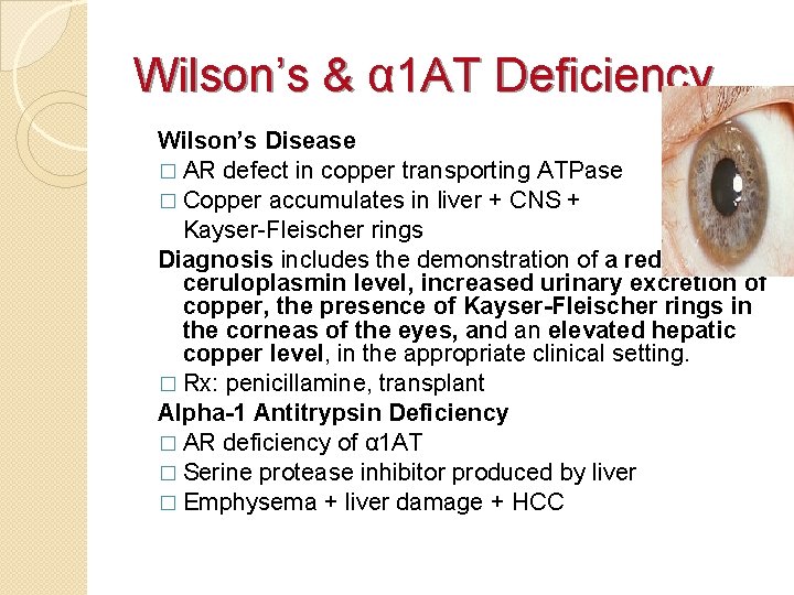
Wilson’s & α 1 AT Deficiency Wilson’s Disease � AR defect in copper transporting ATPase � Copper accumulates in liver + CNS + Kayser-Fleischer rings Diagnosis includes the demonstration of a reduced ceruloplasmin level, increased urinary excretion of copper, the presence of Kayser-Fleischer rings in the corneas of the eyes, and an elevated hepatic copper level, in the appropriate clinical setting. � Rx: penicillamine, transplant Alpha-1 Antitrypsin Deficiency � AR deficiency of α 1 AT � Serine protease inhibitor produced by liver � Emphysema + liver damage + HCC
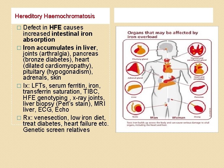
Hereditory Haemochromatosis Defect in HFE causes increased intestinal iron absorption � Iron accumulates in liver, joints (arthralgia), pancreas (bronze diabetes), heart (dilated cardiomyopathy), pituitary (hypogonadism), adrenals, skin � Ix: LFTs, serum ferritin, iron, transferrin saturation, TIBC, HFE genotyping , x-ray joints, liver biopsy (Perl’s stain), MRI liver, ECG, Echo � Rx: venesection, low iron diet, treat diabetes, heart failure etc. Genetic screen relatives �
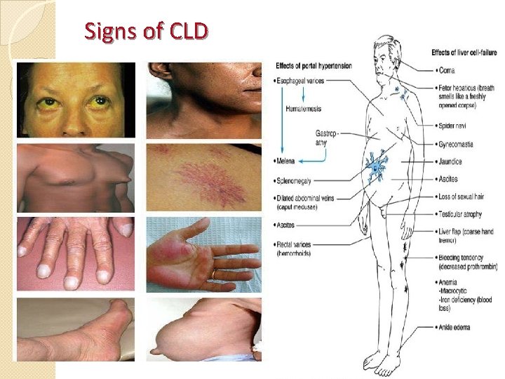
Signs of CLD
Cirrhosis �Cirrhosis represents a late stage of progressive liver fibrosis and disease and results in a distortion of liver architecture �Usually changes are irreversible �Liver transplantation may be the only option
�Cirrhosis is a histological diagnosis and is defined by the presence of nodules of regenerating hepatocytes surrounded by fibrous tissue � 1) Fibrous scarring and disruption of hepatic architecture distort vascular bed leading to Portal hypertension and intrahepatic shunting � 2) Disturbed hepatocellular function
Causes of Cirrhosis � 1) Alcohol and HCV are major causes in western world � 2) HBV is the commonest cause in developing countries � 3) Other causes include: �o Biliary obstruction (Intrahepatic and extrahepatic) leading to Biliary cirrhosis �o Autoimmune hepatitis
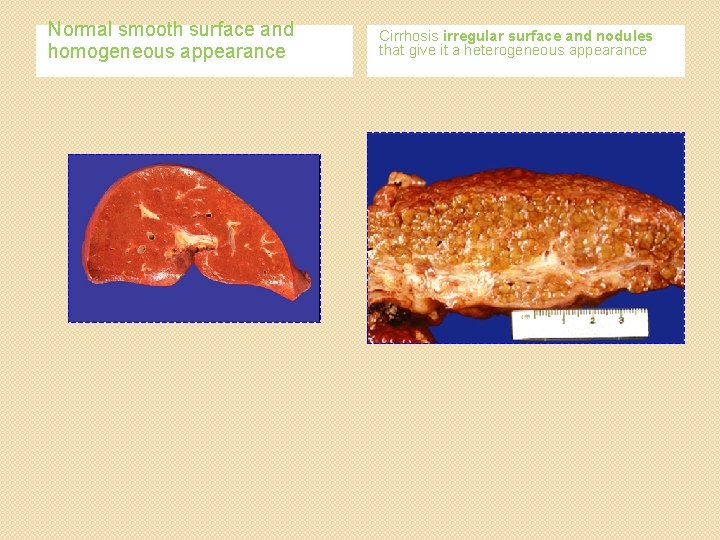
Normal smooth surface and homogeneous appearance Cirrhosis irregular surface and nodules that give it a heterogeneous appearance
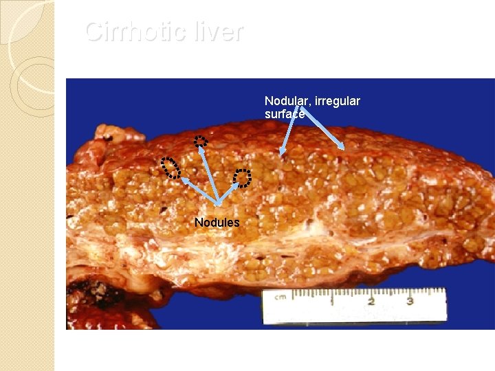
Cirrhotic liver Nodular, irregular surface Nodules
· Clinically there are two types of cirrhosis · Compensated · Decompensated
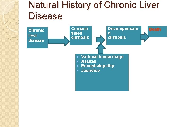
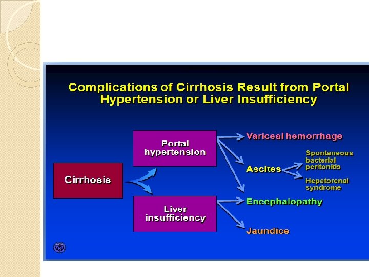
Natural History of Chronic Liver Disease Chronic liver disease Compen sated cirrhosis · · Decompensate d cirrhosis Variceal hemorrhage Ascites Encephalopathy Jaundice Death
Cirrhosis - Diagnosis · Cirrhosis is a histological diagnosis · However, in patients with CLD the presence of various clinical features suggests cirrhosis · The presence of these clinical features can be followed by non-invasive testing, prior to liver biopsy
In Whom Should We Suspect Cirrhosis · Any patient with chronic liver disease · Chronic abnormal aminotransferases and/or alkaline phosphatase · Physical exam findings · Stigmata of chronic liver disease (muscle wasting, vascular spiders, palmar erythema) · Palpable left lobe of the liver · Small liver span · Splenomegaly · Signs of decompensation (jaundice, ascites, asterixis)
In Whom Should We Suspect Cirrhosis? Laboratory · Liver insufficiency · Low albumin (< 3. 8 g/d. L) · Prolonged prothrombin time (INR > 1. 3) · High bilirubin (> 1. 5 mg/d. L) · Portal hypertension · Low platelet count (< 175 x 1000/ml) · AST / ALT ratio > 1
Clinical and laboratory features � Resulting from Hepatocellular dysfunction ◦ Hypoalbuminemia ◦ Decreased coagulation factors resulting in prolonged prothrombin time ◦ Hyperbilirubinemia ◦ Increased blood ammonia level ◦ Hepatic encephalopathy ◦ Ascites � Portal HTN ◦ Ascites. ◦ Varices ◦ Splenomegaly causing thrombocytopenia and leucopenia. ◦ Hepatic encephalopathy
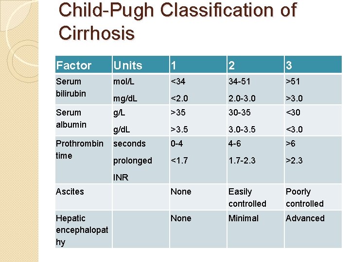
Staging system of cirrosis � Cirrhosis can also be staged clinically. � A reliable staging system is the modified Child- Pugh classification with a scoring system of 5– 15: � scores of 5 and 6 being Child-Pugh class A (consistent with "compensated cirrhosis"), � scores of 7– 9 indicating class B, and � 10– 15 indicating class C � The Child-Pugh score is calculated by adding the scores of the five factors and can range from 5 to 15. � Decompensation indicates cirrhosis with a Child. Pugh score of >7 (class B). � This level has been the accepted criterion for listing liver transplantation
Child-Pugh Classification of Cirrhosis Factor Units 1 2 3 Serum bilirubin mol/L <34 34 -51 >51 mg/d. L <2. 0 -3. 0 >3. 0 Serum albumin g/L >35 30 -35 <30 g/d. L >3. 5 3. 0 -3. 5 <3. 0 0 -4 4 -6 >6 <1. 7 -2. 3 >2. 3 None Easily controlled Poorly controlled Hepatic encephalopat hy None Minimal Advanced Prothrombin seconds time prolonged INR Ascites
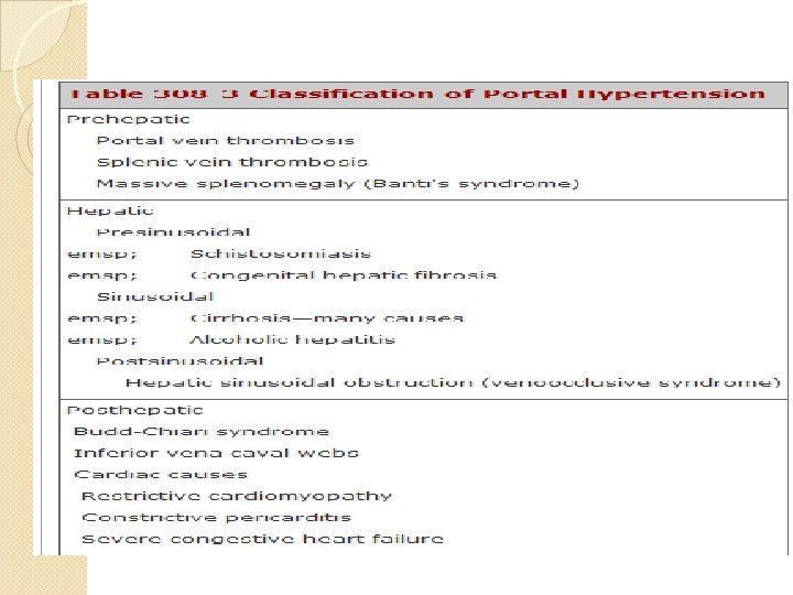
Major complications of cirrhosis 1) Portal Hypertension (HPN) �Normal portal pressure gradient 10 -15 cm of saline � Portal hypertension is said to exist when the portal pressure gradient is over 30 cm of saline � it leads to the formation of venous collateral vessels between the portal and systemic circulation. �the most important clinically being those connecting the portal vein to the azygous vein that form dilated, tortuous veins (varices) in the submucosa of the gastric fundus and esophagus
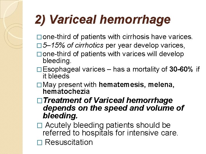
2) Variceal hemorrhage � one-third of patients with cirrhosis have varices. � 5– 15% of cirrhotics per year develop varices, � one-third of patients with varices will develop bleeding. � Esophageal varices – has a mortality of 30 -60% if it bleeds � May present with hematemesis, melena, hematochezia �Treatment of Variceal hemorrhage depends on the speed and volume of bleeding. � Acutely bleeding patients should be referred to hospitals for intensive care. � Resuscitation
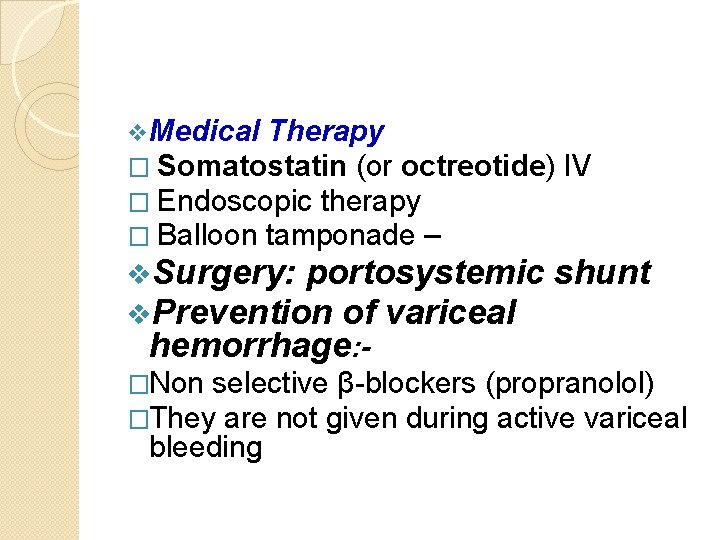
v. Medical Therapy � Somatostatin (or octreotide) IV � Endoscopic therapy � Balloon tamponade – v. Surgery: portosystemic v. Prevention of variceal shunt hemorrhage: - �Non selective β-blockers (propranolol) �They are not given during active variceal bleeding
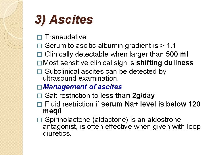
3) Ascites � Transudative � Serum to ascitic albumin gradient is > 1. 1 � Clinically detectable when larger than 500 ml � Most sensitive clinical sign is shifting dullness � Subclinical ascites can be detected by ultrasound examination. � Management of ascites � Salt restriction to less than 2 g/day � Fluid restriction if serum Na+ level is below 120 meq/l � Spirinolactone (aldactone) is an aldostrone antagonist, is often effective when given with loop diuretics.
�Duiresis should be monitored because vigorous duiresis leads to hypovolemia and hypokalemia and precipitate hepatic encephalopathy. �The goal of duiresis should be dependent on the extent of edema and be monitored by daily body weight measurement i. e. �o Decrease weight by 0. 5 kg/day if no peripheral edema and �o Decrease weight by 1 kg/day if peripheral edema is present.
Complications of ascites �Spontaneous bacterial peritonitis � Hepatopulmonary syndrome � Hepatorenal syndrome � Rupture of the umbilicus
Spontaneous bacterial peritonitis(SBP �Usually due to enterobacteriacae or pneumococci �Characterized by fever, abdominal pain and tenderness. �Hepatic encephalopathy may be precipitated by the infection. Diagnosis �is suspected when ascitic polymorphonuclear cell count is over 250/μl. and �confirmed by ascitic fluid culture. Treatment �is with third generation cephalosporin for 5 -7 days.
Prophylaxis for SBP Indications �Previous history of SBP � Patient admitted with gastrointestinal hemorrhages. � Ascitic protein less than 1 gm/dl �Candidates of liver transplantation Drugs � Norfloxacin – 400 mg/day �Ciprofloxacin – 750 mg/once weekly or �Co-trimoxazole - 960 mg/d for 5 days ( Monday – Friday)
4) Hepatic encephalopathy (Hepatic coma or portosystemic encephalopathy � It is a complex neuropsychiatric syndrome that may complicate advanced liver disease and/or extensive portosystemic shunt. � It manifests in two major forms: acute and chronic. Acute � Occurs in the setting of fulminant hepatitis � Cerebral edema plays a more important role � Mortality rate is very high Chronic � Occurs in chronic liver disease � Often reversible
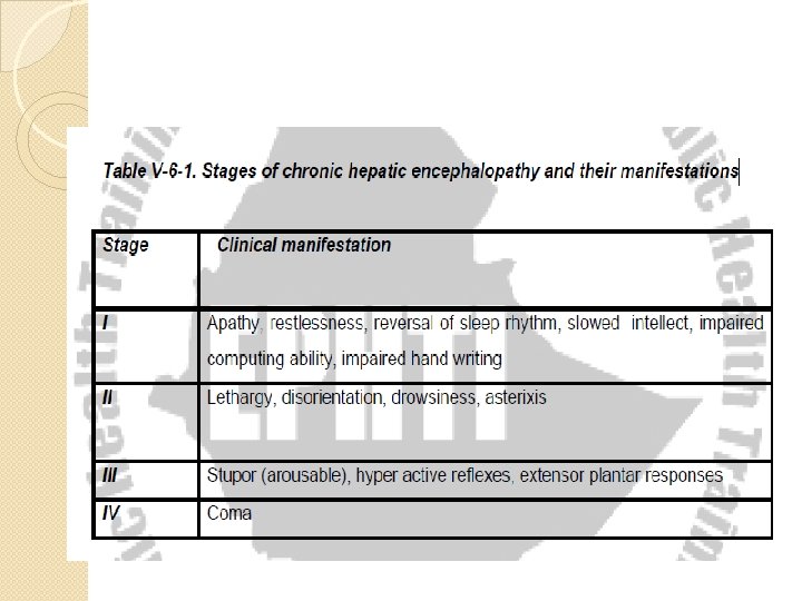
� � � � � The compounds commonly incriminated in causing hepatic encephalopathy include ◦ ο Ammonia is very well known and studied ◦ ο GABA Clinical Features may involve Disturbance of higher neurologic function Intellectual and personality disorder Dementia, inability to copy simple diagrams (constructional apraxia) Disturbance of consciousness Disturbance of neuromuscular function (asterixis, hyperreflexia, myoclonous) The findings are usually asymmetrical, The manifestations depend on the stage of HE
Treatment of chronic hepatic failure is based on the following principles Identify and treat precipitating factors including � GI bleeding � Increased dietary protein � Constipation � Alkalosis � Hypovolemia � Infection � Azotemia � Hypokalimia from overduiresis � CNS depressant drugs
Treatment cont… �Restrict dietary protein �For patients in coma no protein should be given �Cleanse the bowel with enema in patient with gastrointestinal hemorrhage �Give oral preparations of Neomycin or Metronidazole �Administering oral lactulose
C. Hepato cellular carcinoma (Hepatoma) �One of the most frequent malignancies and �important cause of mortality particularly in middle aged men in developing countries. �The incidence is less in developed countries �Arises in cirrhotic liver and is closely associated with chronic hepatitis B or C. �More common in males (m: f ratio 4: 1 to
Hepato cellular carcinoma (Hepatoma). . . Clinical and Laboratory findings �Common symptoms and findings ◦ ◦ Abdominal pain Weight loss Abdominal mass Derangement of liver function �Unusual manifestations ◦ Bloody ascites ◦ Tumor emboli ◦ Jaundice ◦ Gyneacomastia ◦ Hepatic bruit or friction rub
Hepato cellular carcinoma (Hepatoma). . . Metabolic effect � Erythrocytosis � Hypercholesterolemia � Hypoglycemia � Hypercalcemia � High serum α fetoprotein level (> 400 mg/dl) �Ultrasonography : Mass lesion with varying echogenicities but usually hypoechoic �Tissue specimen: may be required to confirm the diagnosis
Hepato cellular carcinoma (Hepatoma). . . � Median survival rate is less than 6 months from the time of diagnosis Treatment: � is usually unsatisfactory but some options include: �Chemotherapy �Radiotherapy � Liver transplantation
10 Q