CELLS ARE ALIVE Cytology the study of cells


- Slides: 34

CELLS ARE ALIVE Cytology – the study of cells (pg. ? ? )

Day 1

Cells Alive – www. cellsalive. com Brain. POP – www. brainpop. com username: edhpop password: edhpop 1 Check out these videos: - Cells - Cell specialization

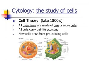
Cell Theory The Cell Theory states that: 1. All living things are composed of a cell or cells. 2. Cells are the basic (smallest) unit of life. 3. All cells come from preexisting cells. 4. Cells contain hereditary information which is passed from cell to cell during cell division 5. All cells are basically the same in chemical composition 6. All energy flow of life occurs within cells

Microscopes “Seeing is Believing” The light microscope or compound microscope allows us magnify objects up to 1000 times! Female egg cell is the largest cell in the body & can be seen without a microscope!

A microscope can be used to view animal and plant cells. Do you see any visible organelles? Plant cells Magnification 10 X 40 = 400 X Animal cells Magnification 10 X 40 = 400 X

Parts of a Microscope Match the names of microscope parts with the correct letter from the microscope diagram. _____1 Arm _____2 Body Tube _____3 Base _____4 Coarse Adjustment Knob _____5 Diaphragm _____6 Fine Adjustment Knob _____7 Light Source _____8 Objective (Lenses) _____9 Ocular Lens (Eyepiece) _____10 Revolving Nosepiece _____11 Stage _____12 Stage Clips

Observation of Plant Cells Materials: Forceps, Microscope Slide, Onion, Scalpel Onion Cells Procedure 1. Using a scalpel and forceps, remove a small piece of ONE LAYER of onion skin off of an onion and place it on a microscope slide. Avoid wrinkling the specimen. 2. View the onion cells on low power. Center the group of onion cells in field of vision. 3. View the onion cells on medium power. Only use the fine adjustment knob. If needed, center cells in field of view. 4. Use colored pencils to draw what you see. Label your drawing. Labels should include the name of the specimen, magnification level, and cell parts that can be seen. 5. Observe onion cells on high power. Only use the fine adjustment knob. As you focus through the group of cells you might see different layers of cells.

Day 2

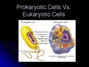
2 major types of cells Prokaryotic cell • No nucleus • No membrane-bound organelles • Circular chromosome • Eubacteria Kingdom • Archeabacteria Kingdom Eukaryotic cell • • Have nucleus Membrane-bound organelles Evolved - like u & me Linear chromosomes Protista Kingdom Fungus Kingdom Plant Kingdom Animal Kingdom

Organelles s Very small size, can only be viewed with a microscope. s Have specific functions. s Found throughout cytoplasm in eukaryotic cells.

Cell Structures and Functions Structure Cell Wall Centriole Chloroplast Cytoplasm Endoplasmic Reticulum (ER) Golgi Bodies Lysosome Mitochondrion Nucleus Plasma Membrane Ribosome Vacuole Cilia Plant, Animal, Both Function City Comparison

Organelles Found in Cells s. Endoplasmic reticulum (rough & smooth) – canals for movement s. Golgi Bodies – wrap & export proteins s. Nucleolus – makes ribosomes s. Lysosomes – digests & gets rid of wastes s. Ribosomes – makes proteins

Golgi Bodies Stacks of flattened sacs s Have a shipping side & a receiving side s Receive & modify proteins made by ER s Transport vesicles with modified proteins pinch off the ends s Transport vesicle

Lysosome Contain digestive enzymes s Break down food and worn out cell parts for cells s Programmed for cell death (lyse & release enzymes to break down & recycle cell parts) s

Nucleolus s s Cell may have 1 to 3 nucleoli Inside nucleus Disappears when cell divides Makes ribosomes that make proteins

Smooth & Rough Endoplasmic Reticulum s. Smooth ER lacks ribosomes & makes proteins USED In the cell s. Rough ER has ribosomes on its surface & makes proteins to EXPORT

Cell Powerhouse Mitochondrion ( mitochondria ) s. Site of Cellular respiration s. Active cells like muscles have more of these s. Burn sugars to produce energy ATP

Surrounding the Cell membrane s Lies immediately against the cell wall in plant cells s Made of protein and phospholipids s Selectively permeable

Cell or Plasma Membrane Cell membrane s Living layer s Controls the movement of materials into and out of the cell s Selectively permeable

Cytoplasm of a Cell Cytoplasm s Jelly-like substance enclosed by cell membrane s Provides a medium for chemical reactions to take place

Nucleus/Inner membrane Nucleus s Each cell has fixed number of chromosomes that carry genes s Genes control cell characteristics

Plant Cell wall s Made of cellulose which forms very thin fibers s Strong and rigid

Stations lab activity

Station 1: Label Prokaryotic Cell Bacteria All bacteria Ribosomes Label the 6 most important organelles!

Station 2: Label Eukaryotic Cell Plant Label the 13 most important organelles!

Station 3: Label Eukaryotic cell - Animal Label the 13 most important organelles!

Station 4: Create a Venn Diagram on LEFT PAGE to show organelles in common and organelles unique to each.

Bacteria Cell

Example Venn Diagram Animal Plant Unique Organelles Shared organelles Bacteria

Study your labeled cells – Quiz to follow!

Quiz

Quiz: 15. What kind of cell is cell A? 16. What kind of cell is cell B? 17. What type of cells are both A and B?

Answers to quiz 1. 2. 3. 4. 5. 6. 7. 8. Cell wall Cell membrane Mitochondrion Vacuole Golgi apparatus Cytoplasm Nuclear membrane Nucleolus 9. 10. 11. 12. 13. 14. 15. 16. 17. Nucleus Chromatin Rough endoplasmic reticulum Chloroplast Centriole Lysosome Plant Animal Eukaryotic
 Jesus is alive forevermore
 Mikael ferm
 He's alive he's alive frankenstein
 Bethesda thyroid cytology
 Abrasive cytology definition
 Proteinaceous background
 Exfoliative cytology
 Schawan cell
 Exfoliative cytology
 Gadolinium mrt
 Summary of meiosis
 Paranasal sinus at birth
 Medullary portion of collecting duct
 Thyroid parafollicular cells
 Gametes vs somatic cells
 Why dna is more stable than rna?
 Red blood cells and white blood cells difference
 Prokaryotic and eukaryotic cells worksheet
 Venn diagram animal and plant cell
 Prokaryotic vs eukaryotic cells venn diagram
 Cell organelle jeopardy
 Masses of cells form and steal nutrients from healthy cells
 Younger cells cuboidal older cells flattened
 What cell type
 Which organisms are prokaryotes
 Nondisjunction in meiosis
 Cells cells they're made of organelles meme
 Hát kết hợp bộ gõ cơ thể
 Slidetodoc
 Bổ thể
 Tỉ lệ cơ thể trẻ em
 Gấu đi như thế nào
 Tư thế worms-breton
 Chúa yêu trần thế alleluia
 Môn thể thao bắt đầu bằng từ đua


