Vaginal Cytology Evaluation Clinical Pathology Vaginal Cytology Indications


 l Parabasal Cells")


 l May become cornified l NO WBC’S EXCEPT")


- Slides: 29
Vaginal Cytology Evaluation Clinical Pathology
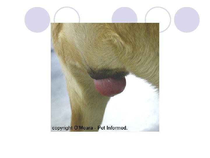
Vaginal Cytology Indications Examine exfoliated cells from the vagina l Vaginal epithelium is ovarian hormonal influenced- stages estrus for optimum breeding time. l ¡ ¡ l Knowledge of the onset of vaginal discharge Character of the discharge Degree of vulvar swelling Attitude of female towards male dog Also useful in detecting inflammation and neoplasia of the genital tract.
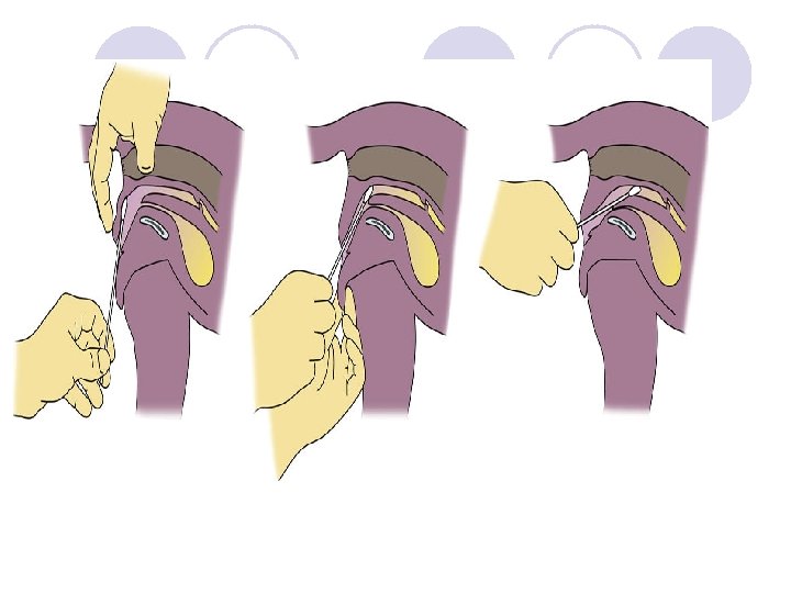
Vaginal Cytology Technique l May or may not want to use a speculum (laryngoscope may work as well). l Carefully part labia l Direct swab craniodorsally l Swab the vaginal wall l Avoid the vestibule and clitorial fossa, superficial cells may alter results l Roll on glass slide, let dry and stain (Diff. Quik)
l http: //www. vetmed. lsu. edu/eilts/Multimedia files/Sound/Video/Canine%20 Cytology. mp g
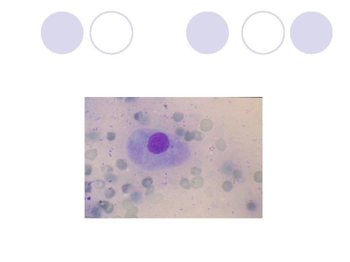
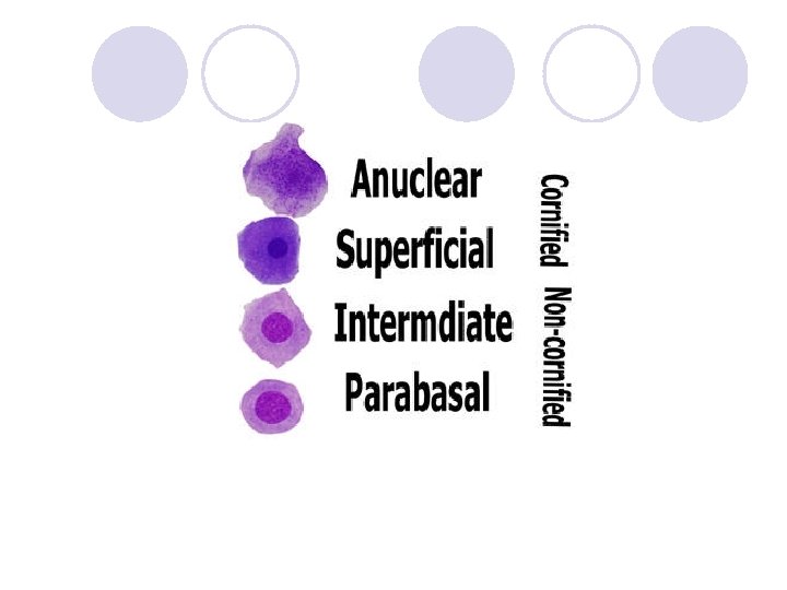
Vaginal Epithelial Cell Types l Basal Cells (may not be seen) l Parabasal Cells l Intermediate l Superficial Cells
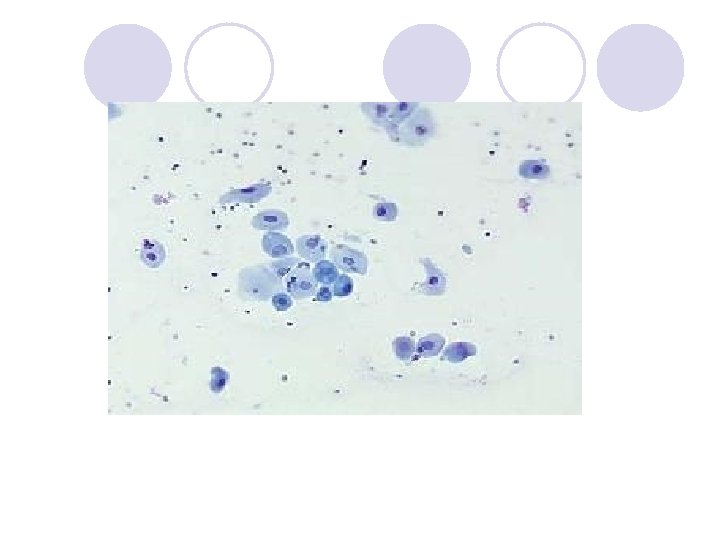
Parabasal Cells l Small round cells with round nuclei and small amount of cytoplasm l Uniform in size and shape
Intermediate Cells l May be small or large l Round nuclei, nucleus similar in size as parabasal cells l Entire cell approximately twice the size of parabasal cells l Cytoplasm becomes angular, irregular and folded as cell enlarges
Superficial Cells l Largest epithelial cell l As they age and degenerate, the nuclei becomes small, pyknotic and fades. l Cytoplasm may contain vacuoles with age
Superficial Cells Continued l Cornification is the degeneration process l Superficial cells are commonly called cornified cells l Once nucleus is lost become Anuclear cells
Staging the Canine Estrus Cycle l Stages of Estrus ¡ Proestrus ¡ Estrus ¡ Diestrus ¡ Anestrus
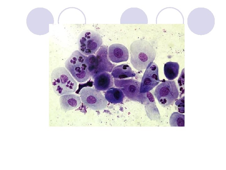
Proestrus l l l l Bitch has a swollen vulva, reddish vulvar discharge Will not accept the male during this time Duration is 9 days (with possibility of 2 -15) Parabasal cells and intermediate cells gradually decrease in number as the cycle nears estrus Superficial cells appear by 2 -3 day and increase in number over time Erythrocytes are numerous and gradually decline Neutrophils are also present and decline
Early Proestrus l Parabasal and intermediate cells predominate l As proestrus progresses, parabasal cells disappear as superficial cells increase.
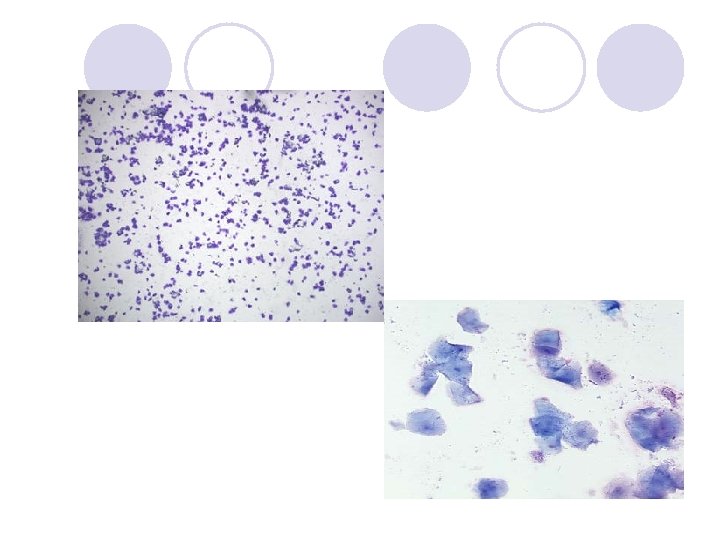
Late Proestrus l l l White Blood cells decrease in number Large intermediate and superficial cells predominate No parabasal cells or small intermediate cells Red blood cells or present or absent Bacteria is often present
Estrus l Lasts an average of 9 days l Female accepts male l Vulvar discharge is less bloody l Vulva is softer l Sometimes bloody discharge may continue through estrus
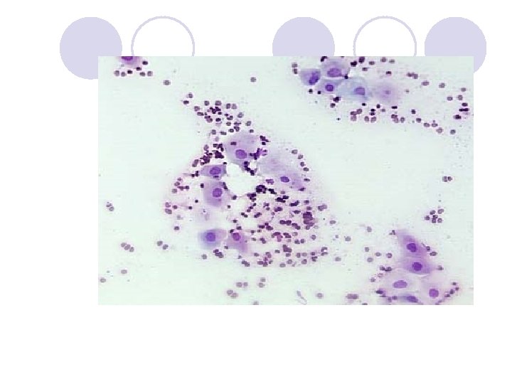
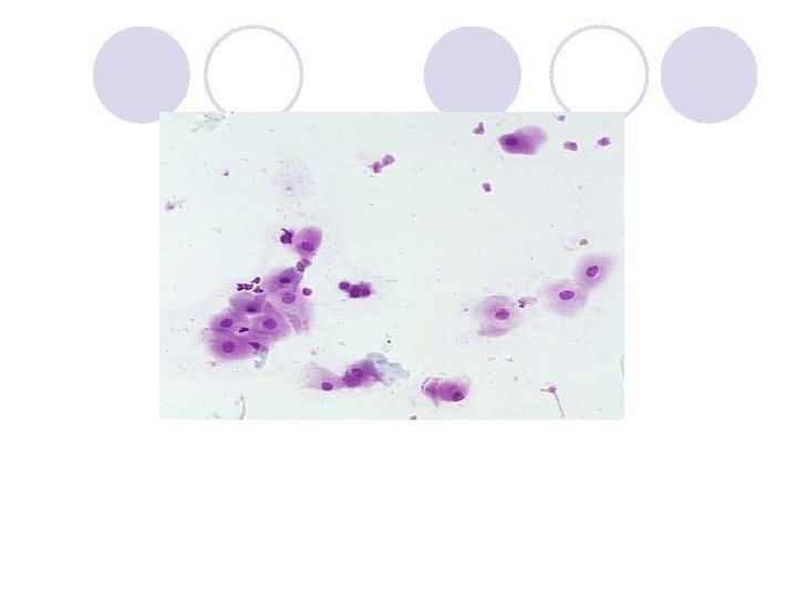
Estrus l Superficial cells predominate (90%) l May become cornified l NO WBC’S EXCEPT AT LAST 1 -2 DAYS OF ESTRUS!!!!!!!!!!!! l Variable RBC’s l Large numbers of bacteria
Estrus-Hormonal Events l Serum progesterone increases above anestrus range l Progesterone rise begins when LH peaks l Ovulation occurs 2 days after LH peak l Eggs take an additional 2 -3 days to mature l Fertile period is 4 -7 days after LH peak l Now do blood tests to check for LH peak
Diestrus l l l l Abrupt decrease in superficial cells Increase in parabasal cells and intermediate cells Many WBC’s, then decrease in late diestrus Variable RBC’s Occurs average 8 days after LH peak Lasts about 2 months Progesterone peaks 15 -30 days post estrus, then declines
Anestrus l The transition period between cycles l 4 -12 months l Parabasa and intermediate cells predominate l Few WBC’s and bacteria
Feline Vaginal Cytology l l l More difficult to collect Ovulation may be induced due to stimulation of cervix during collection Similar to a dog, but develop at a faster rate. Average estrus is 8 days, interval between estrus period is 9 days if ovulation does not occur. Usually no vaginal or scant vaginal discharge may be seen Usually no RBC’s are present during proestrus
Management of Breeding l Do comparison cytology during late proestrus and estrus l Breed every other day beginning on day 9 l Use other hormonal test kits to help stage cycle ¡ Progesterone ¡ LH Assay ELISA Assay