CELL DEATH or NECROSIS What is Necrosis Necrosis


")


- Slides: 29
CELL DEATH or NECROSIS
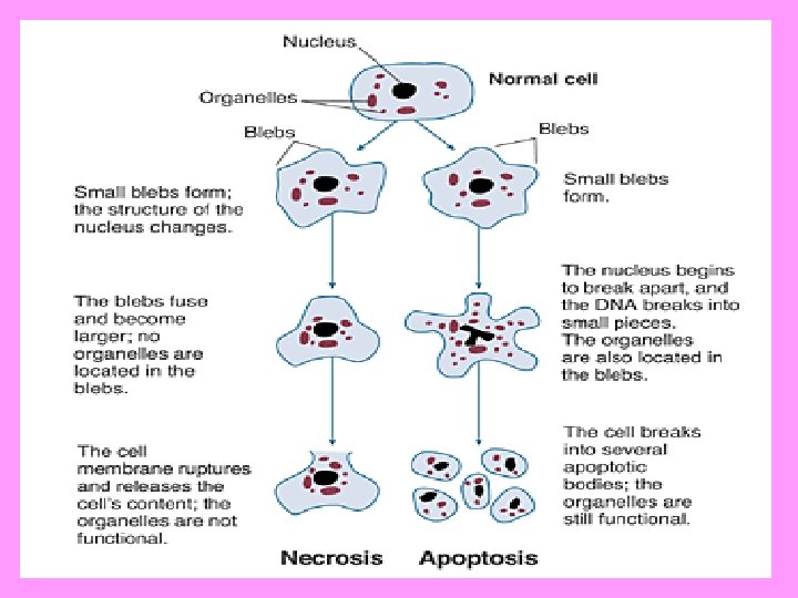
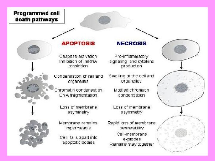
What is Necrosis? • Necrosis is the sum total of morphologic changes that follow cell death in a living tissue or organ • Dead cells usually show changes in both the cytolasm and in the nucleus. • Cytoplasmic changes are: increases eosinohilia, glassy appearance, granular or vacuolated cytolasm, swollen mitochondria, may also show calcification • Nuclear changes: pyknosis, karyorrhexis, karyolysis
What are the types of necrosis? • • • Coagulation Necrosis Liquefactive or Colliquative Necrosis Fat Necrosis Caseous Necrosis Gangrenous Necrosis Fibrinoid Necrosis
COAGULATION NECROSIS A photomicrograph of the heart in a patient with an acute myocardial infarction. In the center, the deeply eosinophillic necrotic cells have lost their nuclei. The necrotic focus is surrounded by paler-staining, viable cardiac myocytes.
COAGULATION NECROSIS-HEART
Coagulation Necrosis-wedge shaped infarct in kidney
Enzymatic and Traumatic Fat Necrosis • Enzymatic fat necrosis is due to the action of lipases on triglycerides resulting to saponification. • Adipose tissue is damaged in pancreatitis and occasionally due to trauma • Traumatic fat necrosis occurs in the female breast, mesenteries and in the omentum. There is no enzymatic lipolysis but there is apparent rupture of the cell membrane with release of neutral fat. Subsequent phagocytosis of the fat follows.
ENZYMATIC FAT NECROSIS
CASEOUS NECROSIS • This is a combination of coagulative and liquefactive necrosis encountered principally in the center of tuberculous infections. • Characteristic appearance is that of a soft, friable, whitish-gray debris resembling clumped cheesy material
CASEOUS NECROSIS
CASEOUS NECROSIS-LUNGS
TUBERCLE • A tubercle or follicle is the diagnostic lesion of both caseous and tuberculous conditions. • It has 4 components: 1. A central caseous necrotic core 2. Concentrically arranged eithelioid-like cells 3. Peripheral zone of lymphocytes 4. Presence of 2 or more Langhan’s giant cells
LIQUEFACTIVE NECROSIS
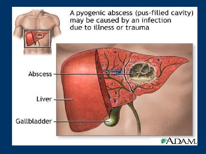
LIQUEFACTIVE NECROSIS • Results from the action of powerful hydrolytic enzymes and occurs when autolysis and heterolysis prevail over conditions that favor denaturation of proteins • This necrosis is characteristic of ischemic destruction of brain tissue. • Commonly encountered in all focal bacterial lesions like abscesses.
LIQUEFACTIVE NECROSISBRAIN ABSCESS
ABSCESS-HEART
DRY GANGRENE (Raynaud’s disease and diabetic gangrene)
GANGRENE • It is produced by ischemia with a superimposed saprophytic bacterial infections • Two types: Dry gangrene and Wet or moist gangrene • Dry gangrene or mummification occurs when bacterial infection does not supervene. • Dry gangrene may be cause by: arteriosclerosis, Buerger’s disease, Raynaud’s disease, Ergot poisoning
GANGRENOUS NECROSIS
Examples of Dry and Wet Gangrene • Raynaud’s disease is more common in females and is caused by spasmodic contraction of the arteries. • Ergot poisoning – is caused by ergot from certain plants that often cause dry gangrene of the extremities • Escharotic drugs like strong acid or alkali cause dry and moist gangrenes respectively. • Frost bite – may cause gangrene of the fingers, toes, nose and ears • Carbolic acid application to a finger can cause cell death followed by gangrene. • Wet gangrene is seen when there is obstruction to the venous return flow. Seen in moist areas like infections of the mouth or the vagina. It may also result when the mesentery of the intestines becomes twisted at the roots.
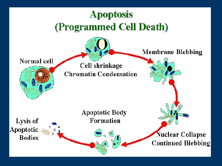
Apoptosis in Health • In embryogenesis and development: 1. Loss of autoreactive response of T cells in the thymus preventing auto-immune attack; 2. in atrophy and involution, often on withdrawal of hormones, e. g. menstrual breakdown of endometrium
APOPTOSIS-LIVER
POSTMORTEM CHANGES • • 1. 2. 3. 4. Somatic death is the death of the organism or the human body as a whole. Changes that occur after somatic death are: Algor mortis – first demontrable change. It is the cooling of the body. Rigor mortis – rigidity or stiffening of the skeletal muscles. It occurs approxiamtely 6 -10 hours after death Livor mortis – is the reddish discoloration of the dependent portions of the body to a red blue color. Blood slowly flows by gravity into the dependent vessels which dilate because of the loss of their muscular tone. Also caused by postmorten hemolysis. Autolysis and putrefaction – or softening of the body. Eventually all the tissues of the body undergo selfdigestion at varying rates of speed called autolysis.
“Your career is entirely in your hands. The decisions you make and the opportunities you choose ultimately shape your career path. You just need to take action to get where you want to go! Your Intensive Course is an investment. Take charge and give it the attention and focus it deserves!”