
Gangrene DR BINO Associate Prof DEPT OF SURGERY
Gangrene DR. BINO Associate Prof. DEPT OF SURGERY
Gangrene • A form of necrosis of tissue with superadded putrefaction. • All types of gangrene, necrosis undergoes liquefaction by the action of putrefactive bacteria. • It may be caused either ischemic or inflammatory • Coagulative Necrosis due to ischaemia – gangrene of the bowel, – gangrene of limb • Gangrenous or necrotising inflammation: primarily inflammation provoked by virulent bacteria resulting in massive tissue necrosis. – Gangrenous appendicitis, – Gangrenous stomatitis (noma, cancrum oris)
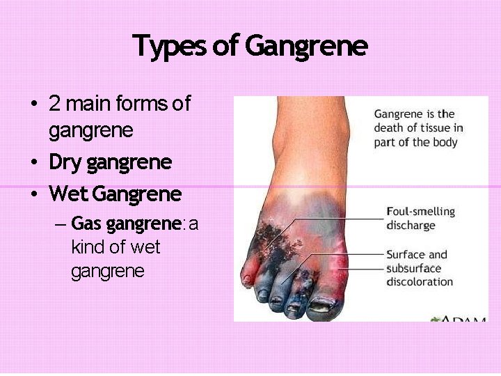
Types of Gangrene • 2 main forms of gangrene • Dry gangrene • Wet Gangrene – Gas gangrene: a kind of wet gangrene
Dry Gangrene • begins in the distal part of a limb due to ischaemia. • The gangrene spreads slowly upwards until it reaches a point where the blood supply is adequate to keep the tissue viable. • A line of separation is formed at this point between the gangrenous part and the viable part. – – – Toes and feet of an old patient due to arteriosclerosis. Thromboangiitis obliterans (Buerger’s disease), Raynaud’s disease, Trauma Ergot poisoning
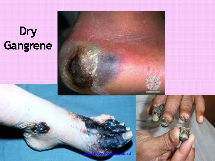
Dry Gangrene Dry gangrene www. facebook. com/notesdental
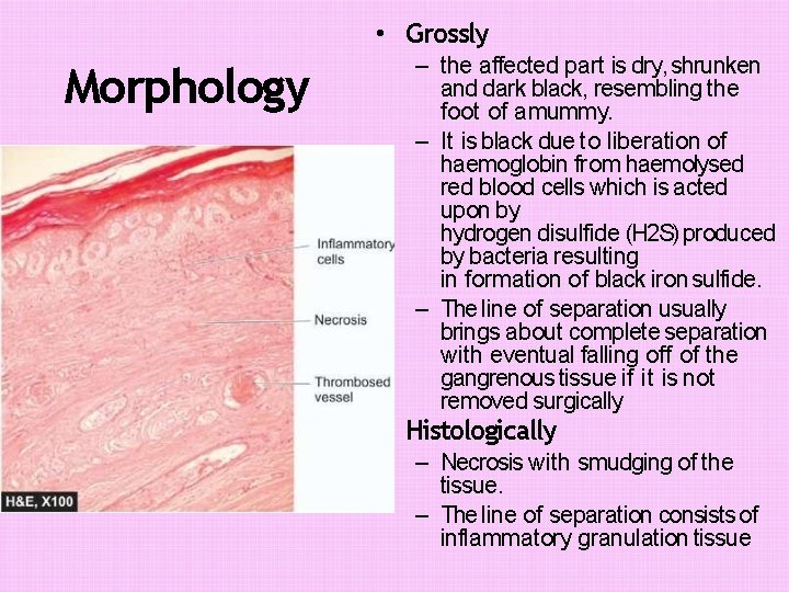
• Grossly Morphology – the affected part is dry, shrunken and dark black, resembling the foot of a mummy. – It is black due to liberation of haemoglobin from haemolysed red blood cells which is acted upon by hydrogen disulfide (H 2 S) produced by bacteria resulting in formation of black iron sulfide. – The line of separation usually brings about complete separation with eventual falling off of the gangrenous tissue if it is not removed surgically • Histologically – Necrosis with smudging of the tissue. – The line of separation consists of inflammatory granulation tissue
Wet Gangrene • Naturally moist tissues and organs such as the mouth, bowel, lung, cervix, vulva. • develops rapidly due to blockage of venous, and less commonly, arterial blood flow from thrombosis or embolism. • The affected part is stuffed with blood which favours the rapid growth of putrefactive bacteria. • The toxic products formed by bacteria are absorbed causing profound systemic manifestations of septicaemia, and finally death.
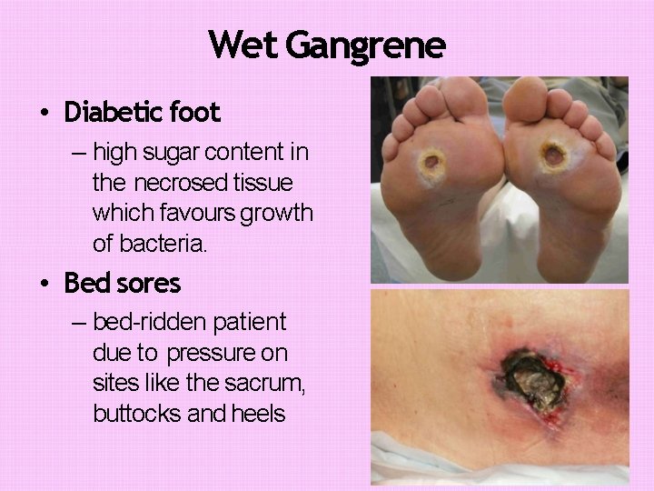
Wet Gangrene • Diabetic foot – high sugar content in the necrosed tissue which favours growth of bacteria. • Bed sores – bed-ridden patient due to pressure on sites like the sacrum, buttocks and heels
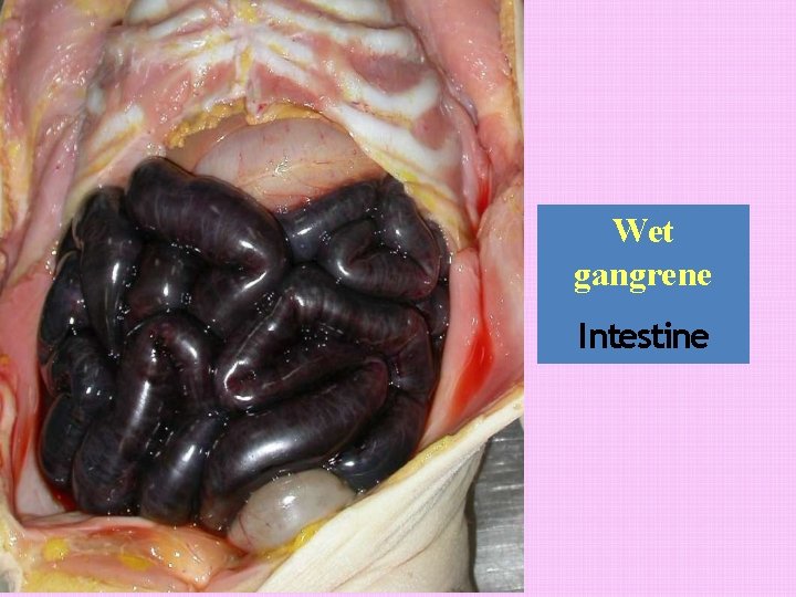
Wet gangrene Intestine
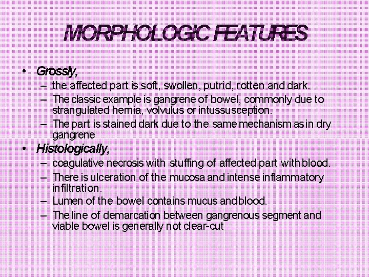
MORPHOLOGIC FEATURES • Grossly, – the affected part is soft, swollen, putrid, rotten and dark. – The classic example is gangrene of bowel, commonly due to strangulated hernia, volvulus or intussusception. – The part is stained dark due to the same mechanism as in dry gangrene • Histologically, – coagulative necrosis with stuffing of affected part with blood. – There is ulceration of the mucosa and intense inflammatory infiltration. – Lumen of the bowel contains mucus and blood. – The line of demarcation between gangrenous segment and viable bowel is generally not clear-cut
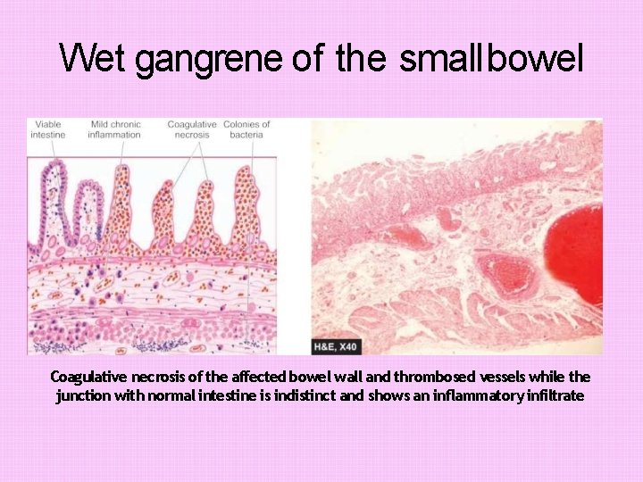
Wet gangrene of the small bowel Coagulative necrosis of the affected bowel wall and thrombosed vessels while the junction with normal intestine is indistinct and shows an inflammatory infiltrate
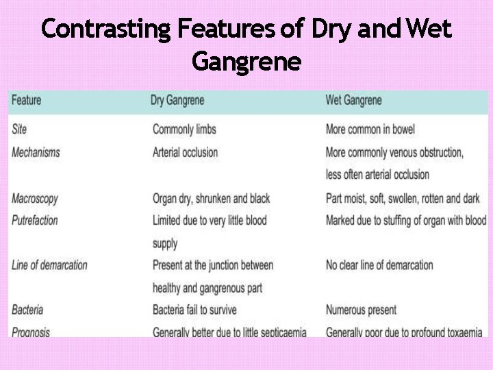
Contrasting Features of Dry and Wet Gangrene
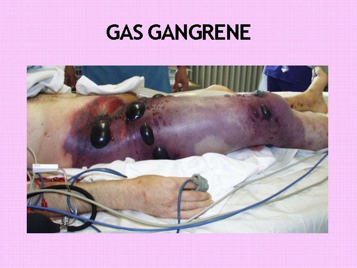
GAS GANGRENE • Special form of wet gangrene caused by gas-forming clostridia (gram-positive anaerobic bacteria). • gain entry into the tissues through open contaminated wounds, • especially in the muscles, or as a complication of operation on colon which normally contains clostridia. • It produce various toxins which produce necrosis and oedema locally • Also absorbed producing profound systemic manifestations.
GAS GANGRENE
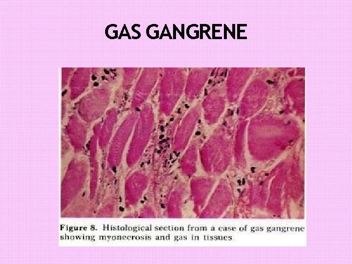
MORPHOLOGIC FEATURES • Grossly – the affected area is swollen, oedematous, painful and crepitant due to accumulation of gas bubbles within the tissues. – Subsequently, the affected tissue becomes dark black and foul smelling. • Microscopically – the muscle fibres undergo coagulative necrosis with liquefaction – Large number of gram-positive bacilli can be identified. – At the periphery, a zone of leucocytic infiltration, oedema and congestion are found. – Capillary and venous thrombi are common.
GAS GANGRENE
References • Robbinson's basic pathology 8 ed • Harsh Mohan - Textbook of Pathology 6 th Ed. • Color atlas of pathology

THANKS…… www. facebook. com/notesdental
- Slides: 18