CardioRespiratory Exercise Physiology Chapter 2 Cardiovascular System CARDIOVASCULAR


 is transported in the blood in")


 Left Atriumreceives oxygen rich blood from")
 Bicuspid (Mitral) Valve- One way valve")
")

 – found in the wall of")


: the amount of blood pumped from the")


- Slides: 44
+ Cardio-Respiratory Exercise Physiology – Chapter 2 Cardiovascular System
+ CARDIOVASCULAR SYSTEM n Made up of: n The heart – pumps blood throughout the body n Vascular System – comprised of the blood and circulatory vessels n Lungs – site of gas exchange (alveoli) n Each system works together to ensure adequate amounts of oxygen are delivered to the muscles and tissues of the body
+ BLOOD n PLATELETS - <1% : used to assist in repair after injury n WHITE BLOOD CELLS (WBC’s) – <1% known as leucocytes : immune function, protects the body from infection n RED BLOOD CELLS (RBC’s) – known as erythrocytes, make up 40 -45% of blood volume n Amount of RBC’s in the body is dependent on fitness and gender
+ BLOOD FUNCTIONS n Transports gases, nutrients, waste products, hormones, heat n Average blood volume: n ~5 -6 L in men n ~4 -5 L in women n 55% of blood fluid is plasma, 45% is blood cells and platelets
+ BLOOD FUNCTIONS n Carbon Dioxide (CO 2) is transported in the blood in the form of bicarbonate, a buffer that keeps blood from becoming too acidic n O 2 is less soluble in plasma, but easily attaches to hemoglobin – an iron-rich pigment n Protects us from bleeding to death, via clotting, and from disease, by destroying invasive microorganisms
+ BLOOD PLASMA n Contains 90% water along with blood proteins, salt, food substances, waste products, gases (carbon dioxide, oxygen), enzymes and antibodies n Main Function of plasma is to transport blood cells through the body
+ HEMATOCRIT n Hematocrit: percentage of total blood volume composed of platelets, red, and white cells. n Normal range is 4150% in men and 36 -44% in women
+ BLOOD n. Q & A: n What is EPO? And what does it do? n Why is it advantageous for an endurance athlete to have a higher concentration of RBC’s? n How can an athlete naturally increase their RBC stores? n What are some ways athletes are illegally increasing their RBC count? n https: //www. youtube. com/watch? v=G 7 KZx. IR 1 t-o
+ CIRCULATION n ARTERIES: thick muscular vessels that transport blood away from the heart n Arteries pass blood to smaller vessels called arterioles, and then to capillaries where gas exchange occurs n CAPILLARIES: narrow vessels with thin walls; site of exchange between blood and tissue n VEINS: carry deoxygenated blood toward the heart, less muscular, flexible and have valves to prevent back flow
+ CIRCULATION PATHWAY ARTERIES ARTERIOLES CAPILLARIES VENUOLES VEINS
+ THE HEART n Pear-shaped organ found in the thoracic cavity n Positioned slightly to the left of the body deep to the sternum, between the lungs n Involuntary muscle with striated muscle fibres n Comprised of 4 chambers, 4 valves, and several veins and arteries
+ HEART CIRCULATION n PULMONARY CIRCULATION: delivers deoxygenated blood from the right side of the heart to the lungs n SYSTEMIC CIRCULATION: delivers the oxygenated blood from the left side of the heart to the body
+ FUNCTIONS OF THE HEART 1. 2. Generates Blood Pressure Routes blood around the body 3. Regulates blood supply
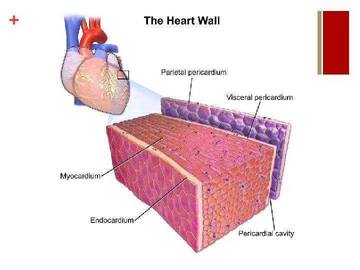
+ ANATOMY OF THE HEART n Heart consists of three layers: 1. Pericardium- outer double layer bag or membrane containing fluid. Functions to reduce friction and maintain heart shape 2. Myocardium (Cardiac Muscle)- contracts the same way as skeletal muscle. Receives stimulus from the CNS to the hearts pacemaker. Works in a all or none principle) 3. Endocardium- smooth inner membrane. Functions to prevent friction between heart muscle and blood
+ HOW THE HEART WORKS VIDEO n https: //www. youtube. com/watch? v=7 Xaftd. E_h 60
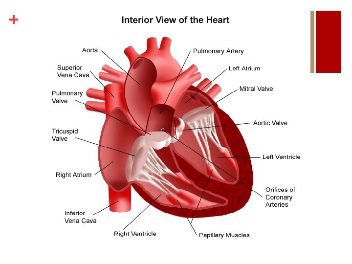
+ ANATOMY OF THE HEART: Heart Chambers 1) Left Atriumreceives oxygen rich blood from the lungs through the pulmonary veins. 2) Left Ventriclereceives oxygenated blood from left atrium through the bicuspid valve. Pumps blood to the body through the aortic valve to aorta 3) Right Atrium- receives de-oxygenated blood from the body through the superior and inferior vena cava. 4) Right Ventriclereceives de-oxygenated blood from the right atrium through the tricuspid valve. Send blood to lungs through the pulmonary artery
+ ANATOMY OF THE HEART: Heart Valves 1) Bicuspid (Mitral) Valve- One way valve allowing blood flow from the left atrium to the left ventricle 3) Tricuspid valve- one way valve allowing deoxygenated blood to move from the right atrium to the right ventricle 2) Aortic Valve- One way valve allowing blood flow from the left ventricle to the aorta 4) Pulmonary Valve- one way valve allowing deoxygenated blood to move from the right ventricle to the lungs
+ ANATOMY OF THE HEART: Veins and Arteries n Vena Cavae (Superior and Inferior) – deliver deoxygenated blood from the body into the right atrium n Pulmonary Arteries- deliver de-oxygenated blood from the right ventricle to the lungs n Pulmonary Veins – deliver oxygenated blood from the lungs into the left atrium n Aorta – delivers oxygenated blood from the left ventricle to the body
+ HEART CONTRACTION n The heart can actually beat on its own, outside the body of an individual for several hours if supplied with proper nutrients and salts… n The reason for this is the heart makes itself contract and this process is called a MYOGENIC contraction n The contractions of the heart chambers contract in a sequence initiated from the pacemaker
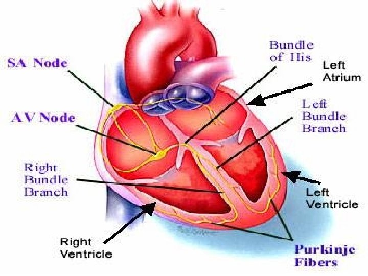
+ CARDIAC IMPULSES n SINOATRIAL NODE (SA NODE) – found in the wall of the right atrium and acts as the hearts pacemaker because it initiates each heart beat n ATRIOVENTRICAL NODE (AV NODE) – found near the bottom of the right atrium. Receives electrical signal from the SA node and passed onto the Bundle of His and Purkinje Fibres n PURKINJE FIBRES – deliver electrical signal to the cardiac muscles which signals the ventricles to contract
+ CARDIAC CYCLE n Defined as the order of events that make up one heart beat. n Cycle lasts for ~0. 8 seconds and occurs ~72 times a minutes n Consists of a period of relaxation, known as diastole (0. 5 sec), followed by a period of contraction, known as systole (0. 3 seconds)
+ DIASTOLE: RELAXATION n Period of relaxation when the heart, specifically the atria refill with blood n Tricuspid and bicuspid valves are closed at this time n The one way valves are pushed open due to increased atrial pressure n As a result, ventricles begin to fill with blood
+ SYSTOLE: CONTRACTION n ATRIAL SYSTOLE: n SA node sends an electrical impulse to the atrium walls signaling them to contract n Resulting contraction causes atrioventricular valves (bicuspid and tricuspid) close once all blood has been forced into the ventricles n VENTRICULAR SYSTOLE: n Pressure inside the ventricles pushes open the semilunar valves (Pulmonary and aortic) n Electric signal travels down the Purkinje Fibers stimulates contraction of the ventricular myocardium n Blood flows into the pulmonary (lungs) and systemic (around the body) systems.
+ CARDIAC CYCLE VIDEO n https: //www. youtube. com/watch? v=f. ZT 9 vlb 2 ua
+ FACTORS AFFECTING HEART RATE n Heart contraction rate is affected by hormones and the nervous system n PARASYMPATHETIC (PNS): PNS is part of the Autonomic Nervous System (ANS) that functions to bring the body systems to rest. (REST and DIGEST). It helps to bring the heart rate down during recovery by acting on the SA node. n SYMPATHETIC NERVOUS SYSTEM (SNS): SNS prepares your body for action (FIGHT or FLIGHT). SNS functions by sending a signal from the cardiac centre to the hearts pace maker in response to increased demand of oxygen.
+ FACTORS AFFECTING HEART RATE n Cardiac centre is found in the MEDULLA OBLONGATA n SNS: during exercise, sympathetic nerves release noradrenalin, a neurotransmitter onto the SA node which increases heart rate n PNS: during recovery the PNS sends impulses down the vagus nerve, which slows the heart rate and helps to bring the body back to rest levels and homeostasis
+ Nervous System Control of the Heart
+ FACTORS AFFECTING HEART RATE n During intense exercise, adrenaline affects the heart rate in the body n When the body is preparing for action, adrenaline is released n This release reduces blood flow to the organs that are not needed or important for the activity and increases the blood flow necessary to skeletal muscle, cardiac muscle and lungs
+ BE SURE TO KNOW! n Name the function of the following: n Sinoatrial Node n Atrioventricular Node n Purkinje Fibres n Describe what takes place during different stages of the cardiac cycle n Diastole n Atrial and Ventricular Systole n Understand be able to distinguish between the sympathetic and parasympathetic nervous systems, and the affect they have on heart rate
+ BE SURE TO KNOW! n Describe the make up of our blood n Describe the function of the following: n Erythrocytes (RBC’s) n Leukocytes (WBC’s) n Platelets
+ SOUNDS OF THE HEART n Through a stethoscope, the heart sounds are generally described as “lub-dub” n The “lub” sound comes from the closing of the tricuspid and bicuspid valves n The “dub” sound is caused by the closing of the pulmonary and aortic valves (semilunar) n TO DO: Page 40 n Heart Rate according to stethoscope: ________ bpm
+ BLOOD DISTRIBUTION n Blood flow distribution is based on which tissues are in greatest need n During exercise, blood flow to working muscles increases up to 80% of all blood circulating through the body (Cardiac Output) n At rest, kidney and liver receive ~ half of cardiac output while skeletal muscles only receive 15 -20% of cardiac output
+ BLOOD FLOW AT REST vs. EXERCISE
+ BLOOD PRESSURE n BP is defined as the amount of pressure exerted by blood on the vessel walls n Expressed by 2 numbers: n Systolic Blood Pressure n Diastolic Blood Pressure n Also referred to as ARTERIAL BLOOD PRESSURE
+ BLOOD PRESSURE n Resting, healthy adult range for blood pressure is generally 120 mm. Hg(systolic) to 80 mm. Hg(diastolic), and is read as “ 120 over 80 mm. Hg” n Blood pressure within this range is considered to allow efficient emptying and filling of the heart, but still maintaining enough pressure to send blood to the working tissues in the body. n Low blood pressure: 90 -100/50 -60 mm. Hg n High blood presure: 140/100 mm. Hg
+ BLOOD PRESSURE: Systolic vs. Diastolic n SYSTOLIC PRESSURE: process of ventricle contracting and forcing blood into the aorta n Systolic pressure represents highest pressure n DIASTOLIC PRESSURE: ventricle relaxes allowing for chambers to fill up with blood n Diastolic pressure represents lowest pressure n Important to note that systolic blood pressure will increase with exercise and training, while diastolic stays consistent
+ BLOOD PRESSURE n Q&A n TO DO: Green box, 41 n What is your blood pressure? n Explain what happens to blood flow distribution during exercise? n Answer questions #1 -5 in green box on 41.
+ CARDIAC OUTPUT n Cardiac Output (Q): the amount of blood pumped from the heart in one minute. Cardiac output is measured in liters and considers all blood except that which goes to the lungs Cardiac Output (Q) = (Stroke Volume x Heart Rate) / 1000 n Stroke Volume (SV): the amount of blood being pumped by each ventricle with each contraction n Heart Rate = 1 Cardiac Cycle
+ CARDIAC OUTPUT DURING EXERCISE n On average, cardiac output is ~5 L per min at rest and can increase to 30 L per min during strenuous exercise n Why does cardiac output increase during exercise? ? n As heart rate increases, so does stroke volume, therefore resulting in an increase in cardiac output. n However, during endurance activities where the intensity and demand remains the same over time, cardiac output does not increase further.
+ CARDIAC DRIFT n During endurance activities where cardiac output stays constant, heart rate will eventually increase slightly and progressively n As the heart rate begins to ‘drift’ upward, the stroke volume decreases, which allows for cardiac output to remain the same n Sweating Thermoregulation Heart rate increase n Complete P. 43 TO DO green box in text