CARDIOPULMONARY SYSTEM Cardio heart Pulmonary lungs Respiratory medium

 O 2 CO 2 Respiratory surface Organismal level Circulatory")


 - head and")

- thicker wall- pumps blood to Pulmonary Artery 5. Pulmonary Semi-lunar")


 : pumps blood into Aorta 11. Left Semilunar Valve :")


 Normal artery Smooth muscle Plaque Endothelium 50 µm (b) Partly")

 MAMMALS AND BIRDS Lung and skin capillaries Lung capillaries FISHES")
- Slides: 32
CARDIOPULMONARY SYSTEM Cardio – heart Pulmonary - lungs
Respiratory medium (air of water) O 2 CO 2 Respiratory surface Organismal level Circulatory system Cellular level Energy-rich molecules from food Cellular respiration ATP
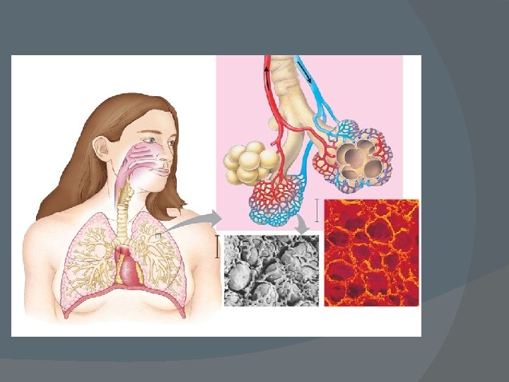
Gas Exchange in Humans 1. Mouth/Nose/Nasal Cavity, Pharynx, Larynx: air entrance Epiglottis open - allows air into trachea Larynx: Voice Box - air passing over vocal chords causes vibration http: //www. youtube. com/watch? v=Kvr 9 LFz. Oo 5 0
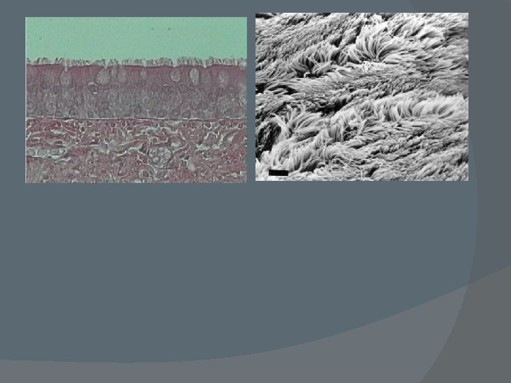
2. Trachea: Wind Pipe - in front of esophagus Cartilage rings - reinforcement 3. Bronchi: Trachea splits into bronchi Each bronchus splits further into more bronchioles
4. Alveolus: air sac - located at the end of bronchioles - enveloped by capillaries for gas exchange 5. Diffusion of Gases Inhale: lots of oxygen - diffuses across membrane into blood - fixed by hemoglobin - binds to the iron at the center of the hemoglobin subunit - oxygen binds less well in acid environments (control mechanism) Blood has lots of CO 2 - diffuses out of RBC into alveoli
Circulation: RBC move throughout body and O 2 diffuses with concentration gradient to cells via the interstitial fluid - CO 2 diffuses opposite direction 6.
RESPIRATION CONTROL 1. Bulk Flow: moving air in and out of lungs Negative Pressure: mammals, birds and reptiles - generated by diaphragm (muscle separating the pulmonary and abdominal cavities) and intercostal muscles (muscles between ribs) - diaphragm contracts and moves downward Result: decreases the pressure in lungs (more volume) - air rushes in - diaphragm relaxes and moves up - air expelled
Rib cage expands as rib muscles contract Air inhaled Rib cage gets smaller as rib muscles relax Air exhaled Lung Diaphragm INHALATION Diaphragm contracts (moves down) Figure 42. 24 EXHALATION Diaphragm relaxes (moves up)
2. Breathing Rate: Balance of p. H - chemoreceptors in carotid artery and medulla oblongata monitor p. H - when p. H drops (excess CO 2) a signal is sent to the breathing control center (medulla oblongata) in the brain which signals the diaphragm to contract more often – CO 2 out/O 2 in/p. H rises
Cerebrospinal fluid 1 The control center in the medulla sets the basic rhythm, and a control center in the pons moderates it, smoothing out the transitions between inhalations and exhalations. 2 Nerve impulses trigger muscle contraction. Nerves from a breathing control center in the medulla oblongata of the brain send impulses to the diaphragm and rib muscles, stimulating them to contract and causing inhalation. Breathing control centers Pons Medulla oblongata The medulla’s control center also helps regulate blood CO 2 level. Sensors in the medulla detect changes in the p. H (reflecting CO 2 concentration) of the blood and cerebrospinal fluid bathing the surface of the brain. 5 Nerve impulses relay changes in CO 2 and O 2 concentrations. Other sensors in the walls of the aorta and carotid arteries in the neck detect changes in blood p. H and send nerve impulses to the medulla. In response, the medulla’s breathing control center alters the rate and depth of breathing, increasing both to dispose of excess CO 2 or decreasing both if CO 2 levels are depressed. Carotid arteries Figure 42. 26 In a person at rest, these nerve impulses result in about 10 to 14 inhalations per minute. Between inhalations, the muscles relax and the person exhales. Aorta Diaphragm Rib muscles 6 The sensors in the aorta and carotid arteries also detect changes in O 2 levels in the blood and signal the medulla to increase the breathing rate when levels become very low.
Circulatory System Function: distribute nutrients and oxygen transport waste products for removal immune system
Structures of Circulatory Systems Vessels: Arteries Veins move blood away from heart blood to heart thick layer of smooth muscle thin layer of smooth muscle Branch into arterioles Formed from converging venules
Vein Artery Basement membrane Endothelium 100 µm Valve Endothelium Smooth muscle Connective tissue Smooth muscle Capillary Connective tissue Artery Vein Venule Arteriole
Capillaries: smallest blood vessels - transfer of nutrients and waste Heart: Pumping mechanism: cardiac muscle tissue Compartments: Atria: receive blood from veins pump blood to ventricles Ventricles: typically larger chamber with thicker wall - pump blood into arteries
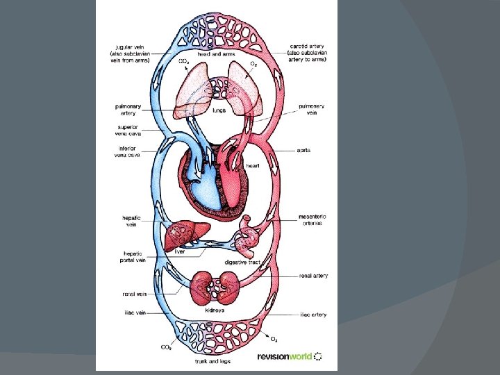
Human Circulatory Systems Parts/Pathway: 1. Vena Cava- largest veins: Superior (anterior) - head and forelimbs and Inferior (posterior) - torso and legs 2. Right Atrium (RA)- receives blood from vena cavas 3. Atrioventricular Valve (AV valve): tricuspid valve - passes blood to RV - separates the right chambers - prevents backflow of blood from RV so blood only moves forward
4. Right Ventricle (RV)- thicker wall- pumps blood to Pulmonary Artery 5. Pulmonary Semi-lunar Valve: gateway to pulmonary artery - prevents blood from flowing into the RV 6. Pulmonary Artery: carries blood to lungs for oxygen - NOTE: blood is leaving the heart through an artery but is O 2 Poor - in lungs the arteries branch into arterioles and then into a capillary net around the alveoli allowing for gas exchange
7. Pulmonary Veins: from lungs to LA carries O 2 rich blood 8. Left Atrium (LA): receives O 2 rich blood and pumps it into the LV 9. Left Atrioventricular Valve - aka mitral valve or bicuspid valve - prevents backflow from LV
10. Left Ventricle (LV) : pumps blood into Aorta 11. Left Semilunar Valve : prevents blood from flowing back into LV 12. Aorta: main artery - branches - sends blood to body systems 13. Arteries: - branch into arterioles and then into capillaries 14. Capillaries/Capillary Nets: - gas and nutrient exchange 15. Venules and veins: capillaries merging into larger vessels like streams into rivers
THE HEART In 1 year, the average human heart circulates from 770, 000 to 1. 6 million gallons of blood through the body. This is enough fluid to fill 200 tank cars, each with a capacity of 8, 000 gallons Beating Heart/Heart Surgery
Blood Pressure: 120/80 Systole: blood being forced into the arteries - larger pressure because of the stronger contraction of the ventricles Diastole: relaxing of the ventricles Sphygmomanometer
1 A typical blood pressure reading for a 20 -year-old is 120/70. The units for these numbers are mm of mercury (Hg); a blood pressure of 120 is a force that can support a column of mercury 120 mm high. 4 The cuff is loosened further until the blood flows freely through the artery and the sounds below the cuff disappear. The pressure at this point is the diastolic pressure remaining in the artery when the heart is relaxed. Blood pressure reading: 120/70 Pressure in cuff above 120 Rubber cuff inflated with air Pressure in cuff below 120 Pressure in cuff below 70 120 70 Sounds audible in stethoscope Artery closed 2 A sphygmomanometer, an inflatable cuff attached to a pressure gauge, measures blood pressure in an artery. The cuff is wrapped around the upper arm and inflated until the pressure closes the artery, so that no blood flows past the cuff. When this occurs, the pressure exerted by the cuff exceeds the pressure in the artery. Figure 42. 12 3 A stethoscope is used to listen for sounds of blood flow below the cuff. If the artery is closed, there is no pulse below the cuff. The cuff is gradually deflated until blood begins to flow into the forearm, and sounds from blood pulsing into the artery below the cuff can be heard with the stethoscope. This occurs when the blood pressure is greater than the pressure exerted by the cuff. The pressure at this point is the systolic pressure. Sounds stop
Increased blood pressure: - higher amounts of water in the blood due to increased solute content - sugar or salt - decreased diameter of blood vessels – atherosclerosis and arteriosclerosis
Atherosclerosis/Arteriosclerosis Connective tissue (a) Normal artery Smooth muscle Plaque Endothelium 50 µm (b) Partly clogged artery Figure 42. 18 a, b 250 µm
Capillary Exchange: - nutrient rich blood from arteries enters capillaries - water, food and gases leave blood to cells - metabolic wastes enter blood and nearly all of the fluid that left the capillary
AMPHIBIANS REPTILES (EXCEPT BIRDS) MAMMALS AND BIRDS Lung and skin capillaries Lung capillaries FISHES Gill capillaries Pulmocutaneous circuit Artery Gill circulation Heart: ventricle (V) A Atrium (A) Systemic circulation Vein Systemic capillaries Right systemic aorta Pulmonary circuit A A V Right V Left Right Systemic circuit Systemic capillaries Figure 42. 4 Pulmonary circuit Left Systemic V aorta Left A Systemic capillaries A V Right A V Left Systemic circuit Systemic capillaries