Respiratory System Lungs Lungs are lateral to the


- Slides: 10

Respiratory System

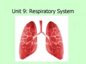
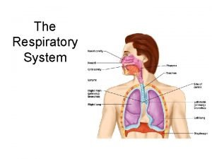
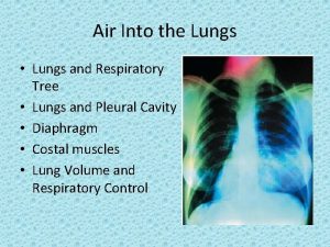
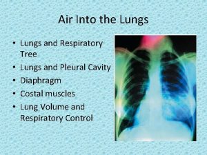
Lungs • Lungs are lateral to the • • • heart Each is located in its own enclosed pleural cavity within the thoracic cavity Each lung is covered by a pleural membrane A pleural space lubricated with fluid also surrounds each lung

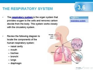
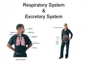
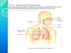
Pathway of Air to the Lungs • Pharynx - throat (extends to • • • larynx) Larynx - houses vocal chords leads to trachea Trachea - air passes through this tube on way to the lungs (windpipe) Bronchi - branches of trachea going to each lung Bronchioles - smaller branches of the bronchi Alveoli - air sacs surrounded by capillaries where oxygen is exchanged for CO 2 - (blood returns to heart from here)

Pathway of Air Into the Lungs Can be divided into 2 zones: • Conducting Zone • Respiratory Zone

Conducting Zone • Nose, nasal cavity, • • pharynx, larynx, trachea, bronchi Function: warm, filter and moisten inspired air Mucous traps airborne pathogens and cilia propel matter into the pharynx to be swallowed

Respiratory Zone • Bronchioles, alveoli • Function: gas exchange (CO 2 enters alveoli from blood and O 2 passes into blood from the alveoli) • Capillaries surround each alveolus for efficient gas exchange * In emphysema, the alveoli are damaged, reducing ability for gas exchange

Mechanics of Breathing • Diaphragm - muscle at the base of the lungs that regulates pressure within the pleural cavities by moving up and down

Inspiration & Expiration (see Figure 22. 13) Inspiration (breathing in) Expiration (breathing out) • Diaphragm contracts and • Diaphragm relaxes and moves down moves upwards • Intercostal muscles contract • Intercostal muscles relax and expand ribcage collapses • Volume of thoracic cavity increased as intrapulmonary decreases as (inside alveoli) pressure intrapulmonary pressure decreases increases • Air moves into lungs because • Air is forced out of lungs atmospheric pressure is because atmospheric greater than intrapulmonary pressure is less than pressure intrapulmonary pressure

Diaphragm movements

Regulation of Breathing • The medulla and pons • • contain respiratory centers that control breathing (figure 22. 24) The depth and rate of breathing are controlled by CO 2, O 2 and p. H (H+) concentrations in the blood CO 2 levels are the strongest stimulus for breathing Chemoreceptors in aorta and carotid arteries monitor blood and send signals to respiratory centers in pons and medulla
 Mikael ferm
 What is the conducting zone of the respiratory system
 Digestive system circulatory system and respiratory system
 Rectangular prism total surface area formula
 Tiny air sacs at the end of the bronchioles
 Circulatory system and respiratory system work together
 Respiratory system bozeman
 Unit 9 respiratory system
 Diagnostic test of respiratory system
 What is the respiratory system
 What is larynx
