Anesthesia for the Pediatric Patient with Epiglottitis Updated


- Slides: 22

Anesthesia for the Pediatric Patient with Epiglottitis Updated 7/2019 Jennifer Chiem, MD Seattle Children’s Hospital Seattle, WA USA GLOBAL

Disclosures No relevant financial relationships

Learning Objectives: • Learners will be able to identify signs and symptoms of epiglottitis • Learners will be able to describe anesthetic techniques for a patient with epiglottitis • Learners will be able to describe antibiotic regimens used to treat epiglottitis

Overview of Epiglottitis Infectious Etiology • Haemophilus influenza Type B (Hi. B), most common • Haemophilus influenza Type A, F, and nontypable • Streptococci, including Group A Streptococci • Staphylococcus aureus

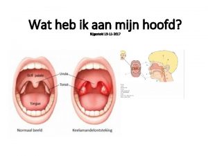
Overview of Epiglottitis Non-infectious Etiology • Trauma: thermal injury • Foreign Body Ingestion • Caustic Ingestion Picture: erythematous oropharynx

Overview of Epiglottitis • Epidemiology - Decreased incidence with Hi. B vaccination, although epiglottitis can still occur - Median age increased from 3 years old to 6 -12 years old in vaccinated patients - Estimated epiglottitis rates: 0. 6 -0. 8 cases per 100, 000 • Risk Factors - Immune deficiency - Lack of Hi. B immunization

Signs and Symptoms of Epiglottitis • Respiratory Distress - • • Stridor Tachypnea Anxiety Refusal to lie down “Sniffing” or “Tripod” posture Dysphagia Drooling Fever Sore Throat Picture: Toddler in “tripod” position (top); Toddler drooling (bottom)

Differential Diagnosis of Epiglottitis • Viral laryngotracheobronchitis (Croup) • Gradual Onset • Low grade fever • Stridor • Hoarseness • Barking Cough

Differential Diagnosis of Epiglottitis • Bacterial tracheitis • Acute onset • Fever • Imaging studies – X-ray • Irregular tracheal wall • Normal epiglottis

Differential Diagnosis of Epiglottitis • Retropharyngeal abscess • Less toxic appearance • Fever may be present • Imaging studies (CT scan) will help determine if abscess is present

Differential Diagnosis of Epiglottitis • Foreign Body • • History Lack of fever Acute onset Can cause partial vs. complete airway obstruction Foreign body in the airway

Differential Diagnosis of Epiglottitis • Diphtheria • • Gradual onset Sore throat Low grade fever Gray, sharply demarcated membrane in the oropharynx Gray membrane in the oropharynx

Diagnosis of Epiglottitis • History and clinical presentation • Radiologic imaging can help to confirm diagnosis, but not always necessary Enlarged epiglottis “Thumb Sign” on lateral neck X-ray Lateral neck X-ray

Airway Management of Epiglottitis • Determine severity of obstruction • Determine if intubation is necessary vs. observation • Involve anesthesiologist and otolaryngologist as soon as possible • The provider with the most airway experience should make first intubation attempt

Airway Management of Epiglottitis If patient is able to maintain airway • Transport to operating room for airway management • Minimize distress to the patient (no awake IV, parental presence if appropriate) • Mask induction with Sevoflurane/Halothane – try to maintain spontaneous ventilation • Propofol, Ketamine, and/or Dexmedetomidine to maintain spontaneous ventilation • Consider Glycopyrrolate to minimize secretions • First intubation attempt with advanced airway equipment (bougie, video laryngoscopy vs. fiber-optic scope) • Back up: tracheostomy tray set up

Airway Management of Epiglottitis If Patient is not able to maintain airway • Bag valve mask • Transport, if appropriate, to operating room for airway management • Mask induction with Sevoflurane/Halothane – try to maintain spontaneous ventilation • Propofol, Ketamine, and/or Dexmedetomidine to maintain spontaneous ventilation • First intubation attempt with advanced airway equipment (bougie, video laryngoscopy vs. fiber-optic scope) • Back up: tracheostomy tray set up

Airway Management Tips • At least a half size smaller than age appropriate endotracheal tube should be used due to tissue swelling • Do NOT use a supraglottic airway (laryngeal mask airway) as this can cause further airway obstruction

Antimicrobial Treatment • Ideally draw cultures prior to starting antibiotics • Empiric treatment • Third generation cephalosporin (ceftriaxone, cefotaxime) AND anti-staphylococcal agent (vancomycin) • Once susceptibility results are available, adjust antibiotic regimen • Duration of treatment: approximately 7 -10 days

Post-Operative Management • All epiglottitis patients should be monitored in an intensive care unit • If the patient is intubated, after 2 -3 days of antibiotics, can assess for possible extubation

Post-Operative Management • Extubation considerations • Resolution of epiglottic/supraglottic swelling • Air leak • Can swallow comfortably

Conclusions: • Haemophilus influenza Type B is the most common cause of epiglottitis • Provider with the most airway experience should make first attempt at intubation • Have all the advanced airway equipment available and prepared, including tracheostomy set up

References: 1. Abdullah, Claude. Acute epiglottitis: Trends, diagnosis, and management. Saudi Journal of Anesthesia, 2012 Jul-Sept; 6(3): 279 -281. 2. Woods, Charles. Epiglottitis (supraglottitis): Clinical features and diagnosis. Up. To. Date. Sept 2018. 3. Woods, Charles. Epiglottitis (supraglottitis): Management. Up. To. Date. Sept 2017. 4. Images from Creative Commons
 Epiglottitis vs croup
 4 d's of epiglottitis
 Epiglottitis vs croup
 Aryepiglotic
 Epiglottitis symptomen
 Epiglottitis nursing interventions
 The symbol tsfa in alu operations include
 Kra 1
Kra 1 Patient 2 patient
 Nyckelkompetenser för livslångt lärande
 Shingelfrisyren
 Personlig tidbok fylla i
 Gibbs reflekterande cykel
 Matematisk modellering eksempel
 Orubbliga rättigheter
 Myndigheten för delaktighet
 Verktyg för automatisering av utbetalningar
 Tack för att ni lyssnade
 Jag har gått inunder stjärnor text
 Vem räknas som jude
 Boverket ka
 Strategi för svensk viltförvaltning
 Vad är verksamhetsanalys


